Introduction
Cerebrospinal nematodiasis (CSN) is a neuropathological disorder resulting in major dysfunctioning of the central nervous system, producing symptoms of motor weakness, ataxia, lumbar paralysis and ultimate death of infected animals (Innes & Shoho, Reference Innes and Shoho1952). Therefore, unsurprisingly, this disease poses a serious threat to livestock farming in tropical countries. Setaria digitata, the main causative agent of CSN, is a filarial nematode that naturally resides in the peritoneal cavity of ungulates such as cattle and buffaloes. Adult S. digitata parasites can live up to 18 months in their natural vertebrate hosts. Female adult worms residing in the peritoneal cavity produce microfilariae, 190 μm in length, which migrate to the bloodstream. This nematode is transmitted via mosquito vectors (intermediate hosts) belonging to the genera Aedes, Culex, Anopheles, Hyrcanus and Armigeres (Tung et al., Reference Tung, Cheng, Lai, Wang, Wang and Lee2004). Within 8–10 days following ingestion by mosquitoes, these microfilariae moult twice and develop into an infective larval stage (L3 larvae) in the thoracic muscles of the vector. Infected mosquitoes then transmit L3 larvae to other hosts during their blood meals. Within 8–10 months these larvae reach sexual maturity, completing the life cycle.
Infection by S. digitata may result in mild fibrinous peritonitis, and is therefore generally non-pathogenic to their natural hosts – cattle and buffaloes. But accidental transmission of infective L3 larvae to aberrant (non-permissive) hosts, such as goats, sheep and horses, causes migration of immature worms to other organs, including the central nervous system, resulting in CSN (Bazargani et al., Reference Bazargani, Eslami, Gholami, Molai, Ghafari-Charati, Dawoodi and Ashrafi2008). Infections in humans are also reported, causing abscesses, allergic reactions, enlarged lymph nodes, eye lesions and lung inflammation (Rodrigo et al., Reference Rodrigo, Dassanayake, Weerasena and Silva Gunawardene2014a).
Lymphatic filariasis (LF), or elephantiasis, is commonly caused by three filaroid nematodes – Wuchereria bancrofti, Brugia malayi and Brugia timori – and is a major health issue in many tropical and subtropical regions (Dissanayake & Jayasekera, Reference Dissanayake and Jayasekera1989; Ojiako & Onyeze, Reference Ojiako and Onyeze2009). Since 1997, when LF was classified as a potentially eradicable disease by the World Health Organization, measures have been taken globally to stop transmission and control morbidity. However, insufficient knowledge of the molecular biological aspects of W. bancrofti, the most prevalent causative agent of human lymphatic filariasis, has hindered the development of effective therapeutic strategies for this disease (Keiser & Nutman, Reference Keiser and Nutman2002). This is due to the limited accessibility of adult parasites residing in the lymphatics and the difficulties in culturing nocturnally periodic microfilariae in vitro for research (Harinath, Reference Harinath1984; Rodrigo et al., Reference Rodrigo, Dassanayake, Weerasena and Silva Gunawardene2014a). Annual mass drug administration has not proved to be completely effective, due to difficulties in targeting adult worms, the development of drug resistance and low patient compliance to treatment (Kalyanasundaram et al., Reference Kalyanasundaram, Mathew, Paily and Prabakaran2009; World Health Organization, 2013). As a result of its morphological, histological and antigenic similarities to W. bancrofti, and its better accessibility and availability, S. digitata has been used in many studies as a model system for the study of lymphatic filariasis and other filarial parasitic diseases (Murugananthan et al., Reference Murugananthan, Karunanayake and Tennekoon2010; Rodrigo et al., Reference Rodrigo, Dassanayake, Weerasena and Silva Gunawardene2014a).
Morphology and histology
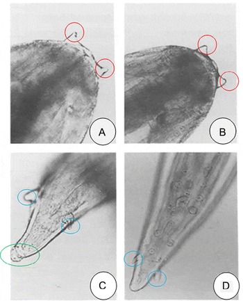
Setaria digitata is generally described as an ivory coloured, slender worm, several centimetres long, with a spirally coiled, tapering tail. The cuticle is smooth and layered with fine transverse striations. At the anterior end, the peri-buccal crown bears notched dorsal and ventral projections with round lateral lips surrounding the mouth opening. Positioning of the vulval opening anteriorly (0.5–0.6 mm from the mouth in S. digitata) is characteristic of the Setaria genus. It has a 6–7 mm long oesophagus, divided into an anterior part (0.5–0.6 mm) and a longer posterior part (5.5–6.5 mm). It possesses well-developed lateral caudal appendages at the upper terminal end. Males are distinguished from females by differences in length (males: 35–45 mm, females: 65–75 mm), breadth (males: 0.2–0.3 mm, females: 0.5–0.7 mm) and appearance of tail (females with smooth and spherical terminal knob), among others (fig. 1) (Liang-Sheng, Reference Liang-Sheng1959; Rhee et al., Reference Rhee, Choi, Park and Jang1994; Kim et al, Reference Kim, Kim, Sim, Ji and Park2010).
Light microscopic examination of S. digitata: lateral view of the anterior end of female (A) and male (B) worms, showing the dorsal and ventral projections (circled in red) at the peri-buccal crown. Lateral appendages (circled in blue) are clearly visible at the posterior end of female (C) and male (D) worms, with females having a smooth, spherical terminal knob (circled in green).

Detailed morphological and histological studies describe its well-developed digestive and reproductive systems. Light microscopy revealed the presence of numerous eggs in different developmental stages in the uteri (Sahu et al., Reference Sahu, Mohapatra, Majumder, Das and Ravindran2005; Kim et al., Reference Kim, Kim, Sim, Ji and Park2010). Unique embryonic interconnections were observed in the reproductive system, which were thought to play a role in nutrient circulation and thus have a possible evolutionary significance (Decruse & Raj, Reference Decruse and Raj1990). Furthermore, electron microscopic analysis of the surface structure of S. digitata showed striations, annulations and bosses, depicting a surface similar to that of W. bancrofti (Madathiparambil et al., Reference Madathiparambil, Kavitha Raj, Linda and Kaleysa Raj2011).
Immunology
Unravelling the mechanism of prolonged survival of parasite in host requires intense studies pertaining to parasite immunology. Moreover, immunological studies enable the discovery of suitable markers for the detection of infection. Filarial infections give rise to various clinical manifestations in patients; symptomatic and microfilariaemic (also known as chronic filariasis (CF)), symptomatic but amicrofilariaemic (SAM), asymptomatic but microfilariaemic (ASM), and asymptomatic and amicrofilariaemic (also known as endemic controls (EC)) (Babu & Nutman, Reference Babu and Nutman2012). Immunological studies in filariasis have been difficult due to varied immune responses to the different clinical manifestations mentioned above. However, accurate characterization of these stage-specific immune responses would no doubt provide valuable insights for eradication measures. Limited correlation between the serum antibody levels and the degree of infection in bancroftian filariasis, and the excessively broad cross-reactivity of filarial antigens with other helminth antigens, are major drawbacks in detecting antibodies as a measure of filarial infection (Ambroise-thomas, Reference Ambroise-thomas1974). Therefore the characterization of S. digitata antigens; their stage-specific origin, immunogenicity and correlation to disease status may help to address the drawbacks in the detection of human filarial antigens for serodiagnostics and immunization.
The surface molecules of S. digitata, along with the excretory–secretory (ES) material they release to the host environment, are highly immunogenic. The ES material of S. digitata, particularly that released during the hatching of microfilariae, is found to be the most immunogenic of the various worm fractions (Decruse & Raj, Reference Decruse and Raj1988). The surface coat of S. digitata is suggested to be comprised of loosely held ES material of the parasite together with molecules of the host, which may be responsible for immune evasion and parasite survival in the host (Madathiparambil et al., Reference Madathiparambil, Kavitha Raj, Linda and Kaleysa Raj2011). Although an antifilarial immune response is generated for the shed antigens, parasites evade this immune response by ‘surface epitope hiding’; grooves and striations on the adult parasite surface may be ideal for antigenic concealment (Bright & Raj, Reference Bright and Raj1994). ES material demonstrated immunosuppression, evident by depleted T-cell levels, and this effect was attributed to lipids and not proteins of ES material (Raj et al., Reference Raj, Pradeep, Devi, John, Rekha and OOmmen1997). Generally, filarial infections mount a T-helper 2 (Th2)-type immune response in mammalian hosts, which constitutes increased production of interleukin (IL)-4, IL-10 and IL-13 cytokines, elevated IgG1, IgG4 and IgE antibody isotypes and other innate-immune cells (Babu & Nutman, Reference Babu and Nutman2012). Although IgE antibodies mediate host immunity against parasites via antibody-dependent cell-mediated cytotoxicity (ADCC), a Th2 response is generally detrimental to the effective removal of these parasites, thus necessitating a Th1-type immune response for complete parasite clearance. However, an inhibition of the Th1 response by the secreted filarial lipid (SFL) fraction of S. digitata has been reported, shown by decreased T-cell proliferation, and reduced IL-12 and interferon-γ (IFNγ) production (Muthian et al., Reference Muthian, Pradeep, Sargapradeep, Kaleysaraj and Bright2006). Therefore, this finding highlights yet another mechanism by which S. digitata evades host immune responses.
Practical difficulties in detecting adult W. bancrofti antigens have diverted studies towards the characterization of S. digitata antigens that interact with specific serum antibodies against W. bancrofti. An approximately 30 kDa glycoprotein fraction of adult S. digitata was found to cross-react with infective larval (L3) antigens of W. bancrofti (Beuria & Das, Reference Beuria and Das1992). Specific antibodies against this S. digitata antigen were found to be present in the sera of W. bancrofti-infected patients, with a significantly enhanced IgG seropositivity in CF patients compared to ASM individuals. This demonstrates that the IgG response may be dependent on the disease severity, thus implying its potential use as a marker for chronic filariasis. EC and other infected groups had equally high levels of S. digitata antigen-specific IgE levels. As a result of the short half-life of IgE in serum, this study suggests that an increased IgE antibody level may indicate early exposure to infection.
Several S. digitata antigens were found to be similar to microfilarial surface antigens of W. bancrofti based on indirect immunofluorescence antibody (IFA) analysis (Dissanayake & Ismail, Reference Dissanayake and Ismail1980). However, the antigen species of S. digitata (SD2-4) which showed the highest reactivity against serum antibodies of W. bancrofti-infected individuals was not found to represent microfilarial surface antigens of W. bancrofti, but was found to correspond to circulating immune-complexes containing antigens originating from its adult form (Dissanayake et al., Reference Dissanayake, Galahitiyawa and Ismail1982). Detection of antigens present on the W. bancrofti adult form provides a hassle-free method of filarial diagnosis as it does not involve the detection of nocturnally periodic microfilariae. Moreover, it enables the accurate detection of amicrofilariaemic patients. Thus this study suggests the use of S. digitata antigen SD2-4 in serodiagnosis of bancroftian filariasis. Comparing the levels of IgG in filarial sera, another study corroborated the utilization of SD2-4 for serodiagnostics by demonstrating that although the sera of CF patients possessed IgG antibodies against microfilarial antigens of W. bancrofti, they contained an even higher level of IgG that was found to react with adult S. digitata antigen SD2-4 (Dissanayake & Ismail, Reference Dissanayake and Ismail1981). Moreover, equal antibody levels against adult and microfilarial antigens were seen in SAM patients. Therefore, this study highlighted that the detection of serum antibodies in filariasis patients using adult S. digitata antigens may serve as a useful indicator of infection in both microfilariaemic and amicrofilariaemic patients, while the detection of antibodies against microfilariae may be relevant in patients with clinically evident disease.
A study aimed at developing an assay based on dot-ELISA (enzyme-linked immunosorbent assay) for the diagnosis of ASM individuals, using antigens of S. digitata, identified two potential antigens from adult S. digitata, with molecular weights 52 kDa and 130 kDa, suitable for this purpose (Wickremanayake et al., Reference Wickremanayake, Ekanayake and Karunanayake2001). These antigens were detected in sera of ASM individuals, but were absent in W. bancrofti microfilariae, suggesting their presence in the adult worm. Development of a diagnostic test using these S. digitata antigens is relatively cost effective. However, of the patients classified as positive for microfilariae by night blood examinations, only 52% and 82% were positively detected by the dot-ELISA test developed with 52 kDa and 130 kDa antigens, respectively.
In another study, five protein bands were observed following separation of a detergent-soluble surface antigen fraction of S. digitata by polyacrylamide gel electrophoresis (PAGE). These bands were analysed for their immunogenicity and diagnostic use (John et al., Reference John, Bright and Raj1995). The electro-eluted major protein of molecular weight 29 kDa showed the highest reactivity with the serum antibodies from W. bancrofti-infected individuals. The sera of patients belonging to both microfilariae-positive but asymptomatic and symptomatic groups were 100% positively detected for specific antibodies using this 29-kDa protein antigen, indicating its suitable diagnostic use. However, this same protein was also found to show maximal immunosuppression, with a 70% decrease in total lymphocyte levels, leading to a quite contradictory finding. This suppressive effect may be beneficial for the prolonged survival of this parasite in its host. It was hypothesized that this 29-kDa protein may exist in two forms – one activating and the other suppressing the immune system – which requires confirmation by further studies. The fact that immunosuppression was displayed by the heat-inactivated 29-kDa protein fraction implies that this effect may not be due to protein antigens alone, but that lipids may also be associated.
Several studies have been conducted on the analysis of filarial parasite-specific antibody isotypes. Of the IgG antibodies, the sera of CF, ASM and EC patients were found to contain high titres of IgG4 isotype (Dalai et al., Reference Dalai, Das and Kar1998). Sera of ASM individuals, but not EC patients, possessed IgG4 against 14–20 kDa antigens of adult S. digitata worms. Furthermore, these antigens were found to produce a predominant Th2 cytokine response in ASM individuals, a Th1 cytokine response in EC individuals, and CF patients demonstrated a mixed response. Microfilariaemic patients were found to have low levels of parasite-specific IgA antibodies, which also correlates with their decreased Th1 response (Sahu et al., Reference Sahu, Mohanty, Sahoo, Satapathy and Ravindran2008). However, compared to CF individuals, sera of EC patients showed increased levels of IgA antibodies, demonstrating that high IgA levels correlate with the absence of infection, and hence the protective role of parasite-specific IgAs. Sex-related differences in IgA levels were also observed; men had relatively lower parasite-specific IgA than women. Setaria digitata antigens have also been employed to detect antibodies against parasite carbohydrates in sera of patients with bancroftian filariasis (Mohanty et al., Reference Mohanty, Satapathy, Sahoo and Ravindran2001). Several other glycoprotein surface antigens of S. digitata were also characterized and enabled stage-specific detection of infection via immunoblotting (Madathiparambil et al., Reference Madathiparambil, Kaleysa and Raghavan2009; Theodore & Kaliraj, Reference Theodore and Kaliraj2009).
The aforementioned immunological studies of filariasis have been focused on immune responses against microfilariae, infective larvae and the adult form of parasites. A recent study employed a flow-cytometry-based approach to assess the progression of embryogenesis of S. digitata and to study the intra-uterine stage-specific immune responses, i.e. by quantifying the binding of antibodies to these different stages (Sahu et al., Reference Sahu, Mohapatra, Majumder, Das and Ravindran2005). The interruption of the reproductive process, in particular, could disrupt disease transmission and lessen morbidity, and therefore further studies on these embryonic stages may lead to previously unidentified key processes that can be targeted for eradication.
Metabolism
Currently, due to the lack of effective vaccines and vector-controlling strategies, filariasis eradication is almost entirely chemotherapy based. However, the development of parasite resistance to antifilarials is a main challenge, and this requires a better understanding of the exact mode of action of currently available drugs, and the discovery of novel targets (Gupta & Srivastava, Reference Gupta and Srivastava2005). Thus extensive studies have been conducted on the biochemical pathways occurring in these nematodes, highlighting their differences from host metabolism.
Unique and diverse metabolic pathways have evolved in parasitic nematodes to enable survival in various host environments. The activities of succinate dehydrogenase (SDH) and fumarate reductase (FR) exhibit a ratio of 1.75, a value characteristic of facultative anaerobes (Banu et al., Reference Banu, Dhandayuthapani and Nellaiappan1991). Oxygen uptake assays and scoring of the wriggling rate of adult worms have been used to demonstrate that aerobic respiration is insensitive to the respiratory poison cyanide (Raj et al., Reference Raj, Puranam, Kurup and Ramasarma1988). This was evident by unchanged rates of oxygen uptake in the presence of cyanide, and by the absence of spectrophotometrically detected cytochrome oxidase – the enzyme inhibited by cyanide. Oxygen consumption is proposed to occur via two pathways. One such pathway is substrate-dependent, involving a readily auto-oxidizable quinone, Q6, which mediates electron transfer from flavoprotein dehydrogenases to oxygen in the electron transport chain (Santhamma & Raj, Reference Santhamma and Raj1990). The presence of a quinone that links electron transfer from both aerobic and anaerobic pathways was found to be unique to S. digitata (Sivan & Raj, Reference Sivan and Raj1992). The reduction of oxygen would produce H2O2 which is detoxified by catalase. An adult-specific quinone, Q8, having no involvement in the electron transport chain, was also thought to function as an antioxidant (Abhilashkumar et al., Reference Abhilashkumar, Mohan, Jayakumar and Raj2001). The second pathway is substrate-independent and occurs through lipid peroxidation.
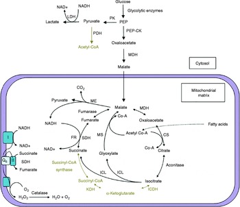
The activities of several enzymes were assayed to determine the metabolism in adult S. digitata worms. An early study characterized the enzyme β-d-galactosidase, which was abundant in the digestive tract of S. digitata, suggesting its role in hydrolysing galactosidase bonds of substrates obtained from the peritoneal fluid of the host (Banu et al., Reference Banu, Kalyani and Nellaiappan1990). The activity ratio of pyruvate kinase (PK) to phosphoenolpyruvate carboxykinase (PEP-CK) was intermediate, indicating that S. digitata utilizes both carbon dioxide fixation and lactate fermentation for energy production (Banu et al., Reference Banu, Dhandayuthapani and Nellaiappan1991). The absence of several tricarboxylic acid (TCA) cycle enzymes, such as isocitrate dehydrogenase (ICD), α-ketoglutarate dehydrogenase (KDH) and succinyl-CoA synthase, in mitochondrial isolates suggests that carbon dioxide fixation is via the glyoxylate cycle (Rafi & Raj, Reference Rafi and Raj1991). As pyruvate dehydrogenase (PDH) was not detected, the acetyl-CoA that enters the glyoxylate cycle should originate from lipid and amino acid catabolism. Together, these findings proposed the presence of a PEP–succinate–glyoxylate pathway in S. digitata (fig. 2).
Carbohydrate metabolism and energy generation in Setaria digitata, highlighting the incomplete Kreb's cycle, a proposed PEP–succinate–glyoxylate pathway, lactic acid fermentation and malate dismutation pathways, and also showing the classical pathways that are not detected in the parasite (represented in grey). PK, Pyruvate kinase; LDH, lactate dehydrogenase; PDH, pyruvate dehydrogenase; PEP-CK, phosphoenolpyruvate carboxykinase; MDH, malate dehydrogenase; CS, citrate synthase; ICDH, isocitrate dehydrogenase; KDH, α-ketoglutarate dehydrogenase; SDH, succinate dehydrogenase; ICL, isocitrate lyase; MS, malate synthase; ME, malic enzyme; FR, fumarate reductase.

Under anaerobic conditions, the pyruvate produced by glycolysis in S. digitata is converted to lactate via lactate dehydrogenase (LDH), which has been characterized (Banu et al., Reference Banu, Nellaiappan and Dhandayuthapani1989). LDH plays an important role in maintaining the cytoplasmic NAD/NADH redox potential in this nematode, and it is thus linked to energy production in S. digitata (Sivan & Raj, Reference Sivan and Raj1994). In addition to this anaerobic fermentation, many parasitic nematodes, like S. digitata, utilize the malate dismutation pathway: the malate (produced by the reduction of oxaloacetate, which is generated by glycolytically produced PEP) is transported to mitochondria, where a portion of it is oxidized to pyruvate while the rest is reduced to succinate (reversal of steps of the TCA cycle). The reducing equivalents of NADH produced by malic enzyme (ME) drives the reduction of malate to succinate. Detailed studies on the fumarate reductase (FR) system of S. digitata displayed its key role in malate dismutation and energy generation, with the involvement of transhydrogenases linking the FR/SDH, ME and NADH–coenzyme Q reductase systems (Unnikrishnan & Raj, Reference Unnikrishnan and Raj1992, Reference Unnikrishnan and Raj1995). Several of these enzymes have been characterized as possible drug targets (Banu et al., Reference Banu, Nellaiappan and Dhandayuthapani1992).
Although S. digitata is devoid of typical cytochrome systems, several other haem proteins, such as catalases and peroxidases, were found to be abundant in S. digitata along with appreciable amounts of enzymes required for haem biosynthesis (Kumar & Raj, Reference Kumar and Raj1998). A haemoglobin-like molecule was also detected in S. digitata as a possible target for chemotherapy, but was not fully characterized. The isoprenoid biosynthesis pathway has also been studied in S. digitata and has been demonstrated to be valuable for drug targeting, mainly due to the differences in parasite (Q6 and Q8) and host (Q10) ubiquinone systems (Kumari et al., Reference Kumari, Santhamma and Raj1994).
Genomics, characterization of proteins and their genes
Cytological studies of S. digitata using the acetocarmine squash technique revealed a diploid chromosome number of 12 in females and 11 in males, which is suggestive of a XX–XO mechanism of sex determination (Decruse & Raj, Reference Decruse and Raj1992). Several genes of S. digitata, having the potential to serve as effective targets for eradication, have been sequenced and characterized (Jayasena et al., Reference Jayasena, Chandrasekharan and Karunanayake1999; Murugananthan et al., Reference Murugananthan, Karunanayake and Tennekoon2010; Rodrigo et al., Reference Rodrigo, Dassanayake, Weerasena and Silva Gunawardene2014a). Sequence data unveil a genome that is relatively AT-rich; a characteristic feature of nematode genomes. The relative gene structure of the characterized genes of S. digitata, in terms of exons, introns, splice-sites (with universal GT–AG splicing) and regulatory elements, appears to be fairly conserved with their corresponding counterparts in other filarial nematodes (Jayasena et al., Reference Jayasena, Chandrasekharan and Karunanayake1999; Murugananthan et al., Reference Murugananthan, Karunanayake and Tennekoon2010; Rodrigo et al., Reference Rodrigo, Dassanayake, Weerasena and Silva Gunawardene2014a). However, more genes need to be sequenced before consensus sequences can be built up and for clear patterns to be identified.
Effective eradication of filariasis necessitates accurate detection of nematode-defining genetic elements, and genomic studies enable the identification of these elements to be used in such detection. Of these, repetitive DNA sequences have been useful in the development of DNA probes. Two repetitive sequences from the genome of S. digitata have been identified and used as probes for the accurate detection of S. digitata in cerebrospinal nematodiasis, proving to be much more efficient than the common microscopic detection of this parasite (Wijesundera et al., Reference Wijesundera, Chandrasekharan, Karunanayake and Dharmasena1996). When used as a probe, the relative abundance of these tandemly repeated sequences ensures the minimal detection of ~100 pg genomic DNA, corresponding to a single L3 larva in mosquitoes, and a single microfilaria in 50 μl of blood, thus providing a sensitive means of detection of different stages in both host and vector.
Studies on the mitochondrial genome of S. digitata have revealed important aspects of their biology, and have also provided ample gene markers for the study of population genetics and systematics of these socio-economically important parasites. The structure and organization of the mitochondrial genome of S. digitata was analysed and compared with those of other nematodes (Yatawara et al., Reference Yatawara, Wickramasinghe, Rajapakse and Agatsuma2010). The complete mitochondrial genome (13,839 bp) contains 36 genes, which include 12 protein-coding genes (cox1–cox3, nad1–nad6, nad4L, atp6 and cytb), 22 tRNA genes and genes for small and large subunits of rRNA (rrnS and rrnL). Although the gene content is similar, there are variations in the gene arrangement compared with other nematodes studied thus far. As with other nematode genomes, the mitochondrial genome of S. digitata is relatively AT-rich (75.1% AT); it consists of a tandemly repeated AT-rich region, multiple poly(T) tracts, and its codons are T-biased with TTT used frequently for translation initiation. A recent phylogenetic analysis of filarial worms based on cytochrome c oxidase subunit I (cox1), 12S rDNA and combined cox1+12S rDNA nucleotide sequences showed a fair level of proximity between W. bancrofti and S. digitata, to warrant studies of the former using the latter as a model organism (Eamsobhana et al., Reference Eamsobhana, Lim and Yong2013).
In an attempt to identify novel targets for anti-filarial therapeutics, studies have been conducted on Wolbachia – the bacterial endosymbiont residing in Wolbachia-dependent parasites whose absence results in growth, development and fertility defects followed by ultimate death of its host. Our recent study using polymerase chain reaction (PCR)-based multilocus sequence typing, whole-mount immunofluorescence staining and transmission electron microscopy confirmed the absence of Wolbachia in S. digitata (Voronin et al., Reference Voronin, Abeykoon, Gunawardene and Dassanayake2015). Previous reports have highlighted the impact of Wolbachia on energy requirements of its host, W. bancrofti, and its strong influence on mitochondrial evolution due to the vertical co-transmission of mitochondria and Wolbachia (Strubing et al., Reference Strubing, Lucius, Hoerauf and Pfarr2010). Based on this, the mitochondrial genomes of filarial parasite S. digitata, which is Wolbachia-independent, and of Wolbachia-dependent W. bancrofti were analysed and compared (McNulty et al., Reference McNulty, Mullin, Vaughan, Tkach, Weil and Fischer2012). However, the content, organization and sequence of mitochondrial DNA were found to be similar among both Wolbachia-dependent and -independent nematodes, thus requiring further studies to elucidate the basis of their functional differences.
A number of genes of S. digitata and their products have been characterized as potential targets for antifilarial drug development. The lack of cytochrome-P450 enzymes in this nematode highlights the importance of glutathione S-transferase (GST), which primarily undertakes this detoxification function. Prolonged survival of parasites is mainly attributed to the protective function of their GSTs against oxygen radicals generated by the host immune system. This rightly led to the biochemical characterization of the GST of S. digitata (Srinivasan et al., Reference Srinivasan, Mathew, Karunan and Muthuswamy2011). Studies on subcellular localization of GST revealed a high content in the cytosolic fraction, with lesser activities detected in mitochondrial and microsomal fractions. The 54 kDa native protein was determined to be a homodimer of 27 kDa subunits. The kinetic parameters K m and V max were determined to be 0.45 mm and 0.105 μmol min− 1ml− 1, respectively, against the substrate glutathione (GSH). These preliminary studies provide a strong platform for prospective antifilarial drug screening against GST. An earlier enzymatic study showed the stage-specific expression of GST, where high GST activity was observed in adult S. digitata but with apparently no activity in the other stages, highlighting its importance in the immunodiagnosis of filariasis (Bal & Das, Reference Bal and Das1996).
Heat-shock proteins (HSPs) are known to play an indispensable role in the migration and survival of parasites in hosts. Transmission between different host and vector environmental conditions produces a classical heat-shock response in filarial parasites to which HSPs provide protection. This crucial function of HSPs along with their strong antigenic potential led to studies on the characterization and regulation of the HSP-70 gene in S. digitata (Jayasena et al., Reference Jayasena, Chandrasekharan and Karunanayake1999). Several HSP-70-related genes were identified from the S. digitata genome via Southern hybridization. The HSP-70-2 gene encodes a protein of 645 amino acids and was also found to be expressed constitutively (i.e. not heat-shock induced). Nucleotide and amino acid sequence analysis showed HSP-70-2 of S. digitata to have strong homology to its B. malayi counterpart. Its upstream sequence is rich in cis-acting regulatory elements, including functional heat-shock elements (HSEs).
Immunoscreening of the cDNA library of S. digitata with W. bancrofti-infected sera led to the discovery of a gene encoding an alkali-myosin protein (MLC-3) which was then characterized (Murugananthan et al., Reference Murugananthan, Karunanayake and Tennekoon2010). Filarial myosin proteins have been previously identified to induce strong immune responses (Verma et al., Reference Verma, Bansal, Vedi, Saxena, Katoch and Bhattacharya2008). The encoded MLC-3 protein is suggested to consist of 149 amino acids, having a molecular weight of 17 kDa. Further analysis in silico suggested the presence of cytoplasmic localization signals in the protein, and that it also possesses conserved domains characteristic of calcium-binding proteins. More than 80% sequence similarity was demonstrated in the amino acid sequence of this protein in S. digitata and Caenorhabditis briggsae, and a much lesser sequence similarity in other vertebrates and invertebrates. This low level of sequence conservation among taxonomic groups, and its presence in the parasite cuticle, provide means of selective therapeutic targeting of this protein. However, its antigenic potential has not been studied thus far.
Nematodes possess multiple unique parasite genes that enable their invasion and survival in the host. The prime focus of recent research has aptly been on the characterization of nematode-specific genes to provide an understanding of the mechanism of parasitism and then to use these genes as targets for eradication of the parasites. SXP-1, a protein belonging to the SXP/RAL2 family of nematode-specific proteins, was also characterized in S. digitata as a promising therapeutic target (Sasisekhar et al., Reference Sasisekhar, Suba, Sindhuja, Sofi and Narayanan2005). Phylogenetic analysis displayed a close resemblance of SXP-1 of S. digitata (SdSXP-1) to that of W. bancrofti (WbSXP-1). Two SdSXP-1 protein bands of 34 and 66 kDa were identified following immunoblot analysis of the crude extract of S. digitata probed with rabbit anti-WbSXP-1. The SdSXP-1 was found to be mainly contained in the sheath protein fraction of the worm. Immunofluorescence analysis of frozen cryosections of the worm localized the protein in the hypodermal region of the parasite. The proposed role of SXP-1 as an active developmental protein may be ideal for targeting; however, further characterization of the structural and functional aspects of this protein are required for this purpose.
Expressed Sequence Tag (EST) analysis of S. digitata led to the discovery of a previously unidentified parasitic nematode-specific protein in S. digitata (SdNP), and its open reading frame (ORF) and genomic regions were sequenced and characterized bioinformatically (Dassanayake et al., Reference Dassanayake, Rodrigo, Karunanayake, Weerasena and Chandrasekharan2009). It was found to encode a protein of 204 amino acids, with a molecular weight of 22.8 kDa. SdNP was found to have homologous counterparts in human filarial parasitic nematodes, including W. bancrofti, and was later determined to share 79% sequence similarity with them. The SdNP gene is apparently expressed ubiquitously in all stages of the life cycle of S. digitata: similar EST sequences were observed in all stages of the nematode life cycle; reverse transcription (RT)-PCR analysis of RNA from adult male and female worms, and microfilariae, showed transcription of the SdNP gene; and Western blot analysis with anti-SdNP revealed the expression of the protein in all life stages examined (Rodrigo et al., Reference Rodrigo, Dassanayake and Voronin2014c). Southern hybridization revealed SdNP to be a single-copy gene. Analysis of the genomic region of SdNP was found to display a conserved gene structure with that of W. bancrofti and other human filarial nematodes. The intron regions of the gene encoding SdNP are rich in di- and trinucleotide microsatellites, which may be useful markers to study population genetics of S. digitata. Secondary structural analysis of the protein predicted abundant β-sheeted regions, but the absence of transmembrane β-barrel regions suggests that it is globular (Dassanayake et al., Reference Dassanayake, Rodrigo, Karunanayake, Weerasena and Chandrasekharan2009). Cellular functional analysis suggested that the protein is non-enzymatic and growth-factor-like. This growth-factor-like protein of S. digitata was successfully expressed in its soluble form in a pET expression system in Escherichia coli, along with chaperone co-expression, and later in a more successful Pichia pastoris expression system (Rodrigo et al., Reference Rodrigo, Dassanayake, Karunanayake, Silva Gunawardene and Weerasena2014b). The presence of nuclear localization signals in the C-terminus suggested that SdNP is localized mainly in the nucleus, which was later confirmed by fluorescent microscopic analysis of SdNP–GFP fusion proteins in yeast cells and Western Blot analysis of their cellular fractions (Rodrigo et al., Reference Rodrigo, Dassanayake and Voronin2014c). Further, the possibility that this protein translocates between the nucleus and cytoplasm is suggested, through a reversible phosphorylation–dephosphorylation reaction in the presence of the protein nuclear transport factor 2-like domain and potential sites for kinases. Immunohistochemical staining demonstrated that SdNP is highly localized in longitudinal muscle layers and the endodermis, thus revealing a possible locomotive function of the protein in the adult worm. It is also thought to play a vital role in reproduction, embryogenesis and development, as such staining showed this protein to be significantly localized in the male and female reproductive tissues and developing embryos.
In a recent preliminary study, yet another putative protein of S. digitata was discovered and characterized bioinformatically by the analysis of amino acid sequences. Similarity searches detected 100% amino acid sequence identity with RNA-binding protein genes of B. malayi and W. bancrofti, indicating its importance as a drug target (Nagaratnam et al., Reference Nagaratnam, Karunanayake, Tennekoon, Samarakoon and Mayan2014). RNA-binding protein of S. digitata (SdRBP) showed a 54% amino acid sequence identity with the RNA recognition motif (RRM) of the human cleavage stimulation factor 64 kDa subunit (CstF-64). Moreover, homology modelling using the RRM of CstF-64 as template revealed a RRM topology in SdRBP. Thus, SdRBP is assumed to have a role in pre-mRNA polyadenylation, similar to that of CstF-64.
Conclusion and future directions
Filarial infections still remain a major cause of morbidity and mortality in humans and livestock, despite the eradication efforts that are currently underway. The limited knowledge of parasite biology and host–parasite relationships hinders major breakthroughs in diagnostic and therapeutic discoveries for their eradication. The biology of S. digitata, used as an experimental model for filariasis, has been studied in the past, although there still remain areas that are unexplored. New research strategies are required for filariasis, with the intentions of improving detection and assessing disease status, and discovering more efficacious targets for therapeutic intervention. Since most treatment measures are chemotherapy-based, research into vaccines that would prevent filarial infection, or at least prevent transmission, needs greater focus. For the implementation of effective vector-controlling strategies, detailed studies on the lifecycle progression of filarial nematodes, specifically with respect to the development stages in mosquito vectors, would aid in the identification of key processes for intervention. Development of diagnostics and vaccination strategies would be accelerated by genomic research on S. digitata, giving focus to the discovery of nematode-specific genes using the EST approach. Recombinant antigen-based detection methods can be used for improved sensitivity and specificity of diagnosis. Several genes, including their protein products, have been characterized based on their potential to be targeted for therapeutics. Immunogenic proteins are characterized in order to provide insight into regulation of host immune responses by S. digitata and to unravel the mechanism of their prolonged survival in hosts. Biochemically, only a limited number of pathways have been elucidated in S. digitata. Therefore, for S. digitata to be used as a model organism for the control of human filariasis, particularly bancroftian filariasis, there is a pressing requirement for further studies on S. digitata, focusing on well-identified filarial targets, such as proteins involved in isoprenoid metabolism and collagen biosynthesis in addition to DNA topoisomerases and retinol-binding proteins, to name a few. Furthermore the aforementioned studies should be analysed en masse for the effective development of novel research strategies targeting multiple aspects of the biology of S. digitata.
Financial support
This work was supported by a grant (AP/3/2011/CG/03) awarded to Professor R.S. Dassanayake by the University of Colombo, Sri Lanka, to promote collaborative research.
Conflict of interest
None.