Alveolar capillary dysplasia with misalignment of the pulmonary veins is a rare autosomal dominant condition typically presenting within the first 24–48 hours of life with symptoms of respiratory failure.Reference Bishop, Stankiewicz and Steinhorn1 The alveolar-capillary underdevelopment with disproportionate relationship and malposition between the pulmonary vasculature and dilated alveoli results in the development of persistent refractory pulmonary hypertension.Reference Bishop, Stankiewicz and Steinhorn1 Most of the reported cases have been fatal within the neonatal period, with few atypical late presentations that survived beyond this period.Reference Towe, White and Grady2 It is often associated with extrapulmonary anomalies that involve the cardiac, gastrointestinal, genitourinary, and/or musculoskeletal systems.Reference Alturkustani, Li and Byers3 The association of cardiovascular anomalies has been found in approximately 10%–13% of the affected infants of which left-sided obstructive CHDs have been the most common presentation.Reference Stark, Schneider and Biermann4,Reference Laux, Malan and Bajolle9 We report two cases with different genetic variants in the FOXF1 gene associated with left-sided CHDs that presented with persistent supra-systemic pulmonary hypertension despite surgical correction.
Case 1
A full-term female born without prenatal concerns developed signs of respiratory distress and persistent hypoxia in the immediate postnatal period. An echocardiogram was performed and revealed severe discrete aortic coarctation with mildly hypoplastic transverse arch, an apex-forming hypoplastic left ventricle, hypoplastic mitral valve, and bicuspid aortic valve. Due to worsening in clinical status, the patient underwent emergent aortic arch reconstruction on day 3 of life but was unable to be weaned off cardiopulmonary bypass due to evidence of supra-systemic pulmonary arterial pressures requiring transition to veno-arterial extracorporeal membrane oxygenation. Due to unusual clinical presentation, the decision was made to obtain a lung biopsy that showed dysplastic capillaries, dilated bronchial veins within the broncho-arterial bundles (formerly known as misaligned pulmonary veins), and lymphatic dilation. Additional atypical histopathologic features included eosinophilic disease (eosinophilic pneumonia and eosinophilic infiltrates), focal but extensive extramedullary haematopoiesis, and glycogen-rich immature alveolar lining cells. Whole-genome sequencing was ordered reporting a point mutation of c.179C > T (p. Ala60Val) variant in the FOXF1 gene located on chromosome 16q24.1; further analysis of the parental samples was negative indicating a de novo mutation of the patient. These findings confirmed the diagnosis of alveolar capillary dysplasia with misalignment of the pulmonary veins. Although she was decannulated from extracorporeal membrane oxygenation on day 18 of life, and despite aggressive pulmonary vasodilator therapy, she had an acute hypoxic episode three days later with severe respiratory acidosis leading to cardiopulmonary arrest and death despite aggressive resuscitation measures.
Case 2
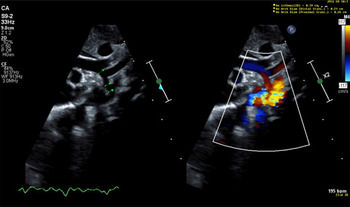
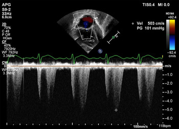
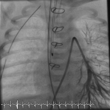
A full-term male born with prenatal concern for coarctation of the aorta and borderline hypoplastic left ventricle showed no signs of respiratory distress in the immediate postnatal period. Echocardiogram confirmed coarctation of the aorta, mild to moderate transverse arch hypoplasia, small peri-membranous VSD, and a mildly hypoplastic aortic and mitral annulus with dysplastic valves (Fig 1). He remained stable and underwent diverting sigmoid colostomy on day three of life due to associated imperforate anus followed by aortic arch reconstruction on day seven. Post-operative course was complicated by severe persistent pulmonary hypertension with subsequent echocardiograms demonstrating right ventricular dilation with supra-systemic right ventricular pressures (Fig 2). Whole-genome sequencing revealed a deletion of approximately 25.187 Kb, located at 16q24.1 encompassing the FOXF1 gene; analysis of the parental samples showed that the mother was heterozygous for this deletion. He was started on combined vasodilator therapy with inhaled nitric oxide and epoprostenol; a right cardiac catheterisation was performed for haemodynamic assessment showing severe pre-capillary pulmonary arterial hypertension, about 150% systemic, main pulmonary artery 100/42/62 mmHg, systemic 67/31/44 mmHg, with normal capillary wedge pressures, mean 12 mmHg, PVR 8 WU/m2, and no evidence of coarctation of the aorta or pulmonary vein stenosis (Fig 3). Therefore, the decision was to proceed with a lung biopsy that reported dilated bronchial veins and lymphatic vessels, underdeveloped acini, and dysplastic capillaries, confirming the diagnosis of alveolar capillary dysplasia. The patient remained on medical cardiorespiratory support without any improvement and family elected for withdrawal of care on day 21 of life.
Two-dimensional transthoracic echocardiogram suprasternal notch view (2-D on the left; color flow Doppler on the right) done prior surgical intervention. Demonstrating moderate transverse arch hypoplasia, aortic coarctation with distal displacement of the left subclavian artery, a patent ductus arteriosus with flow acceleration and retrograde flow into aortic arch.

Two-dimensional transthoracic echocardiogram apical view (continuous wave spectral Doppler) on post-operative day 4 after aortic arch reconstruction. The tricuspid regurgitant jet predicted a systolic gradient from the right ventricle to the right atrium of 101 mm of Hg + mean right atrial pressure with a systemic blood pressure at the same time of 75/50 mm of Hg; findings suggestive of supra-systemic pulmonary artery pressures.

Right cardiac catheterisation performed for haemodynamic assessment coupled with planned lung biopsy. Antero-posterior angiogram of distal left lower pulmonary artery, demonstrating patent first and second order branches with distal pruning (loss of the smaller vessels).

Discussion
The presentation of alveolar capillary dysplasia with misalignment of the pulmonary veins represents a diagnostic challenge, especially if associated with extrapulmonary malformations. Left heart obstruction may be the aetiology of persistent pulmonary hypertension, as well as abnormal lung development leading to acute episodes of respiratory failure with low cardiac output, making the diagnosis of alveolar capillary dysplasia even more difficult.Reference Stark, Schneider and Biermann4 Some expert consensus has recommended to consider alveolar capillary dysplasia as a possible cause of persistent pulmonary hypertension and to proceed with biopsy when there is no resolution in the first two weeks after appropriate management has been established.Reference Hilgendorff, Apitz and Bonnet8 Our approach to do further investigation took place within the first days after surgical correction of the CHDs for both of our patients because of the persistent supra-systemic pulmonary hypertension resistant to vasodilator therapy and haemodynamics that are not typically found in individuals with this type of corrected heart lesions.Reference Laux, Malan and Bajolle9 It is paramount for clinicians to have a high index of suspicion of alveolar capillary dysplasia in the setting of refractory or supra-systemic pulmonary hypertension despite adequate intervention of associated congenital defects.Reference Jourdan-Voyen, Touraine and Masutti5 An early recognition of this disease will provide guidance for therapeutic decisions and appropriate counseling for the affected families, identifying those that will have an increased risk of subsequent children that may inherit the condition.Reference Bourque, Fonseca and Staines6 Histopathological analysis remains the diagnostic gold standard, although there are atypical cases with differences in their histological features, as with case 1, in which the biopsy had atypical features that have not been previously reported in association with alveolar capillary dysplasia.Reference Alturkustani, Li and Byers3,Reference Jourdan-Voyen, Touraine and Masutti5 Hence, it is important to consider complementing with chromosomal microarray or whole-genome sequencing to identify individuals with FOXF1 gene mutations.Reference Jourdan-Voyen, Touraine and Masutti5 Szafranski et al reported in a large collection of samples 86 pathogenic variants in the FOXF1 locus that included 38 deletions, 1 complex rearrangement, and 47 point mutations.Reference Szafranski, Gambin and Dharmadhikari7 As for our patients, they had different genetic variants: Case 1 had a point mutation in c.179 C > T (p. Ala60Val), and case 2 had a 25.187 Kb deletion encompassing the FOXF1 gene. Further data collection from multiple centres would be beneficial to create a large registry for future references.Reference Jourdan-Voyen, Touraine and Masutti5 To conclude it is important to develop a strategic approach for the affected individuals as a multidisciplinary team and consider FOXF1 gene mutation testing in the setting of persistent supra-systemic pulmonary hypertension after surgical repair. In patients with left heart obstructive lesions and post-operative, unrelenting, supra-systemic pulmonary hypertension, we suggest obtaining FOXF1 gene testing prior to any further surgical intervention to guide families and the provider team, avoiding futile interventions.
Acknowledgements
The authors would like to thank Bennett P. Samuel MHA/BSN/RN our clinical research nurse for all the advice and useful suggestions.
Financial support
This research received no specific grant from any funding agency, commercial, or not-for-profit sectors.
Conflicts of interest
None.
Ethical standards
Not applicable.