INTRODUCTION
Today we are witness to remarkable progress in schistosome research. Supported by the improvement of automated sequencing technologies, comprehensive genome and transcriptome data have been generated (Oliveira et al. Reference Oliveira, Franco and Verjovski-Almeida2008; Berriman et al. Reference Berriman, Haas, LoVerde, Wilson, Dillon, Cerqueira, Mashiyama, Al-Lazikani, Andrade, Ashton, Aslett, Bartholomeu, Blandin, Caffrey, Coghlan, Coulson, Day, Delcher, DeMarco, Djikeng, Eyre, Gamble, Ghedin, Gu, Hertz-Fowler, Hirai, Hirai, Houston, Ivens, Johnston, Lacerda, Macedo, McVeigh, Ning, Oliveira, Overington, Parkhill, Pertea, Pierce, Protasio, Quail, Rajandream, Rogers, Sajid, Salzberg, Stanke, Tivey, White, Williams, Wortman, Wu, Zamanian, Zerlotini, Fraser-Liggett, Barrell and El-Sayed2009; The Schistosoma japonicum Genome Sequencing and Functional Analysis Consortium, 2009). This marks the beginning of the post-genomic era, which necessitates the interpretation and exploitation of the amassed data. In this context it is essential to have technologies available allowing functional genetics. Heterologous expression systems such as C. elegans, mammalian cell lines or frog oocytes have been used to characterize parasite genes of interest in the past to overcome limitations associated with some parasite systems (Brooks and Isaac, Reference Brooks and Isaac2002; Boyle and Yoshino, Reference Boyle and Yoshino2003; Britton and Murray, Reference Britton and Murray2006). Although new data on gene regulation and functional evidence were obtained, heterologous systems are only partially able to provide indications for the function of a gene because it is expressed in a different genomic environment. Conclusive interpretations about gene functions can only be made upon its analysis in a homologous genomic environment. Therefore, it is of vital importance to establish transformation protocols for each parasite of interest.
For all plathyhelminths, transformation protocols have been developed demonstrating the possibility of generating genetically modified flatworms (Aboobaker and Blaxter, Reference Aboobaker and Blaxter2004; Grevelding, Reference Grevelding, Maule and Marks2006; Spiliotis et al. Reference Spiliotis, Lechner, Tappe, Scheller, Krohne and Brehm2008). The methods used are largely based on existing techniques and can be applied to any organism of interest if in vitro culture systems exist allowing the maintenance and manipulation of parasite life stages ex vivo. Premium prerequisites are met if accessible life stages can be repatriated into the life cycle, which is the case for schistosomes. To this end, miracidia can be harvested from eggs in vitro, transduced with nucleic acids by physical methods, such as particle bombardment (Wippersteg et al. Reference Wippersteg, Kapp, Kunz and Grevelding2002a,Reference Wippersteg, Kapp, Kunz, Jackstadt, Zahner and Greveldingb, Reference Wippersteg, Ribeiro, Liedtke, Kusel and Grevelding2003), and used for snail infection afterwards (Beckmann et al. Reference Beckmann, Wippersteg, El-Bahay, Hirzmann, Oliveira and Grevelding2007). Furthermore, miracidia can be transformed in vitro and the emerging mother sporocysts transplanted into snails (Jourdane and Theron, Reference Jourdane and Theron1980; Jourdane et al. Reference Jourdane, Liang and Bruce1985; Cohen and Eveland, Reference Cohen and Eveland1988). Even daughter sporocysts can be generated in vitro from mother sporocysts (Bayne and Grevelding, Reference Bayne and Grevelding2003) and then transplanted into snails. Finally, schistosomula can be generated in vitro from cercariae and transplanted into final hosts such as mice (Nollen et al. Reference Nollen, Floyd, Kolzow and Deter1976; Basch and Humbert, Reference Basch and Humbert1981; Clough, Reference Clough1981). What approaches have been performed to generate transgenic schistosomes?
SCHISTOSOME TRANSFECTION
In principle, several biochemical or physical methods are available for transfection and transformation. The most common ones are lipofection, microinjection or electroporation which were successfully used for a variety of cells or organisms including protozoan parasites (Clayton, Reference Clayton1999; de Koning-Ward et al. Reference de Koning-Ward, Janse and Waters2000; Meissner et al. Reference Meissner, Agop-Nersesian and Sullivan2007). Consequently, these methods were also tested in different laboratories to evaluate their potential for transient transformation of schistosomes.
In our laboratory, experiments with lipofection were performed with in vitro cultured adults or sporocysts and different commercially available lipofection reagents. Using plasmid-based DNA constructs with GFP (green fluorescent protein) as the reporter gene, no GFP fluorescence could be detected following lipofection regardless of the reagent used (K. Kapp, unpublished). In parallel, using fluorescence-labeled reagents for lipofection, it was shown that these reagents stick to the surface of adult schistosomes after treatment and no indications of uptake into subtegumental areas were observed (J. Kusel, Glasgow, Reference Kusel, McVeigh and Thornhill1999, personal communication). However, Nabhan and colleagues (Reference Nabhan, El-Shehabi, Patocka and Ribeiro2007) successfully used a lipofection reagent to introduce small interfering RNAs (siRNAs) into freshly transformed schistosomula. This indicates that either the stage used for lipofection or the combination of stage and reagent may be pivotal. The siPORT lipid transfection reagent (Ambion) used in their study had not been used by us before. Due to the soft structure of the tegument microinjection was found to be difficult. Approaches with adults and sporocysts showed the possibility to inject dyes which diffuse through the body after treatment (V. Wippersteg, unpublished). However, this technique is time consuming with a low efficiency. Only a very low number of individuals could be manipulated within one day, and most of these did not survive in culture afterwards. Electroporation was first successfully used by Correnti and Pearce (Reference Correnti and Pearce2004) introducing luciferase mRNA into Schistosoma mansoni schistosomula. The electroporation targeted the majority of the schistosomula, and immunolocalization studies indicated that the RNA was delivered to tegumental and subtegumental tissues. However, the RNA was unstable: luciferase activity declined by 24 h post-electroporation, and it was not detectable by 72 h. Nevertheless, these results opened the way to deliver DNA constructs as well as double stranded RNAs (dsRNAs) or siRNAs for RNA-silencing approaches (RNAi) into adult and larval schistosomes by electroporation. Shortly thereafter, electroporation was also established for transformation of S. japonicum (Yuan et al. Reference Yuan, Shen, Wang, Wu, Liu, Dong and Jiang2005). In the following years, electroporation was used to introduce plasmid-based DNA constructs into schistosomula (Correnti et al. Reference Correnti, Jung, Freitas and Pearce2007; Morales et al. Reference Morales, Mann, Kines, Gobert, Fraser, Kalinna, Correnti, Pearce and Brindley2007). For adult schistosomes, electroporation was found to be inefficient for the introduction of DNA constructs, and this method may cause dysregulated transcription of reporter genes (Dvořák et al. Reference Dvořák, Beckmann, Lim, Engel, Grevelding, McKerrow and Caffrey2010). Electroporation was also tested for miracidia but, depending on the conditions, miracidia either died or they were biologically inactive after treatment. Currently, electroporation is mainly used for the transient transformation of schistosomula with mRNAs or DNA constructs as well as for the delivery of dsRNAs and siRNAs into adult and larval S. mansoni or S. japonicum, but also into eggs of S. mansoni (Krautz-Peterson et al. Reference Krautz-Peterson, Radwanska, Ndegwa, Shoemaker and Skelly2007; Ndegwa et al. Reference Ndegwa, Krautz-Peterson and Skelly2007; Zhao et al. Reference Zhao, Lei, Liu, Zhu, Ren, Wang and Shen2008; Kines et al. Reference Kines, Rinaldi, Okatcha, Morales, Mann, Tort and Brindley2010).
Finally, particle bombardment was also tested to transiently transform schistosomes, a biolistic approach that has been shown previously to work when other approaches had not. Particle bombardment was successfully used for different parasites including Leishmania tarantolae (Sbicego et al. Reference Sbicego, Schnaufer and Blum1998), Brugia malayi (Higazi et al. Reference Higazi, Merriweather, Shu, Davis and Unnasch2002), Trypanosoma brucei (Hara et al. Reference Hara, Yasuda and Fukuma2002), and for the free-living nematode C. elegans (Wilm et al. Reference Wilm, Demel, Koop, Schnabel and Schnabel1999; Berezikov et al. Reference Berezikov, Bargmann and Plasterk2004). Compared to microinjection, the biolistic approach allows the manipulation of a higher amount of individuals simultaneously and in a significantly shorter time period. One of the first reports on particle bombardment as a strategy to introduce nucleic acids in multicellular parasites was published more than a decade ago in a landmark study by Davis and colleagues (Reference Davis, Parra, LoVerde, Ribeiro, Glorioso and Hodgson1999). They used embryos of the parasitic nematode Ascaris as a model to develop methods for the introduction of nucleic acids and then successfully applied these methods to adult schistosomes and introduced DNA constructs and mRNA. Using a plasmid containing the luciferase gene under the control of the S. mansoni spliced leader (SL)-RNA promoter, luciferase activity was found to be elevated 20-fold in adults after particle bombardment, indicating that the transgene is expressed in this organism. However, molecular or microscopical data demonstrating the level of transgene expression and the quality of the worms after bombardment were not provided for either Ascaris or for S. mansoni.
Particle bombardment
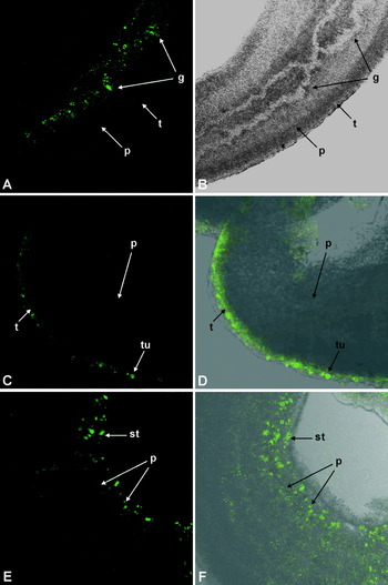
Parallel to the study of Davis et al. (1999) we had started a similar approach with the PDS 1000 particle bombardment system to transform adult and larval S. mansoni. In our experiments a modified form of the green fluorescent protein (GFP) gene from the jellyfish Aequorea victoria (Reichel et al. Reference Reichel, Mathur, Eckes, Langenkemper, Koncz, Schell, Reiss and Maas1996) was used. Its expression generates strong visible bioluminescence (Chalfie et al. Reference Chalfie, Tu, Euskirchen, Ward and Prasher1994), which can be easily detected by fluorescence microscopy. As regulatory elements to control the expression of the GFP reporter-gene we used promoter and terminator regions of a variety of known schistosome genes. Among these was the heat-shock protein (HSP) gene hsp70. Expression studies had demonstrated earlier that the schistosome hsp70 gene is developmentally regulated and inducible in adults by heat stress (Neumann et al. Reference Neumann, Ziv, Lantner and Schechter1993). Thus a plasmid construct was made containing the GFP gene under the control of the promoter and terminator regions of the hsp70 gene. This construct was introduced by particle bombardment in adult S. mansoni males. Molecular analyses demonstrated the presence of the hsp70-GFP vector and its heat-inducible transcription and translation (Wippersteg et al. Reference Wippersteg, Kapp, Kunz, Jackstadt, Zahner and Grevelding2002b). Confocal microscopy finally confirmed correct transgene expression by exhibiting fluorescing signals in different areas of the worms that co-localized with the presence of gold particles. These signals were mainly found in the tegument and the tubercles of the males. Histochemical analyses by methylene-blue staining of 5 μm sections of bombarded worms indicated the presence of gold particles in nearly all tissues. Therefore, this specific signal localization was not due to an insufficient penetrance of gold particles into deeper tissue areas. Additionally, using an antibody against S. mansoni HSP70, its expression was detected predominantly in the tegument of worms after heat shock indicating a predominant role of HSP70 in the tegumental area following stress (Grevelding, Reference Grevelding, Maule and Marks2006). Since the biolistic approach worked well with adults, it was also tried to perform bombardment experiments with larval schistosomes using sporocysts as targets. Establishing a more sensitive protocol with lower pressures assured the survival of the larvae after bombardment. Again, the presence and the transcription of the hsp70-GFP vector after bombardment were confirmed, and fluorescing signals were detected in different tissues of the sporocysts (Wippersteg et al. Reference Wippersteg, Kapp, Kunz, Jackstadt, Zahner and Grevelding2002b). Following up this work, a second ‘proof of principle’ vector was built consisting of GFP fused to the regulatory elements of the ER60 gene. It codes for a cysteine protease in schistosomes and was shown to be expressed in excretory/secretory tissues such as the gastrodermis or the protonephridia (Finken-Eigen and Kunz, Reference Finken-Eigen and Kunz1997). After particle bombardment of adult schistosomes with an ER60-GFP vector, significant GFP signals were detected in the gastrodermis (Fig. 1 A, B). In addition, fluorescence was also observed as stripe-like structures within the parenchyma (Wippersteg et al. Reference Wippersteg, Ribeiro, Liedtke, Kusel and Grevelding2003). Using a colocalization approach of biolistic transformation with ER60-GFP and Texas Red (TxR)-BSA, a fluorescent dye that enters the excretory/secretory system and especially the excretory tubules as part of the protonephridium (Tan et al. Reference Tan, Thornhill, Al-Adhami, Akhkha and Kusel2003), the occurrence of GFP and TxR-BSA in the same tissue was confirmed (Wippersteg et al. Reference Wippersteg, Ribeiro, Liedtke, Kusel and Grevelding2003). Also in sporocysts, ER60 promoter-induced expression of GFP was localized in excretory/secretory tissues such as the lateral gland and the ridge cytons (Wippersteg et al. Reference Wippersteg, Ribeiro, Liedtke, Kusel and Grevelding2003). The same combination of biolistic transformation and co-localization with TxR-BSA was used by Rossi and colleagues (2003) to investigate the expression of calcineurin A from S. mansoni. Similar to the results of ER60, calcineurin A was also expressed in the excretory/secretory system of adult parasites. Using promoters of gut-specific genes it was later shown, in collaboration with James McKerrow's group at the University of California at San Francisco, that tissue-specific reporter gene activity can also be visualized in deeper tissue layers. To this end the promoters of the S. mansoni gut protease gene cathepsin L1 induced GFP expression in the gut following bombardment (Wippersteg et al. Reference Wippersteg, Sajid, Walshe, Khiem, Salter, McKerrow, Grevelding and Caffrey2005). In the same study, the promoter of the protease gene cathepsin B2, whose protein product was known to be localized in the tegumental tubercles of males (Caffrey et al. Reference Caffrey, Salter, Lucas, Khiem, Hsieh, Lim, Ruppel, McKerrow and Sajid2002), was tested in a similar approach. Under the control of the regulatory elements of the cathepsin B2 gene fluorescence was observed in the tegument (Fig. 1 C, D), as expected, confirming the tissue-specificity of reporter gene expression after particle bombardment.
Adult S. mansoni males following particle bombardment with the plasmid construct ER60-GFP-ER60 (A, B; Wippersteg et al. Reference Wippersteg, Ribeiro, Liedtke, Kusel and Grevelding2003), CB2-GFP-HSP70 (C, D; Wippersteg et al. Reference Wippersteg, Sajid, Walshe, Khiem, Salter, McKerrow, Grevelding and Caffrey2005), or Act-GFP-Act (E, F; Beckmann et al. Reference Beckmann, Wippersteg, El-Bahay, Hirzmann, Oliveira and Grevelding2007) (A, C, E: fluorescence images; B: bright field image; D, F: overlay of fluorescence and bright field image). Fluorescence signals were detected by confocal laser scanning microscopy (Leica TCS NT) 24–48 hours after particle bombardment using a wavelength of 488 nm for excitation of GFP. The promoter regions of the cysteine protease gene SmER60, the cathepsin gene SmCB2, and the actin gene SmAct1 induced GFP expression as expected in the gastrodermis around the gut lumen (A, B), the tegument (C, D), and the subtegumental/muscle region as well as the parenchyma (E, F), respectively [g: gastrodermis, p: parenchyma, st: subtegument, t: tegument, tu: tubercle].

As well as particle bombardment works as a transformation method for adult and larval schistosomes, disadvantages are the relatively low number of transgenic worms after bombardment and/or the intensity of transgene expression in the parasite. To improve these parameters, Dvořák and colleagues (2010) recently modified the protocol. First, they created new constructs by fusing the GFP reporter gene with signal sequences of proteases and achieved tissue-specific GFP expression. Second, they introduced mCherry as new fluorescent reporter gene, which can serve as an alternative, spectrally distinct reporter besides GFP. However, mCherry signals were observed less frequently compared to GFP in the parasites and the rate of transgenic worms could not be increased. To consider electroporation as an alternative for the delivery of DNA constructs and transgenes into adult schistosomes, Dvořák et al. (Reference Dvořák, Beckmann, Lim, Engel, Grevelding, McKerrow and Caffrey2010) also tested this method. Their results indicated that electroporation, in contrast to particle bombardment, could lead to a non-specific expression of the reporter genes in adult schistosomes. Electroporation was also tested by Correnti et al. (Reference Correnti, Jung, Freitas and Pearce2007) for schistosomules. Using the promoter sequence of the S. mansoni actin 1 (SmAct1.1) gene and luciferase as reporter, they were able to detect transgene expression in growing schistosomula (Correnti et al. Reference Correnti, Jung, Freitas and Pearce2007). In parallel, we tested the SmAct1.1 promoter to drive GFP expression in bombarded adult S. mansoni males and sporocysts (Beckmann et al. Reference Beckmann, Wippersteg, El-Bahay, Hirzmann, Oliveira and Grevelding2007). In adults, we detected GFP signals in the tegument including the tubercles, subtegument, parenchyma and in muscle cells (Fig. 1 E, F). This pattern corresponded perfectly to previous immunolocalization data showing the presence of actin in these tissues (MacGregor and Shore, Reference MacGregor and Shore1990). Besides GFP and firefly luciferase, also a Gaussia luciferase was proven by Cheng and Davis (Reference Cheng and Davis2007) in biolistic and electroporation experiments as a suitable reporter gene. The authors showed that, besides significantly higher levels of luciferase activity in schistosomes compared to other tested luciferases, the Gaussia luciferase can be secreted into culture media allowing non-invasive analysis of reporter gene activity (Cheng and Davis, Reference Cheng and Davis2007).
TRANSIENTLY TRANSFORMED SCHISTOSOMES
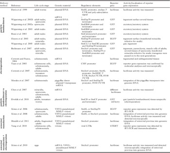
After the establishment of transient transformation in schistosomes, a number of studies have been performed using different regulatory elements and reporter genes introduced by particle bombardment (Davis et al. Reference Davis, Parra, LoVerde, Ribeiro, Glorioso and Hodgson1999; Wippersteg et al. Reference Wippersteg, Kapp, Kunz and Grevelding2002a,Reference Wippersteg, Kapp, Kunz, Jackstadt, Zahner and Greveldingb, Reference Wippersteg, Ribeiro, Liedtke, Kusel and Grevelding2003, Reference Wippersteg, Sajid, Walshe, Khiem, Salter, McKerrow, Grevelding and Caffrey2005; Heyers et al. Reference Heyers, Walduck, Brindley, Bleiss, Lucius, Dorbic, Wittig and Kalinna2003; Rossi et al. Reference Rossi, Wippersteg, Klinkert and Grevelding2003; Beckmann et al. Reference Beckmann, Wippersteg, El-Bahay, Hirzmann, Oliveira and Grevelding2007; Dvořák et al. Reference Dvořák, Beckmann, Lim, Engel, Grevelding, McKerrow and Caffrey2010; Table 1). All these studies proved the reliability of the method. However, the main disadvantage of this approach so far has been the transient nature of transgene expression. To further develop transgenesis in schistosomes, it has been necessary to explore methods for stable germline transformation which would allow genetic as well as phenotypic studies in subsequent generations. Towards this end, we developed a modified particle bombardment protocol that allowed the introduction of transgenes into the germline using miracidia as targets (Beckmann et al. Reference Beckmann, Wippersteg, El-Bahay, Hirzmann, Oliveira and Grevelding2007). After bombardment, the miracidia were still biologically active and able to infect snails to continue the life cycle – a critical step for the establishment and monitoring of stable transgenesis. As proofs of principle miracidia were bombarded with ER60-GFP-ER60 or hsp70-GFP-hsp70 constructs and afterwards used for the snail infection. Using molecular tools, we were able to detect the presence of the transgenes in cercariae and adults of the F0 and the F1 generation. These data demonstrated that transgenes can be passed on from one life stage to the next within one generation and, furthermore, from one generation to the next. Since the germ cells are considered to be the only constant cell line during schistosome development we could indirectly demonstrate the presence of transgenes in the germline and a successful germline-transformation approach (Beckmann et al. Reference Beckmann, Wippersteg, El-Bahay, Hirzmann, Oliveira and Grevelding2007). However, the presence of the transgenes could not be detected from generation F2 on. Therefore, it seemed likely that the constructs occured extrachromosomally as episomes, which failed to integrate into the genome of the germ cells and thus were lost during cell divisions. In an independent study Heyers and colleagues (2003) confirmed the suitability of miracidia as starting stages for germ line transgenesis. In addition to demonstrating that bombarded miracidia could still infect snails, gold particles were detected in the germ balls of parasites within the snail tissue in which also reporter gene expression was also detected (Heyers et al. Reference Heyers, Walduck, Brindley, Bleiss, Lucius, Dorbic, Wittig and Kalinna2003).
Approaches towards transgenic schistosomes (S. mansoni, S. japonicum)

[EGFP: enhanced green fluorescent protein; GFP: green fluorescent protein; hTERT: human telomerase reverse transcriptase].
FROM TRANSIENT TO STABLE TRANSFORMATION
Particle bombardment is a suitable method for adult and larval schistosomes to introduce vector constructs into the germline. However, strategies need to be developed that allow their integration into the genome of germ cells to achieve a heritable transgenesis. Approaches using mobile genetic elements as well as virus-based strategies have achieved the integration of transgenes into schistosome chromosomes, and the principle applicability of these approaches has been successfully shown (reviewed in Mann et al. Reference Mann, Morales, Kines and Brindley2008 and Hagen et al. Reference Hagen, Lee, Fairlie and Kalinna2011).
Mobile genetic elements – endogenous retroposons and DNA-transposons
Transposable elements (TEs) are subdivided into two groups according to their regulatory components and mechanistic features of their mode of action. TEs of class I represent retroposons which are generated and disseminated by reverse transcription of an RNA intermediate. Class II TEs transpose themselves or via a DNA copy (DNA-transposons). Depending on their physical integrity, class II TEs can be autonomous or non-autonomous with respect to their mobility. TEs are ancient vestiges of prokaryotic and eukaryotic genomes including those of parasites (Thomas et al. Reference Thomas, Macias, Alonso and Lopez2010; Venancio et al. Reference Venancio, Wilson, Verjovski-Almeida and DeMarco2010). In schistosomes, class I and II TEs have been described. Among the class I TEs are Boudicca and fugitive, gypsy-like long terminal repeat (LTR) retrotransposon or Sinbad, a Pao/BEL-type retrotransposon (Copeland et al. Reference Copeland, Brindley, Heyers, Michael, Johnston, Williams, Ivens and Kalinna2003, Reference Copeland, Mann, Morales, Kalinna and Brindley2005). Molecular characterization of these TEs provided strong evidence for their transcriptional activity in larval and adult stages of schistosome development, and they occur in different schistosome species. Therefore, it has been debated whether they may have the potential to become useful tools for the establishment of a retroposon-based transgenesis system (Mann et al. Reference Mann, Morales, Kines and Brindley2008). Additionally, the Boudicca and Sinbad LTRs were tested for promoter function, and it was shown that the Sinbad LTRs but not the Boudicca LTRs were able to drive transgene activity in human cell culture (Copeland et al. Reference Copeland, Mann and Brindley2007).
SmMerlin is a class II TE of S. mansoni and may represent another vehicle candidate for transgenesis. Due to its similarity to bacterial insertion sequences Merlin belongs to the superfamily of Merlin/IS1016 DNA elements, which occur also in higher eukaryotes. More than 500 Merlin sequences were identified in the genome of S. mansoni, many of these are deletion variants (Feschotte, Reference Feschotte2004). This indicates that the Merlin family consists of autonomous and non-autonomous members. BlastN-analyses in EST databases of S. mansoni and S. japonicum resulted in many hits indicating not only transcriptional activity, but also the occurrence of Merlin in different schistosome species. Two kinds of transcripts were found, those with and those without flanking host sequences. This shows that Merlin family members can integrate in transcriptional active regions of the genome, but it also indicates a potential influence of transcriptionally active schistosome genes in the neighbourhood on the expression of Merlin family members. Consequently, typical Merlin footprints were identified in the genome of S. mansoni (Feschotte, Reference Feschotte2004) consisting of 8 bp target site duplications, which occur during the integration process and remain after the transposon has left this target site again. All these results suggest that Merlin is an active mobile element tramping through the schistosome genome. Its relatively small size of 1·4 kb makes SmMerlin a ‘lab-friendly’ candidate for vector construction. SmMerlin has two exons separated by a 32 bp intron. The exons code for a protein of 294 amino acids which reveals significant homology to transposases (Feschotte, Reference Feschotte2004). The ends of Merlin are characterized by 24 bp terminal inverted repeats. To start cloning vectors for transgenesis on the basis of SmMerlin we amplified Merlin transposase sequences recently and detected that all of these contained the 32 bp intron (Beckmann et al., unpublished). Its sequence predicts that presence of this intron may lead to a premature stop of translation and an incomplete transposase. This finding indicates that SmMerlin activity may be controlled in the schistosome genome in a similar way as known from P-elements in Drosophila (Bachmann and Knust, Reference Bachmann and Knust2008). Here, an intron between the open reading frames 2 and 3 is not spliced in somatic cells, which prevents any mobilization, whereas in the germline splicing occurs leading to transposition in this cell line (Laski et al. Reference Laski, Rio and Rubin1986). By cloning Merlin-based constructs containing the Merlin transposase without the 32 bp intron it seems feasible to develop a potent transformation system. Using particle bombardment we were able to introduce these Merlin constructs into adult and larval schistosomes and the transcription of the transgene Merlin transposase was detected afterwards by RT-PCR analyses. In future studies we will focus on the detection of excision and integration events to evaluate this system for its suitability to stably transform schistosomes. Besides Merlin, a member of the CACTA (also called En/Spm) superfamily of DNA transposons was also described for schistosomes (DeMarco et al. Reference DeMarco, Venancio and Verjovski-Almeida2006). It was called SmTRC1 and suggested to be a potential new tool for insertional mutagenesis because CACTA elements had been successfully used for this purpose in plants (Tissier et al. Reference Tissier, Marillonnet, Klimyuk, Patel, Torres, Murphy and Jones1999). As for Merlin, future studies have to show whether SmTRC1 is another candidate for DNA transposon-based transformation.
Exogenous DNA-transposons
PiggyBac, a class II TE originating from the genome of the cabbage looper moth Trichoplusia ni, has been successfully used as a vehicle for transformation in diverse eukaryotic organisms such as mosquitoes, planarians, Plasmodium falciparum, but also in human or other mammalian cells. Morales and colleagues (2007) demonstrated that piggyBac is able to deliver reporter transgenes into the genome of S. mansoni. After electroporation of schistosomula with a piggyBac donor plasmid containing the firefly luciferase gene under the control of schistosome gene promoters together with piggyBac transposase mRNA, numerous transposon integrations into the parasites’ chromosomes were detected. This result represented substantial progress in somatic transgenesis, although the inheritance of piggyBac-based vector constructs has yet to be demonstrated.
Virus-based approaches
Since retroviruses have been used successfully for transgenesis in other organisms, Kines and colleagues (2006) established a transduction system for S. mansoni, using replication incompetent Moloney Murine Leukaemia Virus (MMLV) virions that were pseudotyped with Vesicular Stomatitis Virus Glycoprotein (VSVG) carrying EGFP or luciferase reporter genes under the control of the Sl or hsp70 promoter. After co-cultivation of larval schistosomes (schistosomules or spororcysts) with the virions, the authors showed virus binding and uptake into the parasite tegument. Thus, the retroviruses seemed able to transduce cultured schistosomes. Finally, evidence of proviral integration into genomic DNA as well as the presence of transcripts encoding reporter transgenes were obtained. The same group later used Southern blot analysis and an anchored PCR-based approach to demonstrate integration of proviral MMLV retroviruses into schistosome chromosomes, proving somatic transgenesis. Furthermore, reporter gene/luciferase activity in transduced schistosomula and adult schistosomes was measured (Kines et al. Reference Kines, Morales, Mann, Gobert and Brindley2008). The anchored PCR approach detects the transgenes in the chromosomes and also determines the efficiency of transduction after the exposure of schistosomes to virions (Rinaldi et al. Reference Rinaldi, Suttiprapa and Brindley2011). Furthermore, the transduction approach with VSVG-pseudotyped retroviruses also works for schistosomules of S. japonicum (Yang et al. Reference Yang, Brindley, Zeng, Li, Zhou, Liu, Liu, Cai, Zeng, Wei, Lan and McManus2010). Besides larval and adult schistosomes, also S. mansoni eggs can serve as targets for VSVG-pseudotyped MMLV virions (Kines et al. Reference Kines, Rinaldi, Okatcha, Morales, Mann, Tort and Brindley2010). After exposure of schistosomes eggs to virions by either soaking or electroporation, proviral transgenes were detected by PCR within the genomic DNA of miracidia arising from them. Although the integration of proviral forms and transgenes into the genome of schistosomes has been achieved (Kines et al. Reference Kines, Morales, Mann, Gobert and Brindley2008; Rinaldi et al. Reference Rinaldi, Suttiprapa and Brindley2011; Table 1), the heredity of such integrated transgenes has not yet been demonstrated. Since it is possible to transduce eggs and to obtain viable miracidia carrying the transgenes, it seems feasible to obtain germ line integration provided that the virions integrate into chromosomes of germ cells. Detecting transgenes in subsequent life cycle stages and subsequent generations would provide strong evidence for stable transformation and the general applicability of this approach.
RNAi: ELUCIDATING SCHISTOSOME GENE FUNCTION BY SMALL INTERFERING RNAs
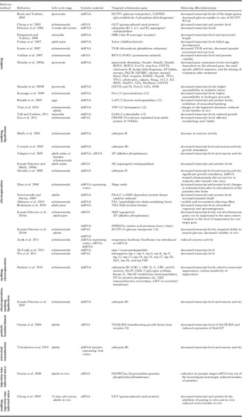
Since classical genetic approaches analyzing gene function are not feasible in schistosomes, RNAi has become a powerful tool for functional gene analysis in this parasite (reviewed in Bhardwaj et al. Reference Bhardwaj, Krautz-Peterson and Skelly2011 and Hagen et al. Reference Hagen, Lee, Fairlie and Kalinna2011). Since the first reports of the successful application of this post transcriptional gene-silencing technique in S. mansoni (Boyle et al. Reference Boyle, Wu, Shoemaker and Yoshino2003; Skelly et al. Reference Skelly, Da'dara and Harn2003) many studies have been published (Table 2) using this method to explore the function of genes with hypothesized roles in physiology (Cheng et al. Reference Cheng, Lin, Shi, Jin, Fu, Jin, Zhou and Cai2005; Correnti et al. Reference Correnti, Brindley and Pearce2005; Delcroix et al. Reference Delcroix, Sajid, Caffrey, Lim, Dvořák, Hsieh, Bahgat, Dissous and McKerrow2006; Krautz-Peterson and Skelly, Reference Krautz-Peterson and Skelly2008a; Morales et al. Reference Morales, Rinaldi, Gobert, Kines, Tort and Brindley2008; Mourão et al. Reference Mourão, Dinguirard, Franco and Yoshino2009a, Reference Mourão, Dinguirard, Franco and Yoshinob; Krautz-Peterson et al. Reference Krautz-Peterson, Simoes, Faghiri, Ndegwa, Oliveira, Shoemaker and Skelly2010b; Kumagai et al. Reference Kumagai, Osada, Ohta and Kanazawa2009; McVeigh et al. Reference McVeigh, Mair, Novozhilova, Day, Zamanian, Marks, Kimber, Day and Maule2011), development (Dinguirard and Yoshino, Reference Dinguirard and Yoshino2006; Freitas et al. Reference Freitas, Jung and Pearce2007; Pereira et al. Reference Pereira, Pascoal, Marchesini, Maia, Magalhaes, Zanotti-Magalhaes and Lopes-Cendes2008; Rinaldi et al. Reference Rinaldi, Morales, Alrefaei, Cancela, Castillo, Dalton, Tort and Brindley2009; Beckmann et al. Reference Beckmann, Buro, Dissous, Hirzmann and Grevelding2010; Taft and Yoshino, Reference Taft and Yoshino2011; Zou et al. Reference Zou, Jin, Liu, Wu, Liu and Lin2011), and other aspects of biology (Tran et al. Reference Tran, Freitas, Cooper, Gaze, Gatton, Jones, Lovas, Pearce and Loukas2010; Yoshino et al. Reference Yoshino, Dinguirard and Mourao2010; Wu et al. Reference Wu, Cai, Chen and Wang2011). For some genes, like the schistosome thioredoxin glutathione reductase (TGR) or cAMP-dependent protein kinase (PKA), RNAi induced lethal phenotypes were observed by simple soaking of schistosomula (Kuntz et al. Reference Kuntz, Davioud-Charvet, Sayed, Califf, Dessolin, Arner and Williams2007) or electroporation of adults (Swierczewski and Davies, Reference Swierczewski and Davies2009) with corresponding dsRNAs indicating a role of these genes in worm survival. Although much was invested to improve the protocols for RNAi in adult and larval schistosomes (Krautz-Peterson et al. Reference Krautz-Peterson, Radwanska, Ndegwa, Shoemaker and Skelly2007, Reference Krautz-Peterson, Bhardwaj, Faghiri, Tararam and Skelly2010a; Ndegwa et al. Reference Ndegwa, Krautz-Peterson and Skelly2007; Stefanić et al. Reference Stefanić, Dvořák, Horn, Braschi, Sojka, Ruelas, Suzuki, Lim, Hopkins, McKerrow and Caffrey2010), there are still some limitations. First, not all schistosomes genes can be silenced by RNAi to the same extent. Second, some genes seem to totally resist silencing and are not “knockable” at all (Krautz-Peterson et al. Reference Krautz-Peterson, Bhardwaj, Faghiri, Tararam and Skelly2010a; Beckmann et al., unpublished observations). This finding is thought not to depend on the expression of the genes in specific cells that can resist RNAi, but rather to be caused by secondary structures of the target mRNAs. The latter may lead to an inaccessibility of the RNAi machinery to these folded transcripts (Krautz-Peterson et al. Reference Krautz-Peterson, Bhardwaj, Faghiri, Tararam and Skelly2010a). Furthermore, in each RNAi experiment a number of adult or larval schistosomes was exposed to dsRNA or siRNA. Not all parasites may take up the same amount of dsRNAs/siRNAs resulting in differences in RNAi pathway activation, and thus in a high variability of gene silencing among individuals but also in different experiments. By simple soaking dsRNA or siRNA can be easily delivered to larval schistosomes (schistosomula). However, additional electroporation seems to deliver dsRNA/siRNA more efficiently into the parasites increasing the efficiency of silencing. In the case of adults (single worms or pairs), it seems that electroporation is more important to achieve a higher level of gene suppression compared to larval stages (Krautz-Peterson et al. Reference Krautz-Peterson, Bhardwaj, Faghiri, Tararam and Skelly2010a). Operational parameters such as taking into in vitro culture may have an additional influence on the results of RNAi approaches since the culture media alone has a baseline effect on, for example, vitality and viability of schistosomula (Stefanić et al. Reference Stefanić, Dvořák, Horn, Braschi, Sojka, Ruelas, Suzuki, Lim, Hopkins, McKerrow and Caffrey2010). Off-target effects, time- and dose-dependency as well as dosing limits seem to be additional critical factors for RNAi experiments in schistosomes (Stefanić et al. Reference Stefanić, Dvořák, Horn, Braschi, Sojka, Ruelas, Suzuki, Lim, Hopkins, McKerrow and Caffrey2010). Furthermore, there is no correlation between the degree of silencing and the appearance of a phenotype. Depending on dosage effects, moderate silencing levels can also produce clear phenotypes (Freitas et al. Reference Freitas, Jung and Pearce2007; Beckmann et al. Reference Beckmann, Buro, Dissous, Hirzmann and Grevelding2010; Krautz-Peterson et al. Reference Krautz-Peterson, Bhardwaj, Faghiri, Tararam and Skelly2010a). In contrast, a significant down-regulation of gene activity is not necessarily associated with an observable phenotype (e.g. Atkinson et al. Reference Atkinson, McVeigh, Kimber, Marks, Eipper, Mains, Day and Maule2010; McVeigh et al. Reference McVeigh, Mair, Novozhilova, Day, Zamanian, Marks, Kimber, Day and Maule2011; Beckmann et al., unpublished observations).
RNAi approaches in schistosomes (S. mansoni, S. japonicum)

[dsRNA: double stranded RNA; siRNA: small interfering RNA; shRNA: small hairpin-RNA].
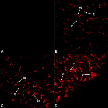
Another question that remains unanswered is how dsRNA or siRNA enter larval or adult schistosomes. For newly transformed schistosomula, simple soaking alone seems to be sufficient for dsRNA delivery. Štefanić and colleagues (2010) investigated whether the gut may serve as a route for dsRNA entry into this larval stage. Using 30 μg/ml Cy5-labeled dsRNA the authors demonstrated that the gut of schistosomula takes up and concentrates the dsRNA within minutes after mechanical transformation of cercariae. Accumulation of the dye was evident along the gut and in the two terminal caecal chambers by 90 min post-transformation, and the signal remained visible during an incubation period of 6 days (Stefanić et al. Reference Stefanić, Dvořák, Horn, Braschi, Sojka, Ruelas, Suzuki, Lim, Hopkins, McKerrow and Caffrey2010). To follow the soaking route of dsRNAs into adult schistosomes, we performed some preliminary experiments in our laboratory and incubated male schistosomes with rhodamin-labeled dsRNAs in vitro. The 5-carboxy-X-rhodamin was covalently linked to the dsRNA using the Label IT® Nucleic Acid Labeling Kit (Mirus Bio; USA). In each experiment, 10 males were cultivated in medium containing 5 μg labeled dsRNA/ml for up to five days. As a control, males were incubated with unlabeled dsRNA. After washing of the males, fluorescence signals were detected with a confocal laser scanning microscope (Leica TCS NT; Heidelberg) with an extinction of 597 nm. After only 2 hours incubation with rhodamin-labeled dsRNAs, weak fluorescence was detected within the excretory tubules and the flame cells (Fig. 2 B). Signal intensity increased with the time of incubation (Fig. 2 C). At day 5, fluorescence was also detected in the parenchyma (Fig. 2 D) and was no longer restricted to the excretory system. This fluorescence pattern in adult schistosomes is congruent with the staining pattern of Texas Red, which specifically stains the excretory tubules (Tan et al. Reference Tan, Thornhill, Al-Adhami, Akhkha and Kusel2003; Wippersteg et al. Reference Wippersteg, Ribeiro, Liedtke, Kusel and Grevelding2003). The excretory system of schistosomes has long been thought of as a route of removal for waste products. However, evidence for further functions including endocytosis was also obtained (Kusel et al. Reference Kusel, McVeigh and Thornhill2009). Our results suggest that the excretory tubules may also be involved in the uptake of dsRNAs into adult schistosomes.
Adult S. mansoni males following in vitro incubation with rhodamin-labeled dsRNAs after 2 hours (B), 3 days (C), and 5 days (D), or as control with unlabeled dsRNA (A). Fluorescence signals were detected by confocal laser scanning microscopy (Leica TCS NT) with an extinction of 597 nm. [et: excretory tubules, fc: flame cells, p: parenchyma].

Once the dsRNA is taken up via the gut or the excretory system of schistosomes, it has to be distributed to other tissues and cells to induce effects. In C. elegans, the multispan transmembrane protein SID-1 (systemic RNAi defective-1) is required for the uptake and transport of dsRNA (Feinberg and Hunter, Reference Feinberg and Hunter2003). Krautz-Peterson and colleagues recently identified in silico the schistosome homologue SmSID-1 (Krautz-Peterson et al. Reference Krautz-Peterson, Bhardwaj, Faghiri, Tararam and Skelly2010a). The authors assume that SmSID-1 might also act as a channel to import dsRNA into schistosomes. Furthermore, since most schistosome tissues are syncytial, the authors speculated that once dsRNA has entered a tissue, it may be able to traverse relatively large distances without the need to cross additional membranes (Krautz-Peterson et al. Reference Krautz-Peterson, Bhardwaj, Faghiri, Tararam and Skelly2010a). Once the dsRNA is taken up by cells, it has to be processed by the RNAi machinery to silence gene function. Up to now, a number of proteins has been identified in schistosomes representing homologues of proteins involved in the RNAi pathway of other organisms, which indicates a similar scenario in schistosomes (Krautz-Peterson et al. Reference Krautz-Peterson, Bhardwaj, Faghiri, Tararam and Skelly2010a).
Vector-based dsRNA and siRNA/shRNA delivery
Vector-based approaches have been successfully used to deliver small RNAs (dsRNAs, siRNAs) in gene silencing experiments. Zhao and colleagues (Reference Zhao, Lei, Liu, Zhu, Ren, Wang and Shen2008) first reported vector-mediated gene silencing in S. japonicum. They used siRNAs delivered from short hairpin-RNAs (shRNAs) expressed in vivo in schistosomules by a mammalian Pol III promoter H1 and successfully suppressed a Mago nashi gene (Zhao et al. Reference Zhao, Lei, Liu, Zhu, Ren, Wang and Shen2008). Similarly to this approach Ayuk et al. (Reference Ayuk, Suttiprapa, Rinaldi, Mann, Lee and Brindley2011) established a vector-based RNAi technique for S. mansoni using a plasmid expressing shRNAs driven by the schistosome U6 gene promoter. They demonstrated that a shRNA targeting reporter firefly luciferase reduced firefly luciferase mRNA and luciferase enzymatic activity in transformed schistosomules (Ayuk et al. Reference Ayuk, Suttiprapa, Rinaldi, Mann, Lee and Brindley2011). Tchoubrieva and colleagues (2010) designed a viral vector expressing a dsRNA hairpin to silence the expression of the schistosome cathepsin B1 (SmCB1) gene and were the first to show that this approach also works in adult schistosomes. The vector-based delivery of dsRNAs or shRNAs could be a preferable technique for the investigation of long-term silencing effects of specific mRNAs compared to the transient suppression of gene expression achieved by soaking or electroporation.
Large-scale RNAi approaches
De Moraes Mourao and colleagues (Reference Mourão, Dinguirard, Franco and Yoshino2009a) made the first attempt to use RNAi for large-scale screening approaches. They selected 32 different genes and analyzed potential morphological changes in sporocysts after application of corresponding dsRNAs. Their results indicated that the efficiency of altered gene expression due to dsRNA-treatment is highly variable and dependent on (1) the selected target gene, (2) the selected dsRNA sequence within the target gene, and (3) the timing of evaluation after treatment. The authors concluded that potential off-target effects, non-specific effects of some dsRNAs, and variable efficiencies of specific gene silencing still are critical points of RNAi in schistosomes (de Moraes Mourao et al. Reference Mourão, Dinguirard, Franco and Yoshino2009a). Optimization is needed as well as careful gene-specific testing as part of RNAi experiments and data interpretation. In addition, improvements of dsRNA delivery methods were discussed as being critical. Regarding large-scale RNAi experiments, Mourão and colleagues concluded that low and inconsistent dsRNA uptake, the low number of parasites that can be processed in a single treatment, the limited phenotype repertoire, and the lack of more sensitive detection tools currently restrict large-scale approaches. Thus RNAi seems presently to be suitable only for small scale or gene-by-gene characterization approaches (Mourão et al. Reference Mourão, Dinguirard, Franco and Yoshino2009a). Stefanić and colleagues (Reference Stefanić, Dvořák, Horn, Braschi, Sojka, Ruelas, Suzuki, Lim, Hopkins, McKerrow and Caffrey2010) tried to define operational parameters, which may facilitate larger RNAi screening and suggested the use of newly transformed schistosomula due to handling advantages.
In vivo RNAi approaches
A few approaches have been undertaken using siRNAs as therapeutical agents. Pereira and colleagues (Reference Pereira, Pascoal, Marchesini, Maia, Magalhaes, Zanotti-Magalhaes and Lopes-Cendes2008) successfully used in vivo RNAi to reduce worm burdens in mice chronically infected with S. mansoni. SiRNAs targeting the hypoxanthine-guanine phosphoribosyl-transferase (HGPRTase) were intravenously injected in infected mice. This led to a significant reduction of the total number of parasites after six days as well as to a reduction of the parasite target mRNA, but not of its host's homologue (Pereira et al. Reference Pereira, Pascoal, Marchesini, Maia, Magalhaes, Zanotti-Magalhaes and Lopes-Cendes2008). Cheng et al. (Reference Cheng, Fu, Lin, Shi, Zhou, Jin and Cai2009) used a similar approach to knock down the gynaecophoral canal protein of S. japonicum (SjGCP) in vivo, which led to a reduction in parasite pairing and total worm burden. These results indicate that in vivo RNAi may also be possible.
UNANSWERED QUESTIONS ON THE WAY TO TRANSGENIC SCHISTOSOMES
Over the last decade, much effort and enthusiasm have been invested into the establishment of systems to generate transgenic schistosomes. Different innovative approaches were performed indicating their suitability for transient transformation/transfection of this multicellular parasite. Although the first transgene integrations into schistosome chromosomes have been achieved, there is still a long way to go towards obtaining stably transformed schistosomes.
Are miracidia and eggs the only life cycle stages useful as targets for transformation approaches with the aim to enter the germ line? Both appear to be preferable stages, because their germ cells seem to be easily accessible using particle bombardment or retroviruses. Furthermore, miracidia, either directly transformed, transfected or hatching from transfected eggs, can be reintroduced into the schistosome life cycle by snail infection. In theory, although they differ in the number of germ cells, sporocysts, schistosomula or adults also represent potential targets, but they must be reintroduced into the life cycle by implanting into intermediate or final hosts, respectively. The implantation of sporocysts into snails (Jourdane and Theron, Reference Jourdane and Theron1980; Jourdane et al. Reference Jourdane, Liang and Bruce1985) or of schistosomula and adults into final hosts (Basch and Humbert, Reference Basch and Humbert1981) is possible, but the approaches are technically demanding and time consuming.
How can we achieve a tissue-specific knock-down of gene activity? If stable transformation of schistosomes could be achieved, the door would open for a number of fundamental analyses in this direction. For example, by integrating a transgene cassette expressing a dsRNA or shRNA under the control of certain schistosome gene promoters, tissue-specific RNAi experiments would be possible in various different life stages. Problems with tissue-dependent off-target effects as well as with the dsRNA/shRNA delivery and its intra-organism transport could be overcome. Concerning this point, it is still not absolutely clear how dsRNAs, shRNas or siRNAs enter the parasite finding their way to different tissues and cells, and whether all tissues and cells can be reached. Especially for adults we cannot exclude that not all tissues are accessible to dsRNAs, shRNAs or siRNAs to the same degree and that in different tissues RNAi effects might be effective to different extents due to variable concentrations of these nucleic acids or other factors.
Are all members of the RNAi machinery expressed to the same degree in all tissues of schistosomes, and in all life stages? With respect to the observation that some genes can be effectively knocked down in contrast to others may hint at the possibility that ‘non-knockable’ genes are expressed in tissues (1) which cannot be reached by dsRNAs/shRNAs/siRNAs, or (2) in which not all components of the RNAi processing pathway are present. Localization experiments employing whole mount in situ hybridization recently showed that transcription of the S. mansoni Argonaute 2 (Smago2), an essential member of the RNAi pathways (Ketting, Reference Ketting2011), is restricted to the ovary, the vitelline glands and testes of adult worms (Cogswell et al. Reference Cogswell, Collins, Newmark and Williams2011). Since the used hybridization probe was specific for Smago2, it cannot be excluded that the S. mansoni homologues of ago1 and/or ago3 transcripts were present in other tissues fulfilling ago2-like, redundant RNAi functions. Thus, further localization experiments are needed to ascertain the expression patterns of relevant members of the predicted schistosome RNAi pathway. Using RT-PCR analyses, the expressions of the schistosome Dicer homologues (SmDicer, SjDicer) and schistosome Argonaute homologues (SjAGO1, 2, 3) were shown in different life cycle stages with the highest expression in larval stages (Krautz-Peterson and Skelly, Reference Krautz-Peterson and Skelly2008b; Luo et al. Reference Luo, Xue, Wang, Sun, Zou and Pan2010) demonstrating that, in principle, all life stages might be susceptible to RNAi, but also indicating that RNAi could be more effective in the larval stages. However, it still has to be elucidated why not all schistosome genes seem to be susceptible to RNAi, and whether factors like the localization or conformation of the target mRNA or as yet unknown factors may additionally influence RNAi in schistosomes.
Stably transformed schistosomes seem to be the most attractive solution to problems with delivery, uptake and variable concentrations of ds/sh/siRNAs in different tissues. Provided that gene cassettes expressing such RNAs are not silenced at the genomic level by epigenetic factors such as methylation post transformation, as observed in plant transgenesis (Matzke et al. Reference Matzke, Mette and Matzke2000; Fischer et al. Reference Fischer, Hofmann, Naumann and Reuter2006), such integrated transgenes would ensure the constant delivery of RNAs and probably continuous silencing effects.
Summarizing we would like to make the point that combining the techniques already available today might be the way to reach this goal in the near future. For example, with a combination of transposon or retrovirus-based systems and particle bombardment of miracidia it should not only be possible to reach the germ cells and to achieve the integration of transgenes into their genomes, but also to reintroduce transgenes via miracidia into the schistosome life cycle and to monitor their heredity during life cycling through subsequent generations.
ACKNOWLEDGMENTS
The authors would like to gratefully acknowledge the enthusiasm, engagement, and valuable inputs of all post-docs, PhD and Diploma students and technical assistants, which have contributed to this project during the last years. Furthermore, the authors would like to point out past and ongoing fruitful collaborations with Jim Mc Kerrow, Connor Caffrey, Jan Dvořák, James Lok, John Kusel, Mo Klinkert, Alessandro Rossi, Guilherme Oliveira, Ed Pearce, Horst Zahner, Peter Jackstadt, Jörg Hirzmann, and Werner Kunz. The authors acknowledge the great contribution of the Welcome Trust Sanger Institute and TIGR for providing sequence data.
FINANCIAL SUPPORT
Financial support was obtained by a grant of the Deutsche Forschungsgemeinschaft (GR 1549/8-2).