




No CrossRef data available.
Article contents
Age does not increase rate of forgetting over weeks—Neuroanatomical volumes and visual memory across the adult life-span
Published online by Cambridge University Press: 28 January 2005
Abstract
The aim of the study was to investigate whether age affects visual memory retention across extended time intervals. In addition, we wanted to study how memory capabilities across different time intervals are related to the volume of different neuroanatomical structures (right hippocampus, right cortex, right white matter). One test of recognition (CVMT) and one test of recall (Rey-Osterrieth Complex Figure Test) were administered, giving measures of immediate recognition/recall, 20–30 min recognition/recall, and recognition/recall at a mean of 75 days. Volumetric measures of right hemisphere hippocampus, cortex, and white matter were obtained through an automated labelling procedure of MRI recordings. Results did not demonstrate a steeper rate of forgetting for older participants when the retention intervals were increased, indicating that older people have spared ability to retain information in the long-term store. Differences in neuroanatomical volumes could explain up to 36% of the variance in memory performance, but were not significantly related to rates of forgetting. Cortical volume and hippocampal volume were in some cases independent as predictors of memory function. Generally, cortical volume was a better predictor of recognition memory than hippocampal volume, while the 2 structures did not differ in their predictive power of recall abilities. While neuroanatomical volumetric differences can explain some of the differences in memory functioning between younger and older persons, the hippocampus does not seem to be unique in this respect. (JINS, 2005, 11, 2–15.)
Keywords
Information
- Type
- Research Article
- Information
- Journal of the International Neuropsychological Society , Volume 11 , Issue 1 , January 2005 , pp. 2 - 15
- Copyright
- © 2005 The International Neuropsychological Society
INTRODUCTION
Reduction of memory capabilities is generally found with increasing age in cognitively healthy individuals (e.g. Small et al., 1999; Tombaugh & Hubley, 2001). Especially, there is a decrement in the ability to acquire new information (Trahan & Larrabee, 1992). In addition, studies have examined forgetting in older persons. However, neither for visually nor for verbally presented material are differential rates of forgetting between younger and older participants established. With the exception of Tombaugh and Hubley (2001), most studies of age effects on memory use retention intervals between learning and delay of 30 min or less. This fits well with the major neuropsychological tests of memory function, where long time memory is defined as memory across 20–30 min. However, present knowledge and theory indicate that memory consolidation takes place over a significantly longer time period of several days (Riedel & Micheau, 2001), and perhaps up to several weeks (e.g., Haist et al., 2001; Rempel-Clower et al., 1996). Thus, it is possible that some of the memory problems experienced by elderly may be caused by less efficient memory consolidation, even though this is not captured by neuropsychological tests. In the present study, we focus on age-dependent differences in memory across weeks, and we relate these differences to neuroanatomical volumetry.
Forgetting Through the Life-Span
When comparing rates of forgetting among groups, initial learning level has to be taken into account. Using saving scores in terms of percentages of what was initially learned or ANOVA procedures are the most common strategies to overcome this problem. When acquisition differences are controlled for in this way, age differences in forgetting rates are usually not found (e.g., Cullum et al., 1990; Geffen et al., 1990; Trahan & Larrabee, 1992; Youngjohn & Crook, 1993). An alternative method is to ensure comparable levels of acquisition by increasing the level of learning for the group with the lowest performance, for example, by increased inspection time or increased number of repetitions during learning (e.g., Huppert & Piercy, 1979). By this procedure, Huppert and Kopelman (1989) found increased forgetting with increased age. However, other researchers have failed to replicate this (Rybarczyk et al., 1987; Spikman et al., 1995). Even though the procedure has been fruitful, theoretical problems exist. Martone et al. (1986) have argued that equal performance at 10 min does not mean equal learning, and Wickelgren (1975) has indicated that an increase of stimulus duration may produce other sources of variation.
The short retention intervals usually employed in studies of age and forgetting make it premature to conclude that storage and forgetting do not contribute to differences in memory capabilities across the adult life-span. The term memory consolidation is central here, taken to mean the processes where encoded information is stored and memory traces established in the brain in such a form that it is potentially retrievable after longer time periods (weeks, months, and years). These consolidatory processes probably work over days and weeks (Haist et al., 2001; Riedel and Micheau, 2001). Thus, differences in memory consolidation or storage between younger and older adults may be evident when considerably longer intervals are employed between learning and retention than in most previous studies. Tombaugh and Hubley's (2001) study of verbal memory is one of the rare studies where longer retention intervals are used. They investigated performance in three different verbal learning tasks at different retention intervals, from 20 min and up to 62 days, in three age groups (from 20–80 years). All groups showed equal rates of forgetting after 20 min, but increasing age was associated with faster rates of forgetting. This increase in forgetting seemed to happen during Day 1, and did not seem to increase further across longer delay intervals. This effect was reduced or eliminated when prompted-recall instead of free recall procedures was used. The authors speculate that much of the problems older people experience in recalling information are due to retrieval deficits, and that decline in the functionality of frontally based mechanisms may partly explain the results. In addition, the authors argue that as time from the initial learning increases, older adults may also fail to retain as much verbal information as do younger participants because of deterioration in long term storage. Which methods of testing memory that are chosen may be important in such studies, since recall and recognition procedures tap partly different memory components (Brown and Aggleton, 2001).
In the present study, focus is on visual recall and visual recognition memory, using procedures that utilize visually complex material that is difficult to encode verbally. Thus, processes quite different from those involved in the Tombaugh and Hubley (2001) study will be involved. Studies suggest that visually presented information is processed in other parts of the brain than verbally presented material, and the associative networks involved when semantically meaningful stimuli are processed are quite different from those involved when meaningless information is processed. Recent results indicate that various neurocognitive processes are differentially affected by age (Dolcos et al., 2002), and that the distribution of such activity changes with age (Albert & Moss, 1988; Brown & Jaffe, 1975; Cabeza, 2002). Daselaar et al. (2003) have demonstrated differential activation of the medial temporal lobe system in younger and older adults during semantic memory tasks. Thus, when nonsemantic material is used instead of semantic, significant differences in neurocognitive processing can be expected. Thus, it is not evident that Tombaugh and Hubley's (2001) demonstration of age-related differences in the rate of forgetting over long retention intervals are generalizable to other types of memory (e.g., for visual non-semantic material). In the present study, this will be explored for recognition and recall memory separately. Recognition tests are known to reduce or eliminate performance differences between young and old persons (Anderson et al., 1998; Raz et al., 1998), probably because recognition places less demand on retrieval processes, which are known to be vulnerable to age (Parkin, 1993). Tombaugh and Hubley (2001) made a similar finding when they found that prompts eliminated or reduced the age-associated effects. Since we have a smaller number of participants than Tombaugh and Hubley, we are not able to divide the sample in groups according to different retention intervals. Therefore, only one long mean retention interval will be employed (see Methods section).
The Neuroanatomy of Memory
Structures within the temporal lobe, including the hippocampus, are central in memory performance in humans. This has been shown in patient studies (e.g., Milner, 1968, 1972; Scoville & Milner, 1957), experimental animal studies (e.g., Alvarez et al., 1995; Zola et al., 2000), and studies using functional scanning methods (e.g., Monk et al., 2002). Evidence suggests that a hippocampal–neocortical dialogue strengthens the memory trace over time, rendering hippocampus irrelevant at the end of the process (Buzsaki, 1996; Ward et al., 1999). Further, it is known that brain regions underlying human long-term memory performance are engaged differentially according to the nature of the material being encoded. For instance, type of processing in terms of spatial versus associative, influences the extent to which hippocampus is involved (Brown & Aggleton, 2001). Also, it is demonstrated that visual, non-nameable stimuli involve right, but not left, medial temporal lobe (de Toledo-Morrell et al., 2000; Kelley et al., 1998; Martin et al., 1997; Milner 1968, 1972; Monk et al., 2002).
As illustrated above, patient studies and functional neuroimaging studies have established the importance of hippocampus in human memory. However, it is not evident which role structural aspects of a neuroanatomical volume (e.g., volume) play. A plausible assertion is that the volumes of different neuroanatomical structures are a function of the number of neurons and the complexity of the interconnections between them. Some evidence for this view exists; for example, it has been demonstrated that hippocampal volume is proportional to neuronal number (Kuzniecky & Jackson, 1995), and that larger brains generally contain more neurons (Pakkenberg & Gundersen, 1997). Further, it is shown that the volumes of different neuroanatomical structures are reduced in normal aging (e.g., Courchesne et al., 2000, Hackert et al., 2002), and that cognitive abilities such as memory decline with age (Lezak, 1995). However, even though some studies have identified negative relationships between neuronal number and age in certain brain regions (Simic et al., 1997), it seems clear that age-related decreases in the number of neurons in the healthy human brain cannot account for the observed reductions in neuroanatomical volumes (Courchesne et al., 2000). Further, contrary to the hypothesis that neuronal death in aging causes age-related changes in cognitive function, studies of memory in aged rats have suggested that age-related cognitive decline can occur in the absence of significant neuron death in any major, cytoarchitectonically defined component of the hippocampal system (Rapp et al., 2002). Thus, alterations in connectivity and other changes are more likely causative factors. Other studies indicate that the same probably is true for cerebral cortex also. Terry et al. (1987) found age-related decrements in brain weight, thickness of certain cortical regions, and a shrinkage of large neurons, but concluded that neuronal density was relatively unchanged. Peters et al. (1998), in a review paper, conclude that, for the human cerebral cortex, there is no strong evidence to support the concept that significant numbers of neurons are lost during normal aging. Discrepant results exist (e.g., Pakkenberg & Gundersen, 1997; Regeur et al., 1994), and Peters et al. keep the possibility open that regional losses of neurons from one architectonic area or cortical layer with age are possible. However, there is presently no strong case that (1) the often-observed reductions in neuroanatomical volumes in normal aging are caused by neuron deaths, or that (2) if neuron death in aging actually occurs in specific regions in the human brain, this causes the cognitive changes that inevitably come with increasing age.
Even though reductions in the volume of different brain areas with age probably have other causes than reduction in neuronal number, it may still be reasonable to expect a positive relationship between the volume of different brain regions and cognitive function throughout the life-span, for example, due to differences in number of interconnections between neurons in a given brain region. However, a relationship between the size of hippocampus and memory performance in healthy samples has not been easy to establish. While dozens of studies link atrophy in hippocampus and the medial temporal lobe to dementias as Alzheimer's disease, and hippocampal volume is related to memory function within such patient groups (Barber et al., 2001; Cahn et al., 1998; deLeon et al., 1996, 1997; Deweer et al., 1995; Fox et al., 1996; Heun et al., 1997; Jack et al., 1997; Mega et al., 2002; Mori et al., 1997; O'Brien et al., 1997; Small et al., 1999), it is not evident how and if individual differences in hippocampal structure contribute to individual differences in memory abilities in healthy persons. Only a few studies have linked hippocampal volume to memory performance in nonpathological samples. In these studies, the relationship between hippocampal volume and psychometric memory performance has been difficult to establish, even when life-span samples are used (Chantôme et al., 1999; Köhler et al., 1998; Petersen et al., 2000; Raz et al., 1998; Tisserand et al., 2000;Ylikoski et al., 2000), although some studies find the expected relationships (e.g., Golomb et al., 1994). The largest study conducted to date is Hackert et al. (2002), who found that hippocampal head size was related to memory test performance in 60–95 year-olds, even when controlling for age, sex, education, and midsagittal area as proxy for intracranial volume.
Evidently, previous research has not been able to establish any robust relationship between hippocampal volume and memory performance. One reason for this may be that previous studies have used short retention intervals, 1-hr or most often less, between the learning trials and the memory test. As argued above, the hippocampus may be involved in memory consolidation over a prolonged time. Dupont et al. (2002), in a recent fMRI study of immediate recall as well as recall after 24 hr, found significant hippocampal activation during recall only after the 24-hr delay. The short-interval studies may not capture the time lag where the hippocampus exerts some of its most important contribution to human memory performance. Supporting this view is a study by Walhovd et al. (2004). Using a retention interval of several weeks, a positive relationship between hippocampal volume and the free recall score on the California Verbal Learning Test was found. This relationship did not exist independently of age when a 30-min retention interval was used. One aim of the present study is to relate long-term visual memory function to neuroanatomical volumetric measures. If the hippocampus is involved in long-term memory consolidation, it is important to investigate whether memory over weeks is more strongly related to hippocampal volume than memory over 20–30 min. Furthermore, current hypotheses about the nature of hippocampal processing indicate that stronger relations should be found for visual material involving spatial material and using recall. Also, it is of interest whether a relationship between hippocampal volume and memory performance is of a linear or nonlinear nature. If nonlinear relationships exist between neuroanatomical volumes and neuropsychological function, this implies that volumetric differences cannot in a straightforward way be related to differences in cognitive performance. Since the right hemisphere is established as the most important for visual, non-nameable stimuli, we will restrict our focus to the right hemisphere. The visual recognition test employed in the present study, The Continuous Visual Memory Test (CVMT), has been shown to be more vulnerable to right than left hemisphere pathology (Trahan et al., 1990). The recall test, the Rey-Osterrieth Complex Figure Test, may be more dependent upon left hemisphere structures in addition to the right, but it has been established that especially right hemisphere injuries affect both visual reconstructive aspects and the visual recall aspects of the test (Taylor, 1979). However, as background information, some analyses will be performed for left as well as right hemisphere.
Specifically, two main questions are asked: (1) Are longer retention intervals more detrimental for the visual memory performance of older people than younger? and (2) Can variations in hippocampal, cortical, and/or white matter volume explain variations in visual memory performance? In addition to direct answers to these questions, supplementary statistical analyses will be provided when appropriate.
METHODS
Research Participants
Participants were recruited by newspaper adverts or because they were participants in an longitudinal research project in Oslo on cognitive aging (see Walhovd & Fjell, 2003, for more details). The original sample consisted of 84 participants, aged 21–88 years, screened for general health problems that could interact with central nervous system function (e.g., hypothyroidism, stroke, diabetes, drinking or drug problems, medications, etc.), psychiatric disorders (e.g., depression), or cognitive dysfunction (e.g., dementia). For these purposes we used a self-report inventory about physical and mental health, Beck Depression Inventory (Beck, 1987), Wechsler Abbreviated Scale of Intelligence (WASI; Wechsler, 1999), and the Mini Mental Status Exam (Folstein et al., 1975). Participants were required to have a MMS score of at least 26, a Beck score of maximum 15 and an IQ score of at least 85. Eleven persons were excluded before, during, or after the examination, due to failure to satisfy the inclusion criteria. Of these, 70 returned the extended memory tests, of which we had full MRI recordings available for 46. Thus, for analyses involving MRI, the sample size is 46, while for all other analyses the sample size is 70. All participants underwent a broad neuropsychological examination, a 2-hr neurophysiological examination (EEG and ERP, which will not be reported here), and an MRI scan, usually on three different occasions within about two weeks. For some analyses, the sample was divided into three different age groups; 20–30 years, 31–60 years, and 61–90 years. Correlation analyses between Beck Depression score, MMS score, age, education, gender, and IQ showed significant relationships between Beck Depression score and age (r = .48, p < .05), MMS score and age (r = −.40, p < .05), MMS score and IQ (r = .24, p < .05), and education and IQ (r = .45, p < .05), while the other 11 correlations were nonsignificant. These relationships were as expected. The significant relationship between depression score and age is due to the fact that a large part of the Beck Depression Inventory consists of questions regarding sleep pattern, eating pattern, sexual function etc., functions that are known to change and decline with advanced age. All in all, we are reasonably sure that possible age effects on the variables of interest are not due to systematic differences in sample characteristics at different ages, or that the oldest in the sample are showing indications of degenerative disease processes. Further sample characteristics are reported in Table 1.
Sample characteristics

Memory Tests
The CVMT (Trahan & Larrabee, 1985) consists of 112 abstract drawings, which are shown sequentially to the participant, each for 2 s. The task is to decide which pictures are new and which have been shown previously. Within the string of 112 pictures, seven different pictures are shown seven times, and these are the targets. The total score is number of correct (old) responses to recurring items plus number of correct (new) responses to nonrecurring items,, a maximum of 96, and this total score was used in the analyses. After this continuous performance learning trial, a delayed recognition tests is administered after 30 min. The participants are shown seven sheets, each with seven drawings from the first trial, and the task is to decide which of the drawings that were repeated seven times. Total possible score is then seven. Generally, acquisition level declines slowly but steadily from age 30, mostly due to an increase of false alarms (Trahan et al., 1990), and gender effects or effects of education are generally not observed (Trahan, 1985; Trahan & Larrabee, 1988). Trahan and Larrabee (1988) have also shown that the CVMT is a measure of visual memory relatively independent of visual–spatial ability. We repeated the delayed recognition task after a mean of 75 days (SD 34.7), when the participants got a version of the test by mail, and sent the answers back. The retention interval varied, because some participants used much time before they filled out their answers, and some were hard to reach (because they were on vacation, etc.). The participants were not told that we would contact them again to administer the additional memory tasks. The mean retention interval was 88.0 (52–242), 71.2 (46–139), and 71.7 (48–148) days for the young, middle-aged and old group, respectively. The retention interval was not significantly related to age group, but a significant correlation between age and retention interval was found (r = −.31, p < .05), which means that the older participants generally responded faster than the younger. Retention interval was not related to variables as gender (r = −.02, n.s.), IQ (r = .14, n.s.), right hippocampal volume (r = .02, n.s.), right hemisphere cortical volume (r = .21, n.s.), right hemisphere white matter volume (r = −.00, n.s.), or education (r = −.07, n.s.).
In the Rey-Osterrieth Complex Figure Test (Osterrieth, 1944; Rey, 1941), the participants are shown a complex drawing, and asked to reproduce it. They are not told to memorize the figure. When they have finished the drawing, the figure is removed, and they are asked to reproduce it by memory (immediate recall). The same procedure is repeated after 20 min. The recall trials are sensitive to age (e.g., Spreen & Strauss, 1991), and the decline in performance is found to begin in the 30's. Recall scores are slightly related to education (Delaney et al., 1988; Rosselli & Ardila, 1991). This test involves spatial ability and motor skills, especially visuospatial and visuomotor integration abilities, to a greater extent than the CVMT. This adds to the complexity and difficulty of the test. We repeated the free recall task by sending the participants a letter after the learning session, and they sent their answers back (the same retention intervals as for the CVMT test).
MRI Scanning
A Siemens Symphony Quantum 1.5 T MR scanner with a conventional head coil was used. The pulse sequences used for morphometric analysis were: Two 3D magnetization prepared gradient echo (MP-RAGE), T1-weighted sequences in succession (TR/TE/TI/FA = 2730 ms/4 ms/1000 ms/7°, matrix = 192 × 256, FOV = 256 mm), with a scan time of 8.5 min per volume. Each volume consisted of 128 sagittal slices with slice thickness = 1.33 mm, and in-plane pixel size of 1 mm × 1 mm. The image files in DICOM format were transferred to a Linux workstation for morphometric analysis.
MRI Volumetric Analyses
The automated procedures for volumetric measures of the different brain structures are described in Fischl et al. (2002). This procedure automatically assigns a neuroanatomical label to each voxel in an MRI volume based on probabilistic information automatically estimated from a manually labeled training set. The manually labeled training set is a result of the validated techniques of the Center for Morphometric Analysis, and the automated technique extracts the information required for automating the segmentation procedure. Since there is a considerable overlap in intensities between different anatomical structures (even cortical gray matter and white matter overlap by more than 12%; Fischl et al., 2002), spatial information is required to disambiguate the classification problem. The classification technique employs a registration procedure that is robust to anatomical variability, including the ventricular enlargement typically associated with neurological diseases and aging. In the present study, the same automated training set as used in Fischl et al. (2002) is employed. Briefly, the segmentation is carried out as follows. First, an optimal linear transform is computed that maximizes the likelihood of the input image, given an atlas constructed from manually labeled images. Next, a nonlinear transform is initialized with the linear one, and the image is allowed to further deform to better match the atlas. Finally, a Bayesian segmentation procedure is carried out, and the maximum a posteriori (MAP) estimate of the labelling is computed. The segmentation uses three pieces of information to disambiguate labels: (1) the prior probability of a given tissue class occurring at a specific atlas location, (2) the likelihood of the image given that tissue class, and (3) the probability of the local spatial configuration of labels given the tissue class. This latter term represents a large number of constraints on the space of allowable segmentations, and prohibits label configurations that never occur in the training set (e.g., hippocampus is never anterior to amygdala). The technique has been previously shown to be comparable in accuracy to manual labeling. In the present paper, volumes for cortical gray matter, white matter, and hippocampus are reported. A sample of the automated labelling is shown in Figure 1. All volumes were regressed on intracranial volume obtained from separate T2-weighted scans, and the standardized residuals were used in the statistical analyses.

Sample of automated labelling (right picture) of a skull-stripped T1 weighted MRI picture (left picture). Hippocampus (yellow areas), cerebral cortex (violet areas), and white matter (white/light green areas) are shown in one slice in the coronal view of the brain of a young female participant.
Statistical Analyses
Two different memory tests were administered. The Continuous Visual Memory Test (CVMT) was used to assess visual recognition, while the Rey-Osterrieth Complex Figure Test was used to assess visual recall (see Methods section). Correlation analyses with age and CVMT learning scores, 30-min recognition scores, and 75-day recognition scores, and Rey-Osterrieth learning, 20-min recall scores, and 75-day recall scores are calculated. Further, correlations between age and the ratios of the measures will be calculated; CVMT learning/CVMT 30-min recognition (CVMT Ratio 1), 30 min recognition/75-day recognition (CVMT Ratio 2), and Rey-Osterrieth learning/Rey-Osterrieth 20-min recall (Rey Ratio 1), Rey-Osterrieth 20-min recall/Rey-Osterrieth 75-day recall (Rey Ratio 2). Linear regression analyses with age and square of age simultaneously as predictor variables are done to check for possible nonlinear components. To test for interaction effects between age and retention intervals, ANOVAs with 3 (age groups) × 2 (test times) are computed for recognition and recall scores separately.
To get an estimate of the relationship between neuroanatomical volumetric measures, memory function, and age, correlation analyses were used. Regression analyses are used to assess possible nonlinear components (see above). Finally, multiple regression analyses with right hemisphere cortical volume and hippocampal volume simultaneously as predictor variables and the different memory variables in turn as dependent variables are done to investigate unique contributions from either of the two neuroanatomical structures to memory performance.
Greenhouse-Geisser corrections of the degrees of freedom will be used when appropriate, as will Bonferroni correction of probability levels to control for multiple comparisons.
RESULTS
Behavioral Results
Age was significantly correlated with aspects of memory performance. The correlations between age and the various memory test variables were r = −.48 for CVMT learning, r = .50 for CVMT learning false alarms, r = −.42 for CVMT 30-min recognition, r = −.57 for CVMT 75-day recognition, r = −.53 for Rey-Osterrieth 5-min recall, r = −.53 for Rey-Osterrieth 20-min recall, and r = −.63 for Rey-Osterrieth 75-day recall (for all rs, p < .0001). For the ratio scores, the correlations with age were r = .31 (p < .01) for CVMT Ratio 1 (learning/30-min recognition), r = .20 (n.s.) for CVMT ratio 2 (30-min recognition/75-day recognition), r = .08 (n.s.) for Rey Ratio 1 (learning/20-min recall), and r = .28 (p < .05) for Rey Ratio 2 (20-min recall/75-day recall). Regression analyses showed that in no cases did a nonlinear component add significantly to the amount of explained variance. Scatterplots illustrating the relationship between age and memory performance are presented in Figure 2. Memory scores as a function of age and retention interval are shown in Figure 3.

Regression plots showing the relationship between age and visual memory, recall and recognition, at different retention intervals. In no cases did a non-linear component add significantly to the amount of explained variance. CVMT learning = 89.724 −.206x, R2 = .26 (p < .0001), CVMT 30 min recognition = 1.971 − .042x, R2 = .23 (p < .0001), CVMT 75 days recognition = 6.601 − .055x, R2 = .36 (p < .0001), Rey-Osterrieth 5 min recall = 31.110 − .173x, R2 = .25 (p < .0001), Rey-Osterrieth 20 min recall = 30.864 − .179x, R2 = .26 (p < .0001), Rey-Osterrieth 75 day recall = 13.662 − .143x, R2 = .37 (p < .0001).

Relationship between memory scores and age group.
ANOVA with 3 (age groups) × 2 (test times) for CVMT yielded significant main effects of age group [F(2,67) = 15.972, p < .0001] and test time [F(1,67) = 25.800, p < .0001], but no significant Test Time × Age Group interaction [F(2,67) = 1.714, p = .188]. Likewise, ANOVA with 3 (age groups) × 2 (test times) for Rey-Osterrieth yielded significant main effects of age group [F(2,67) = 19.529, p < .0001] and test time [F(1,67) = 419.017, p < .0001], but no significant Test Time × Age Group interaction [F(2,67) = 1.239, p = .296].
Behavior–Brain Relationships
Pearson correlations between age and volumetric measures and memory performance and volumetric measures are shown in Table 2. Scatterplots illustrating the relationship between neuroanatomical volume, age, and memory function are presented in Figures 4 and 5. Cortical, white matter, and hippocampal volume, in right and left hemisphere, showed several significant correlations with memory scores. Although there was a slight tendency for right hemisphere volumes to be more strongly correlated with the memory measures, the coefficients were almost identical. However, no significant correlations between neuroanatomical volumes and the ratio scores were identified. Regression analyses showed that the relationship between age and right hemisphere hippocampal volume [y = −1.197 + 0.09353x (p < .01) − 0.001142x2 (p < .01); F(2,42) = 12.92, p < .0001, R2 = .39] and white matter volume [y = −1.003 + 0.08633x (p < .05) − 0.001083x2 (p < .01); F(2,41) = 13.53, p < .0001, R2 = .40] were best explained by a curvilinear function. Thus, we could not use partial correlations or multiple linear regressions to isolate the relationship between the neuroanatomical volumes and memory performance from the influence of age. The neuroanatomical volumes and the different memory variables were all related in a linear fashion.
Pearson correlations between volumetric brain measures of the right (RH) and left (LH) hemisphere, age, and visual memory tests


Regression plots showing the relationship between right hemisphere hippocampal, cortical, and white matter volume and age. The relationships between age and hippocampal and white matter volume were best described as non-linear: Hippocampus = −1.197 + .09353x − 0.001142x2, R2 = .39 (p < .0001), cortex = 2.105 − 0.03848x, R2 = .69 (p < .0001), white matter = −1.003 + 0.08633x − 0.001083x2, R2 = .40 (p < .0001).

Regression plots showing the relationships between visual memory at two different retention intervals (20–30 min or 75 days) and right hemisphere cortical volume (left column) and right hemisphere hippocampal volume (right column). In no cases did the introduction of a non-linear component (represented by the dashed line) add significantly to the amount of explained variance.
To investigate the unique contributions from cortical and hippocampal volume to memory function, we performed multiple regression analyses with each of the memory variables in turn as dependent variables and cortical and hippocampal volume simultaneously as independent variables. For CVMT learning and CVMT 75 days recognition, cortical volume gave unique significant contributions to the amount of explained variance, while hippocampal volume contributed uniquely in the case of Rey-Osterrieth 75-day recall and marginally for Rey-Osterrieth learning (p = .055). In the rest of the analyses, none of the volumes contributed significantly independently of the other.
DISCUSSION
Are Longer Retention Intervals More Detrimental for the Memory Performance of Older People Than Younger?
For both visual recall and visual recognition, the correlations with age were stronger at retention interval over weeks than over minutes. Further, the correlation between age and the ratio between CVMT learning and 30-min recognition was significant, as was the correlation between Rey-Osterrieth 20-min recall and 75-day recall, explaining about 10% of the variance. This gives some indication that the rate of forgetting may be different at different ages. However, ANOVAs failed to show significant interaction effects between age and retention interval for either CVMT or for Rey-Osterrieth. This disconfirms the hypothesis that older people have a steeper rate of forgetting than younger, and does not correspond to the finding by Tombaugh and Hubley (2001) with auditory verbal material. Previous studies using shorter retention intervals have found that recognition abilities generally are preserved in older adults (Anderson et al., 1998; Raz et al., 1997), and that age-related memory problems may be attributed to retrieval deficits, which are less apparent in recognition tests since the retrieval cue is the to-be-remembered material itself. Tombaugh and Hubley (2001) only found the age-acceleration in forgetting for a free recall format and suggested that frontally based retrieval deficits may explain the accelerated rate of forgetting over longer time intervals identified in their study. The present results indicate that older people are able to store information over long time intervals without decay from the long-term memory store. Thus, the memory problems that older people experience in everyday life are hardly caused by actual decay from long-term memory and should be attributed to other cognitive processes (e.g., retrieval skills). As a consequence, reduction in memory capabilities with increasing age seem to be reasonably accurately indexed by neuropsychological tests with retention intervals of less than an hour, at least with regard to the kind of visually presented meaningless stimuli employed in the present study.
Can Variations in Hippocampal Volume Explain Variations in Visual Memory Performance?
Correlation analyses showed moderate relationships between memory performance and neuroanatomical volumes for both hemispheres. Since, as argued in the introduction, right hemisphere structures are theoretically more related to visual memory processing, and the correlations were similar, we will restrict the discussion to right hemisphere volumes only. Right hemisphere hippocampal volume generally correlated higher with the Rey-Osterrieth variables than the CVMT variables, reaching r = .47 in case of the 75-day recall task, thus explaining up to 22% of the variance. This is the most demanding of all the memory tasks, and successful performance requires the participant to be able to encode, store, and retrieve information that cannot be remembered according to semantic or phonological content over long time intervals. Also, the test requires visuospatial and visuomotor integration for successful performance, which makes it more complex than the CVMT where performance is based on recognition alone. From our data, it is reasonable to conclude that hippocampal volume covaries with the ability to perform such demanding memory tasks. However, while hippocampal volume predicted visual recall after 75 days, this was not the case for the recognition task. From electrophysiological animal studies it has been indicated that hippocampal neurons provide a possible substrate for recognition memory processes involving spatial and other associative information, and is less important for familiarity discrimination of individual items (Brown and Aggleton, 2001). Such findings point to a critical role for hippocampus in recognition memory when the familiarity judgment depends on associations between items rather than on the individual items themselves, where perirhinal cortex probably exert more influence. In a recall task as the Rey-Osterrieth complex figure test, the challenge is twofold. One has to remember the individual parts constituting the figure, and one has to remember the spatial relationship between these parts. Thus, structures within the hippocampus are probably critical for successful performance of such tasks. The CVMT task, requiring recognition of visually presented meaningless material, does not involve spatial relationships between individual figures or drawings. Thus, other variables than hippocampal size may be more relevant for this kind of memory task. An argument for the view that hippocampus in more important in recall tasks in general than in recognition tasks is implied in the fMRI study by Eldridge et al. (2000). They showed that activity in the hippocampus increased only when retrieval was accompanied by conscious recollection of the learning episode, and not for items recognized based on familiarity or for unrecognized items. Even though this result was obtained in a recognition task, it is reasonable to suggest that recognition based on both familiarity and recollection has more in common with recall than recognition based on familiarity alone. Thus, the results in the present study may be interpreted within such a framework.
Further, the present findings show that neuroanatomical volumetric changes in hippocampus with increasing age generally are not more related to memory performance than the global reductions of cortical grey matter. One explanation may be that processes important for successful memory performance depends heavily on cortical structures, such as the frontally based retrieval system known to be vulnerable to normal age changes (e.g., Stebbins et al., 2002; Ungerleider, 1995), or the associational neocortical sites which are active during memory acquisition (Zola-Morgan & Squire, 1996). For the present recognition tasks, cortical volume is actually a more powerful predictor of memory performance than is hippocampal volume. Wicket et al. (2000) state that human abilities generally are more strongly related to gross neuroanatomical volumetric measures than more specific ones. In light of this, our finding is not surprising. Further, the age-related reduction in cortical volume is much more prominent than the reduction in hippocampal volume. Nearly 70% of the variance in cortical volume is explainable by age alone, while hippocampal volume is much less related to age than cortical volume, with about 22% shared variance. These results are comparable to those of previous studies (e.g., Hackert et al., 2002; Schuff et al., 1999; Tisserand et al., 2000). Since performance on the various memory tests also is reduced with increasing age, it is obvious that the relationships between the volumetric and the neuropsychological parameters at least partly depend on the variance induced by age. This does not invalidate the relationship between hippocampal and cortical volume and memory performance, but it implies that the relationship is quite dependent on the variance induced by age. This may also contribute to the present finding that cortical volume is more correlated with memory performance than is hippocampal volume.
Volume of hippocampus and white matter exhibit a non-linear relationship with age. This is not the case for the memory tests, which all relate linearly to age. Thus, the two classes of measures relate to age differently, which makes it more difficult to understand the relationship between them. In the present sample, memory performance seem to decrease with increasing age throughout the adult life-span, while some of the neuroanatomical volumes (hippocampus and white matter) increases with increased age at first, before the reduction sets in.
The present study has demonstrated several relationships between memory function and neuroanatomical volume. However, the ones that most strictly index rate of forgetting, are the ratio scores. None of the ratio scores showed significant correlations with the neuroanatomical volumetric measures. Thus, while processes in right hemisphere hippocampus may be crucial for consolidation of visual material in long-term memory, this is not reflected in gross volumetry.
Limitations of the Present Study
Limitations of the present study exist, mainly that the sample size, especially for the volumetric analyses, is relatively small. This will of course reduce the statistical power of the analyses. A larger sample would also have allowed analyses at different retention intervals to more precisely pinpoint differences in memory capabilities between participants at different ages. Further, a relationship (r = −.31, p < .05) between age and retention interval for the extended memory tests exists. The older participants may therefore have gained an advantage in responding faster than the youngest. However, the differences in retention interval are probably too small to explain the lack of difference between the youngest and the two older groups, and the difference was not significant in an ANOVA analysis. Future research with a larger sample size is needed to confirm the present findings across systematically varied retention intervals.
ACKNOWLEDGMENTS
Support for this research was provided by the Norwegian Research Council, the Institute of Psychology at the University of Oslo, the National Institutes of Health (R01-NS39581, R01-RR16594, P41-RR14075, and R01-RR13609), the Mental Illness and Neuroscience Discovery (MIND) Institute, and in part by the Biomedical Informatics Research Network Project (BIRN, http://www.nbirn.net), which is funded by the National Center for Research Resources at the National Institutes of Health (NCRR BIRN Morphometric Project BIRN002). The two first authors have contributed equally to the present paper, and their names are presented in random order.
References
REFERENCES
Albert, M.S. &
Moss, M.B.
(1988).
Geriatric neuropsychology.
New York:
Guilford Press.
Alvarez, P.,
Zola-Morgan, S., &
Squire, L.R.
(1995).
Damage limited to the hippocampal region produces long-lasting
memory impairment in monkeys.
Journal of Neuroscience,
15,
3796–3807.Google Scholar
Anderson, N.D.,
Craik, F.I.M., &
Naveh-Benjamin, M.
(1998).
The attentional demands of encoding and retrieval in younger and
older adults: 1. Evidence from divided attention costs.
Psychology and Aging,
13,
405–425.Google Scholar
Barber, R.,
McKeith, I.G.,
Ballard, C.,
Gholkar, A., &
O'Brien, J.T.
(2001).
A comparison of medial and lateral temporal lobe atrophy in
dementia with Lewy bodies and Alzheimer's disease: Magnetic
resonance imaging volumetric study.
Dementia and Geriatric Cognitive Disorders,
12,
198–205.Google Scholar
Beck, A.T.
(1987).
Beck Depression Inventory.
San Antonio, TX:
The Psychological Corporation.
Brown, M.W. &
Aggleton, J.P.
(2001).
Recognition memory: What are the roles of perirhinal cortex and
hippocampus?
Nature Reviews Neuroscience,
2,
51–61.Google Scholar
Brown, J.W. &
Jaffe, J.
(1975).
Hypothesis on cerebral dominance.
Neuropsychologia,
13,
107–110.Google Scholar
Buzsaki, G.
(1996).
The hippocampal-neocortical dialogue.
Cerebral Cortex,
6,
81–92.CrossRefGoogle Scholar
Cabeza, R.
(2002).
Hemispheric asymmetry reduction in old adults: The HAROLD Model.
Psychology of Aging,
17,
85–100.Google Scholar
Cahn, D.A.,
Sullivan, E.V.,
Shear, P.K.,
Marsh, L.,
Fama, R.,
Lim, K.O.,
Yesavage, J.A.,
Tinklenberg, J.R., &
Pfefferbaum, A.
(1998).
Structural MRI correlates of recognition memory in Alzheimer's
disease.
Journal of the International Neuropsychological Society,
4,
106–114.CrossRefGoogle Scholar
Chantôme, M.,
Perruchet, P.,
Hasboun, D.,
Dormont, D.,
Sahel, M.,
Sourour, N.,
Zouaoui, A.,
Marsault, C., &
Duyme, M.
(1999).
Is there a negative correlation between explicit memory and
hippocampal volume?
NeuroImage,
10,
589–595.Google Scholar
Courchesne, E.,
Chisum, H.J.,
Townsend, J.,
Cowles, A.,
Covington, J.,
Egaas, B.,
Harwood, M.,
Hinds, S., &
Press, G.A.
(2000).
Normal brain development and aging: Quantitative analysis at in
vivo MR imaging in healthy volunteers.
Neuroradiology,
216,
672–682.Google Scholar
Cullum, C.M.,
Butters, N.,
Tröster, A.I., &
Salmon, D.P.
(1990).
Normal aging and forgetting rates on the Wechsler Memory
Scale–Revised.
Archives of Clinical Neuropsychology,
5,
23–30.CrossRefGoogle Scholar
Daselaar, S.M.,
Veltman, D.J.,
Rombouts, S.A.R.B.,
Raaijmakers, J.G., &
Jonker, C.
(2003).
Deep processing activates the medial temporal lobe in young but not
in old adults.
Neurobiology of Aging,
24,
1005–1011.CrossRefGoogle Scholar
Delaney, R.C.,
Prevey, M.L.,
Cramer, L., &
Mattson, R.H.
(1988).
Test-retest comparability and control subject data for the PASAT,
Rey-AVLT, and Rey-Osterreith/Taylor figures [Abstract].
Journal of Clinical and Experimental Neuropsychology,
10,
44.Google Scholar
DeLeon, M.J.,
Convit, A.,
George, A.E.,
Golomb, J.,
de Santi, S.,
Tarshish, C.,
Rusinek, H.,
Bobinski, M.,
Ince, C.,
Miller, D., &
Wisniewski, H.M..
(1996).
In vivo structural studies of the hippocampus in normal aging and
in incipient Alzheimer's disease.
Annals of the New York Academy of Sciences,
777,
1–13.Google Scholar
DeLeon, M.J.,
George, A.E.,
Golomb, J.,
Tarshish, C.,
Convit, A.,
Kluger, A.,
de-Santi, S.,
McCrae, T.,
Ferris, S.H.,
Reisberg, B.,
Ince, C.,
Rusinek, H.,
Bobinski, M.,
Quinn, B.,
Miller, D.C., &
Wisniewkski, H.M.
(1997).
Frequency of hippocampal formation atrophy in normal aging and
Alzheimer's disease.
Neurobiology of Aging,
18,
1–11.Google Scholar
de Toledo-Morrell, L.,
Dickerson, B.,
Sullivan, M.P.,
Spanovic, C.,
Wilson, R., &
Bennett, D.A.
(2000).
Hemispheric differences in hippocampal volume predict verbal and
spatial memory performance in patients with Alzheimer's
disease.
Hippocampus,
10,
136–142.Google Scholar
Deweer, B.,
Lehericy, S.,
Pillon, B.,
Baulac, M.,
Chiras, J.,
Marsault, C.,
Agid, Y., &
Dubois, B.
(1995).
Memory performanve in probable Alzheimer's disease: The role
of hippocampal atrophy as shown with MRI.
Journal of Neurology, Neurosurgery and Psychiatry,
58,
590–597.CrossRefGoogle Scholar
Dolcos, F.,
Rice, H.J., &
Cabeza, R.
(2002).
Hemispheric asymmetry and aging: Right hemisphere decline or
asymmetry reduction.
Neuroscience and Biobehavioral Reviews,
26,
819–825.Google Scholar
Dupont, S.,
Samson, Y.,
Le Bihan, D., &
Baulac, M.
(2002).
Anatomy of verbal memory: A functional MRI study.
Surgical and Radiologic Anatomy,
24,
57–63.Google Scholar
Eldridge, L.L.,
Knowlton, B.J.,
Furmanski, C.S.,
Bookheimer, S.Y., &
Engel, S.A.
(2000).
Remembering episodes: The selective role of hippocampus in episodic
memory.
Nature Neuroscience,
3,
1149–1152.Google Scholar
Fischl, B.,
Salat, D.H.,
Busa, E.,
Albert, M.,
Dieterich, M.,
Haselgrove, C.,
van der Kouwe, A.,
Killiany, R.,
Kennedy, D.,
Klaveness, S.,
Montillo, A.,
Makris, N.,
Rosen, B., &
Dale, A.M.
(2002).
Whole brain segmentation. Automated labeling of neuroanatomical
structures in the human brain.
Neuron,
33,
341–355.Google Scholar
Folstein, M.F.,
Folstein, S.E., &
McHugh, P.R.
(1975).
“Mini-mental state.”
Journal of Psychiatric Research,
12,
189–198.Google Scholar
Fox, N.C.,
Warrington, E.K.,
Freeborough, P.A.,
Hartikainen, P.,
Kennedy, A.M.,
Stevens, J.M., &
Rossor, M.N.
(1996).
Presymptomatic hippocampal atrophy in Alzheimer's disease: A
longitudinal MRI study.
Brain,
119,
2001–2007.Google Scholar
Geffen, G.,
Moar, K.J.,
O'Hanlon, A.P.,
Clark, C.R., &
Geffen, L.B.
(1990).
Performance measures of 16- to 86-year old males and females on the
Auditory Verbal Learning Test.
Clinical Neuropsychologist,
4,
45–63.Google Scholar
Golomb, J.,
Kruger, A.,
De Leon, M.J.,
Ferris, S.H.,
Mittelman, M.,
Cohen, J., &
George, A.E.
(1994).
Hippocampal formation size in normal human aging: A correlate of
delayed secondary memory performance.
Learning and Memory,
1,
45–54.Google Scholar
Hackert, V.H.,
den Heijer, T.,
Oudkerk, M.,
Koudstaal, P.J.,
Hofman, A., &
Breteler, M.M.
(2002).
Hippocampal head size associated with verbal memory performance in
nondemented elderly.
NeuroImage,
17,
1365–1372.Google Scholar
Haist, F.,
Gore, J.B., &
Mao, H.
(2001).
Consolidation of human memory over decades revealed by functional
magnetic resonance imaging.
Nature Neuroscience,
4,
1139–1145.CrossRefGoogle Scholar
Heun, R.,
Mazanek, M.,
Atzor, K.R.,
Tintera, J.,
Gawehn, J.,
Burkart, M.,
Gaensicke, M.,
Falkai, P., &
Stoeter, P.
(1997).
Amygdala hippocampal atrophy and memory performance in dementia of
Alzheimer type.
Dementia and Geriatric Cognitive Disorders,
8,
329–336.CrossRefGoogle Scholar
Huppert, F.A. &
Piercy, M.
(1979).
Normal and abnormal forgetting in inorganic amnesia. Effect of
locus of lesion.
Cortex,
15,
385–390.Google Scholar
Huppert, F.A. &
Kopelman, M.D.
(1989).
Rates of forgetting in normal aging: A comparison with dementia.
Neuropsychologia,
27,
385–390.Google Scholar
Jack, C.R.,
Petersen, R.C.,
Xu, Y.C.,
O'Brien, P.C.,
Smith, G.E.,
Ivnik, R.J.,
Tangalos, E.G., &
Kokmen, E.
(1997).
Rate of medial temporal atrophy in typical aging and
Alzheimer's disease.
Neurology,
51,
993–999.Google Scholar
Jernigan, T.L.,
Archibald, S.L.,
Fennema-Notestine, C.,
Gamst, A.C.,
Stout, J.C.,
Bonner, J., &
Hesselink, J.R.
(2001).
Effects of age on tissues and regions of the cerebrum and
cerebellum.
Neurobiology of Aging,
22,
581–594.Google Scholar
Kelley, W.M.,
Miezin, F.M.,
McDermott, K.B.,
Buckner, R.L.,
Raichle, M.E.,
Cohen, N.J.,
Ollinger, J.M.,
Akbudak, E.,
Conturo, T.E.,
Snyder, A.Z., &
Petersen, S.E.
(1998).
Hemispheric specialization in human dorsal frontal cortex and
medial temporal lobe for verbal and nonverbal memory encoding.
Neuron,
20,
927–936.Google Scholar
Kuzniecky, R.I. &
Jackson, G.D.
(1995).
Magnetic resonance in epilepsy.
New York:
Raven Press.
Köhler, S.,
Black, S.E.,
Sinden, M.,
Szekely, C.,
Kidron, D.,
Parker, J.L.,
Foster, J.K.,
Moscovitch, M.,
Winocour, G.,
Szalai, J.P., &
Bronskill, M.J.
(1998).
Memory impairments associated with hippocampal versus
para-hippocampal-gyrus atrophy: An MR volumetry study in
Alzheimer's disease.
Neuropsychologia,
36,
901–914.Google Scholar
Lezak, M.D.
(1995).
Neuropsychological assessment
(3rd ed.).
Oxford, UK:
Oxford University Press.
Martin, A.,
Wiggs, C.L., &
Weisberg, J.
(1997).
Modulation of human medial temporal lobe activity by form, meaning,
and experience.
Hippocampus,
7,
587–593.3.0.CO;2-C>CrossRefGoogle Scholar
Martone, M.,
Butters, N., &
Traumer, D.
(1986).
Some analyses of forgetting of pictorial material in amnesic and
demented patients.
Journal of Clinical and Experimental Neuropsychology,
8,
161–178.CrossRefGoogle Scholar
Mega, M.S.,
Small, G.W.,
Xu, M.L.,
Felix, J.,
Manese, M.,
Tran, N.P.,
Dailey, J.I.,
Ercoli, L.M.,
Bookheimer, S.Y., &
Toga, A.W.
(2002).
Hippocampal atrophy in persons with age-associated memory
impairment: Volumetry within a common space.
Psychosomatic Medicine,
64,
487–492.CrossRefGoogle Scholar
Milner, B.
(1968).
Visual recognition and recall after right temporal-lobe excision in
man.
Neuropsychologia,
6,
191–209.Google Scholar
Milner, B.
(1972).
Disorders of learning and memory after temporal lobe lesions in
man.
Clinical Neurosurgery,
19,
421–446.Google Scholar
Monk, C.S.,
Zhuang, J.,
Curtis, W.J.,
Ofenloch, I.-T.,
Tottenham, N.,
Nelson, C.A., &
Hu, X.
(2002).
Human hippocampal activation in the delayed matching- and
nonmatching-to-sample memory tasks: An event-related functional MRI
approach.
Behavioral Neuroscience,
116,
716–721.Google Scholar
Mori, E.,
Yoneda, Y.,
Yamashita, H.,
Hirono, N.,
Ikeda, M., &
Yamadori, A.
(1997).
Medial temporal structures related to memory impairment in
Alzheimer's disease: An MRI volumetric study.
Journal of Neurology, Neurosurgery and Psychiatry,
63,
214–221.Google Scholar
O'Brien, J.T.,
Desmond, P.,
Ames, D.,
Schweitzer, I.,
Chiu, E., &
Tress, B.
(1997).
Temporal lobe magnetic resonance imaging can differentiate
Alzheimer's disease from normal aging, depression, vascular
dementia and other causes of cognitive impairment.
Psychological Medicine,
27,
1267–1275.Google Scholar
Osterrieth, P.A.
(1993).
Complex Figure Copy Test.
J. Corwin & F.W. Bylsma, Trans.
The Clinical Neuropsychologist,
7, 9–15.
Reprinted from Osterrieth, P.A. (1944). Le test de copie d'une
figure complexe. Archives de Psychologie, 30, 206–356.Google Scholar
Pakkenberg, B. &
Gundersen, H.J.G.
(1997).
Neocortical neuron number in humans: Effect of sex and age.
Journal of Comparative Neurology,
384,
312–320.3.0.CO;2-K>CrossRefGoogle Scholar
Parkin, A.J.
(1993).
Memory. Phenomena, experiment and theory.
Oxford, UK:
Blackwell.
Peters, A.,
Morrison, J.,
Rosene, D., &
Hyman, B.
(1998).
Are neurons lost from the primate cerebral cortex during normal
aging?
Cerebral Cortex,
8,
295–300.Google Scholar
Petersen, R.C.,
Jack, C.R.,
Xu, Y.C.,
Waring, S.C.,
O'Brien, P.C.,
Smith, G.E.,
Ivnik, R.J.,
Tangalos, E.G.,
Boeve, B.F., &
Kokmen, E.
(2000).
Memory and MRI-based hippocampal volumes in aging and AD.
Neurology,
54,
581–592.Google Scholar
Rapp, P.R.,
Deroche, P.S.,
Mao, Y., &
Burwell, R.D.
(2002).
Neuron number in the parahippocampal region is preserved in aged
rats with spatial learning deficits.
Cerebral Cortex,
12,
1171–1179.Google Scholar
Raz, N.,
Gunning, F.M.,
Head, D.,
Dupuis, J.H.,
McQuain, J.,
Briggs, S.D.,
Loken, W.J.,
Thornton, A.E., &
Acker, J.D.
(1997).
Selective aging of the human cerebral cortex observed in vivo:
Differential vulnerability of the prefrontal grey matter.
Cerebral Cortex,
7,
268–282.CrossRefGoogle Scholar
Raz, N.,
Gunning-Dixon, F.M.,
Head, D.,
Dupuis, J.H., &
Acker, J.D.
(1998).
Neuroanatomical correlates of cognitive aging: Evidence from
structural magnetic resonance imaging.
Neuropsychology,
12,
95–114.Google Scholar
Regeur, L.,
Badsberg Jensen, G.,
Pakkenberg, H.,
Evans, S.M., &
Pakkenberg, B.
(1994).
No global neocortical nerve cell loss in brains from patients with
senile dementia of Alzheimer's type.
Neurobiology of Aging,
15,
347–352.Google Scholar
Rempel-Clower, N.L.,
Zola, S.M.,
Squire, L.R., &
Amaral, D.G.
(1996).
Three cases of enduring memory impairment after bilateral damage
limited to the hippocampal formation.
Journal of Neuroscience,
16,
5233–5255.Google Scholar
Rey, A.
(1993).
Psychological examination of traumatic encephalopathy.
(J. Corwin & F.W. Bylsma, Trans.)
The Clinical Neuropsychologist,
1, 4–9.
(Reprinted from Rey, A. (1941). Archives de Psychologie,
28, 286–340).Google Scholar
Riedel, G. &
Micheau, J.
(2001).
Function of the hippocampus in memory formation: Desperately
seeking resolution.
Progress in Neuro-Psychopharmacology and Biological Psychiatry,
25,
835–853.Google Scholar
Rosselli, M. &
Ardila, A.
(1991).
Effects of age, education and gender on the Rey-Osterrieth Complex
Figure.
Clinical Neuropsychologist,
5,
370–376.CrossRefGoogle Scholar
Rybarczyk, B.D.,
Hart, R.P., &
Harkins, S.W.
(1987).
Age and forgetting rate with pictorial stimuli.
Psychology and Aging,
2,
404–406.Google Scholar
Schuff, N.,
Amend, D.L.,
Knowlton, R.,
Norman, D.,
Fein, G., &
Weiner, M.W.
(1999).
Age-related metabolite changes and volume loss in the hippocampus
by magnetic resonance spectroscopy and imaging.
Neurobiology of Aging,
20,
279–285.Google Scholar
Scoville, W.B. &
Milner, B.
(1957).
Loss of recent memory after bilateral hippocampal lesions.
Journal of Neurology, Neurosurgery and Psychiatry,
20,
11–21.Google Scholar
Simic, G.,
Kostovic, I.,
Winblad, B., &
Bogdanovic, N.
(1997).
Volume and number of neurons of the human hippocampal formation in
normal aging and Alzheimer's disease.
Journal of Comparative Neurology,
379,
482–494.3.0.CO;2-Z>CrossRefGoogle Scholar
Small, S.A.,
Perera, G.M.,
DeLaPaz, R.,
Mayeux, R., &
Stern, Y.
(1999).
Differential regional dysfunction of the hippocampal formation
among elderly with memory decline and Alzheimer's disease.
Annals of Neurology,
45,
466–472.3.0.CO;2-Q>CrossRefGoogle Scholar
Small, S.A.,
Stern, Y.,
Tang, M., &
Mayeux, R.
(1999).
Selective decline in memory function among healthy elderly.
Neurology,
52,
1392–1396.CrossRefGoogle Scholar
Small, S.A.,
Tsai, W.Y., &
DeLaPaz, R.
(2002).
Imaging hippocampal function across the human life span: Is memory
decline normal or not?
Annals of Neurology,
51,
290–295.Google Scholar
Spikman, J.M.,
Berg, I.J., &
Deelman, B.G.
(1995).
Spared recognition capacity in elderly and closed-head injury
subjects with clinical memory deficits.
Journal of Clinical and Experimental Neuropsychology,
17,
29–34.Google Scholar
Spreen, O. &
Strauss, E.
(1991).
A compendium of neuropsychological tests: Administration, norms,
and commentary.
New York:
Oxford University Press.
Stebbins, G.T.,
Carrillo, M.C.,
Dorfman, J.,
Dirksen, C.,
Desmond, J.E.,
Turner, D.A.,
Bennett, D.A.,
Wilson, R.S.,
Glover, G., &
Gabrieli, J.D.
(2002).
Aging effects on memory encoding in the frontal lobes.
Psychology and Aging,
17,
44–55.CrossRefGoogle Scholar
Taylor, M.L.
(1979).
Psychological assessment of neurosurgical patients. In
T. Rasmussen &
R. Marino (Eds.),
Functional neurosurgery (pp.
165–180).
New York:
Raven Press.
Terry, R.D.,
Deteresa, R., &
Hansen, L.A.
(1987).
Neocortical cell counts in normal human adult aging.
Annals of Neurology,
21,
530–539.CrossRefGoogle Scholar
Tisserand, D.J.,
Visser, P.J.,
van Boxtel, M.P.J., &
Jolles, J.
(2000).
The relation between global and limbic brain volumes on MRI and
cognitive performance in healthy individuals across the age range.
Neurobiology of Aging,
21,
569–576.Google Scholar
Tombaugh, T.N. &
Hubley, A.M.
(2001).
Rates of forgetting on three measures of verbal learning using
retention intervals ranging from 20 min to 62 days.
Journal of the International Neuropsychological Society,
7,
79–91.CrossRefGoogle Scholar
Trahan, D.E.
(1985).
Analysis of gender differences in verbal and visual memory
[Abstract].
Journal of Clinical and Experimental Neuropsychology,
7,
640–641.Google Scholar
Trahan, D.E. &
Larrabee, G.J.
(1985).
Visual recognition memory is patients with closed head trauma,
Alzheimer's type dementia, and amnestic syndrome
[Abstract].
Journal of Clinical and Experimental Neuropsychology,
7,
640.Google Scholar
Trahan, D.E. &
Larrabee, G.J.
(1988).
Continuous Visual Memory Test.
Odessa, FL:
Psychological Assessment Resources.
Trahan, D.E. &
Larrabee, G.J.
(1992).
Effects of normal aging on rate of forgetting.
Neuropsychology,
6,
115–122.CrossRefGoogle Scholar
Trahan, D.E.,
Larrabee, G.J., &
Quintana, J.W.
(1990).
Visual recognition memory in normal adults and patients with
unilateral vascular lesions.
Journal of Clinical and Experimental Neuropsychology,
12,
857–872.Google Scholar
Ungerleider, L.G.
(1995).
Functional brain imaging studies of cortical mechanisms for memory.
Science,
270,
769–775.CrossRefGoogle Scholar
Walhovd, K.B.,
Fjell, A.M.,
Reinvang, I.,
Lundervold, A.,
Fischl, B.,
Quinn, B.T., &
Dale, A.M.
(2004).
Size does matter in the long run—Hippocampal and cortical
volume predict recall across weeks.
Neurology,
63,
1193–1197.CrossRefGoogle Scholar
Walhovd, K.B. &
Fjell, A.M.
(2003).
The relationship between P3 and neuropsychological function in an
adult life span sample.
Biological Psychology,
62,
65–87.CrossRefGoogle Scholar
Ward, M.T.,
Oler, J.A., &
Markus, E.J.
(1999).
Hippocampal dysfunction during aging I: Deficits in memory
consolidation.
Neurobiology of Aging,
20,
363–372.Google Scholar
Wechsler, D.
(1999).
Wechsler Abbreviated Scale of Intelligence.
San Antonio, TX:
The Psychological Corporation.
Wickelgren, W.
(1975).
Age and storage dynamic in continuous recognition memory.
Developmental Psychology,
11,
165–169.CrossRefGoogle Scholar
Wicket, J.C.,
Vernon, P.A., &
Lee, D.H.
(2000).
Relationships between factors of intelligence and brain volume.
Personality and Individual Differences,
29,
1095–1122.Google Scholar
Ylikoski, R.,
Salonen, O.,
Mäntylä, R.,
Ylikoski, A.,
Keskivaara, P.,
Leskelä, M., &
Erkinjuntti, T.
(2000).
Hippocampal and temporal lobe athrophy and age-related decline in
memory.
Acta Neurologica Scandinavica,
101,
273–278.Google Scholar
Youngjohn, J.R. &
Crook, T.H.
(1993).
Learning, forgetting and retrieval of everyday material across the
adult lifespan.
Journal of Clinical and Experimental Neuropsychology,
15,
447–460.Google Scholar
Zola-Morgan, S. &
Squire, L.R.
(1996).
Neuroanatomy of memory.
Annual Review of Neuroscience,
16,
547–563.Google Scholar
Zola, S.M.,
Squire, L.R.,
Teng, E., &
Stefanacci, L.
(2000).
Impaired recognition memory in monkeys after damage limited to the
hippocampal region.
Journal of Neuroscience,
20,
451–463.Google Scholar

Sample characteristics

Sample of automated labelling (right picture) of a skull-stripped T1 weighted MRI picture (left picture). Hippocampus (yellow areas), cerebral cortex (violet areas), and white matter (white/light green areas) are shown in one slice in the coronal view of the brain of a young female participant.

Regression plots showing the relationship between age and visual memory, recall and recognition, at different retention intervals. In no cases did a non-linear component add significantly to the amount of explained variance. CVMT learning = 89.724 −.206x, R2 = .26 (p < .0001), CVMT 30 min recognition = 1.971 − .042x, R2 = .23 (p < .0001), CVMT 75 days recognition = 6.601 − .055x, R2 = .36 (p < .0001), Rey-Osterrieth 5 min recall = 31.110 − .173x, R2 = .25 (p < .0001), Rey-Osterrieth 20 min recall = 30.864 − .179x, R2 = .26 (p < .0001), Rey-Osterrieth 75 day recall = 13.662 − .143x, R2 = .37 (p < .0001).

Relationship between memory scores and age group.

Pearson correlations between volumetric brain measures of the right (RH) and left (LH) hemisphere, age, and visual memory tests

Regression plots showing the relationship between right hemisphere hippocampal, cortical, and white matter volume and age. The relationships between age and hippocampal and white matter volume were best described as non-linear: Hippocampus = −1.197 + .09353x − 0.001142x2, R2 = .39 (p < .0001), cortex = 2.105 − 0.03848x, R2 = .69 (p < .0001), white matter = −1.003 + 0.08633x − 0.001083x2, R2 = .40 (p < .0001).
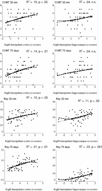
Regression plots showing the relationships between visual memory at two different retention intervals (20–30 min or 75 days) and right hemisphere cortical volume (left column) and right hemisphere hippocampal volume (right column). In no cases did the introduction of a non-linear component (represented by the dashed line) add significantly to the amount of explained variance.
 You have
Access
- 25
- Cited by