Introduction
Generic circumscriptions in Miltogramminae (Diptera: Sarcophagidae) rely almost exclusively on head shape, wing venation, chaetotaxy, and colour patterns. Male terminalia are structurally fairly simple and, although generally species specific, rarely present characters of systematic significance. This is contrary to the grey flesh flies of Sarcophaginae with their pronounced homogeneity in external morphology and remarkable diversity in structures of the male terminalia, which are notoriously difficult to put on descriptive terms.
Perhaps for this reason, authors have in general been more in agreement about miltogrammine than about sarcophagine genera, although a few genera are still only tentatively monophyletic (Pape Reference Pape1996). Some of this uncertainty pertains to taxa showing a reduction of the vibrissa, which most probably is an apomorphic feature relative to the miltogrammine ground plan, but for which the existence of intermediate forms have made it difficult to make clear-cut definitions. The reduced vibrissa is particularly crucial in the circumscription of the genus-group taxon Miltogramma Meigen (Pape Reference Pape1996). Most species of Miltogramma have the vibrissa fully reduced (i.e., it is either totally absent or indistinguishable from the sub- and supravibrissal setulae) and in such cases this feature will often enable the observer to classify a specimen as belonging to Miltogramma by a glance. However, a few Miltogramma-like species possess a vibrissa of intermediate length (i.e., it is neither well developed nor fully reduced) and a few species currently assigned to Miltogramma have a vibrissa that is slightly but unmistakably stronger than the adjacent setae, blurring the distinction between a reduced and a fully developed vibrissa.
Recent field work in Israel has secured material that adds significant pieces of taxonomic information to ongoing studies, allowing us to present a revision of two poorly known species-group taxa, both of which possess a vibrissa of intermediate length, which may have caused earlier authors to differ in opinion about the generic affiliation.
It is a great pleasure for us to dedicate this paper to the coordinators of the Manual of Nearctic Diptera, thereby showing our deep appreciation of their efforts in compiling this dipterological platform.
Materials and methods
Morphological terminology follows Merz and Haenni (Reference Merz and Haenni2000) for adults and Courtney et al. (Reference Courtney, Sinclair and Meier2000) for immatures, with modifications and additional larval terminology as in Szpila and Pape (Reference Szpila and Pape2005, Reference Szpila and Pape2007) and Szpila (Reference Szpila2010). The following abbreviations have been used for morphological structures:
Adult: T1–T5 = abdominal tergites one to five.
First instar larva: AI–AVII, abdominal segments one to seven; APL/PPL, anterior part of labrum/posterior part of labrum; HBS/LBS = height of basal sclerite/length of basal sclerite; LAD/WAD, length of antennal dome/maximum width of antennal dome; LLB/WLR, length of labrum/width of labrum; LIS/WIS, length of intermediary sclerite/width of intermediary sclerite; LPB/LBS, length of parastomal bar/length of basal sclerite; LSB/WSB, length of exposed part of first sensillum basiconicum/width of first sensillum basiconicum; P1–7, papillae one to seven around spiracular field on anal division; TI–TIII, thoracic segments one to three.
Specimens studied in the present paper are deposited in the institutions given by the following abbreviations:

Eremasiomyia macularis (Wiedemann), comb. nov.
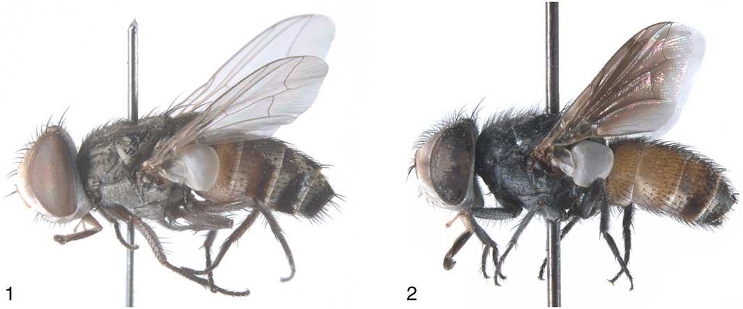
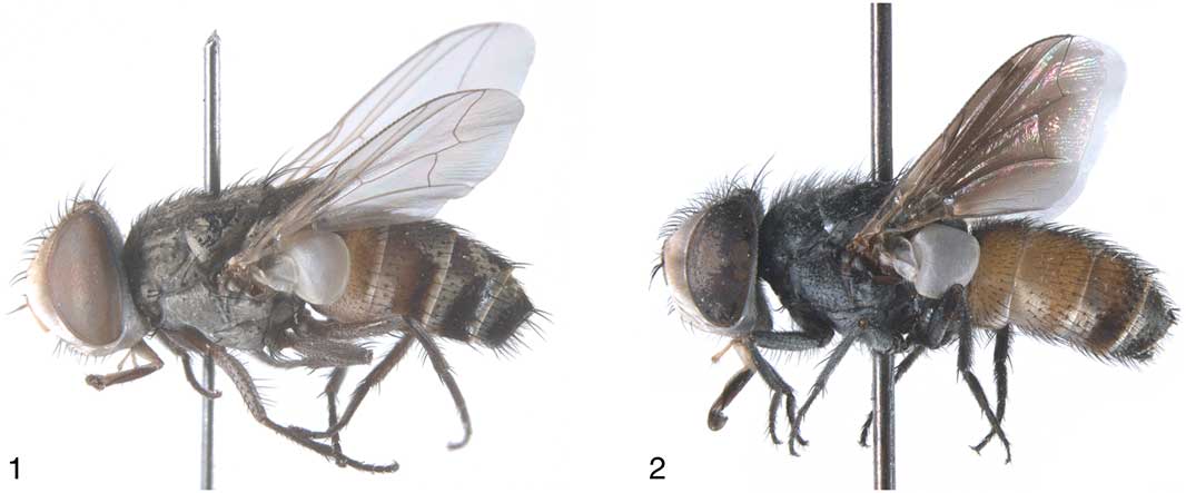
Males of Eremasiomyia macularis (Wiedemann) and Miltogramma maroccana (Séguy). 1, Eremasiomyia macularis, lateral habitus, body length = 6.1 mm; 2, Miltogramma maroccana, lateral habitus, body length = 7.8 mm.
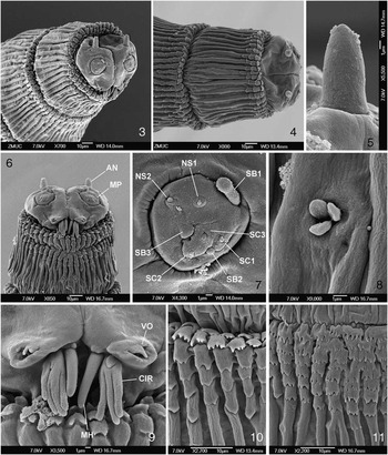
First instar of Eremasiomyia macularis (Wiedemann). 3, Anterior end, antero-lateral view; 4, Anterior end, dorsal view; 5, Antenna; 6, Anterior end, ventral view; 7, Maxillary palpus; 8, Keilin's organ; 9, Functional mouth opening; 10, Thoracic segment II, spines on dorsal surface; 11, Thoracic segment II, spines on ventral surface. AN, antenna; CIR, cirri; MP, maxillary palpus; NS1–2, additional sensilla coeloconica one and two; SC1–3, sensilla coeloconica one to three; SB1–3, sensilla basiconica one to three; VO, ventral organ.

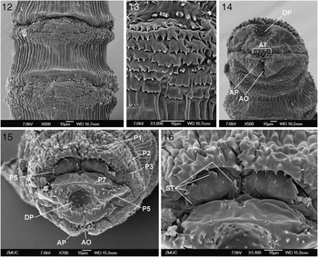
First instar of Eremasiomyia macularis (Wiedemann). 12, Abdomen, dorsal view, showing entire AII and parts of other segments; 13, Spines ventrally on AII; 14, Anal division, ventral view; 15, Anal division, posterior view; 16, Spiracular field. AO, anal opening; AP, anal pad; AT, anal tuft; DP, hemispherical depression; P1–3, 5, 7, papillae one to three, five and seven around spiracular field on anal division; PS, posterior spiracle; ST, spiracular tufts.

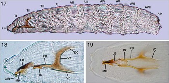
First instar of Eremasiomyia macularis (Wiedemann). 17, Habitus, lateral view, body length = 1.27 mm; 18, Cephaloskeleton, lateral view; 19, Cephaloskeleton, ventral view. AI–VII, abdominal segments one to seven; AD, anal division; CIR, cirri; DC, dorsal cornu; IS, intermediate sclerite; LB, labrum; PB, parastomal bar; PC, pseudocephalon; TI–III, thoracic segments one to three; VC, ventral cornu; VP, ventral plate.

Tachina macularis Wiedemann, Reference Wiedemann1824: 45. SE Asia [“India orient”].
Setulia luteicornis Villeneuve, Reference Villeneuve1933: 256. Egypt. syn. nov. [Valid species of Eremasiomyia in Pape (Reference Pape1996: 85).]
Eremasiomyia setulioides Rohdendorf, Reference Rohdendorf1935: 73. Egypt, Ramleh. syn. nov. [Valid species of Eremasiomyia in Pape (Reference Pape1996: 85.]
Eremasiomyia orientalis Verves, Reference Verves1979: 889. Sri Lanka, Hambantota District, Palatupana. syn. nov. [Valid species of Eremasiomyia in Pape (Reference Pape1996: 85).]
Setulia leuteicornis: Shaumar and Kamal (Reference Shaumar and Kamal1984: 131); incorrect subsequent spelling of Setulia luteicornis Villeneuve, Reference Villeneuve1933.
Eremasioides setuloides: Verves (Reference Verves1979: 889); incorrect subsequent spelling of Eremasioides setulioides Rohdendorf, Reference Rohdendorf1935.
Description
Adult male.Head. Light yellowish, almost whitish ground colour, except for dark grey postgena and occiput and dark vertex. Head covered with thin, silvery microtrichosity except for upper half of frontal stripe, which is dark reddish or brownish, and vertex, which is dark brown to blackish. Eye bare. Frons about as broad as one eye in dorsal view. A very few black setulae at vertex, fronto-orbital plate otherwise bare. Inner and outer verticals well developed. Two proclinate and one reclinate orbital bristles. Lateroclinate ocellars plus a few setulae; small postocellars. Several well-developed, inclinate frontals. Antenna inserted above middle of eye; scape, pedicel and first flagellomere orange; arista orange with distal half blackish. Antenna with a single, short, semi-stout bristle on pedicel, scape and pedicel otherwise bare or with a few black setulae. Arista bare, 0.8× as long as first flagellomere, thickened in about basal half, and with aristomere 2 as broad as long. Lunule bare. Parafacial margin moderately receding giving the head profile a trapezoidal shape; parafacial plate with scattered, short, hyaline setulae. Vibrissa shorter than first flagellomere, situated half-way between tip of antenna and margin of facial plate; vibrissae crossing in midline. Distance between vibrissae longer than one vibrissa and about as long as width of first flagellomere in lateral view. No supravibrissal setae, three to five subvibrissal setae and three to five setae of about equal size along ventral margin of gena, gena otherwise bare or with scattered minute hyaline setae. Postgena and occiput with scattered, short, black setulae. Proboscis with prementum about as long as fore tarsus and about as long as shortest diameter of eye, palpus half as long as prementum; palpus yellow, prementum dark brown.
Thorax. Black ground colour; covered with dense, grey microtrichosity; very faint trace of an olive-coloured median stripe. Chaetotaxy with weak differentiation of most bristles: acrostichals 0(−1) + 1, dorsocentrals 1 + 2, intra-alars 0 + 1, supra-alars 1 + 2, postalars 2; proepisternum (depressed part of propleuron) bare, postpronotum with two bristles, notopleuron with zero to four setulae in addition to the usual two notopleurals; scutellum with two pairs of laterals, one pair of apicals, with or without a pair of weak discals. Legs. Fore tarsus 0.9× as long as fore tibia, first tarsomere 0.75× as long as combined length of tarsomeres 2–5, tarsomeres without specialized setae, claws about half as long as tarsomere 5. Claws of mid and hind tarsi less than half as long as tarsomere 5. Fore tibia with one posterior bristle, otherwise with bristles only apically; mid tibia with one anterodorsal, one ventral, and one weak posterior bristle; hind tibia with three to four anteroventral bristles distal to middle, a row of anterodorsals from base to level of distalmost anteroventral bristle; and one to two posterodorsal bristles. Hind femur with one long subapical anteroventral bristle and one to two posteroventral bristles near base. Wing. Tegula blackish brown, basicosta whitish yellow, veins orange or light brown, membrane hyaline; costal spine not developed; base of vein R4 + 5 with one to two setulae dorsally and ventrally; cell r4 + 5 broadly open; vein M bent in a right angle. Lower calypter arched but not particularly dome-shaped.
Abdomen. Integument of T1 + 2–T4 mostly yellowish, but with a median, oval black spot on all segments and a lateral, rectangular spot on T3–T4; T5 and terminalia black. Tergites with yellowish parts covered by microtrichosity, black parts semi-shining and almost without microtrichosity. Syntergosternite 7 + 8 with grey microtrichosity.
Adult female. Similar to male but with slightly weaker bristles of the head, and vertex and posterior part of frontal stripe light yellowish and with silvery microtrichosity (i.e., not darkened).
First instar larva.Pseudocephalon. Antenna small, antennal dome long (LAD/WAD = 2.1), antennal basal ring entirely reduced; maxillary palpus shaped as flat disc clearly distinguished from surrounding surface of pseudocephalon, sensillum basiconica one long (LSB/WSB = 3.6) and shifted from central cluster of sensilla to dorso-median periphery of maxillary palpus, tip of sensillum basiconica one slightly swollen; ventral organ on flat, fleshy lobe, dorsal margin of median sensillum of ventral organ with one to two small cones; below ventral organ (close to functional mouth opening) with a group of three to four large and long processes; oral ridges absent; surface of pseudocephalon without cuticular ridges. Cephaloskeleton well sclerotized. Labrum not elongated (LLB/WLB = 4.9), anterior part of labrum clearly distinguished, short (APL/PPL = 0.3), narrow and slightly curved downward; mouthhooks widening gradually apically, tip of each mouthhook with single sharp tooth set at a right angle to mouthhook; intermediate sclerite in lateral view partly hidden behind parastomal bars, in ventral view almost 1.5× longer than wide (LIS/WIS = 1.5); transverse junction of intermediate sclerite (crossbeam) wide; parastomal bar long (LPB/LBS = 1.0); basal sclerite higher than long (HBS/LBS = 1.2); vertical plate slightly broader than dorsal cornu and of the same width as ventral cornu; dorsal bridge absent.
Thoracic segments. Anterior spinose band with four (lateral surfaces) to eight (ventral surfaces of TI–TIII) rows of spines; thoracic segments on dorsal surface with scale-shaped spines with serrated or straight posterior margin, on ventral surface with flat multicuspoid spines; middle part of ventral surface of TI with additional transverse rows of spines; remaining surface of TI–TIII with cuticular ridges, arranged densely, with distance between ridges smaller or equal to width of ridges; lateral papilla with slightly elongated sensillum; Keilin's organ with short, rounded sensilla.
Abdominal segments. Anterior spinose band on AI–AVII with from 2 to 12 rows of spines (including spines on ventral creeping welts), bands complete; on dorsal and lateral surfaces spines sparse, blunt and arranged separately, on ventral surface spines of anterior spinose bands occupy about one-third of length of segments, on ventral surface small spines arranged in clusters where several spines are fused basally; posterior spinose band present on AIV–AVII, bands restricted to lateral surface or lateral and dorsal surfaces; spines of posterior spinose band blunt and arranged separately; lateral creeping welts well developed and equipped with a few spines; ventral creeping welts well developed and with several rows of small spines; whole surface of AI–AVII with fine cuticular ridges, the latter sparsely arranged and with distance between ridges greater than width of ridges; lateral papilla with slightly elongated sensillum; prolegs not developed. Anal division with anterior spinose band complete, but narrow on lateral surface; spines of anterior spinose band blunt and arranged separately on dorsal and lateral surfaces; ventral surface with small spines arranged in short irregular rows and reaching from anterior margin of anal division almost to anal pads; remaining surface of anal division with cuticular ridges on dorsal and lateral surfaces; P1–P7 visible as very small cones with sensillum at tip; lower half of spiracular field with scattered small spines, hair-like spines around spiracular field absent; posterior surface of anal division between spiracular field and anal pads with hemispherical depression; posterior spiracles with four spiracular tufts, distal margin of tufts shallowly serrated; anal pads rounded and with numerous spines.
Biology. Unknown except that two females from Egypt were apparently collected from rotting fish and human excrement (see ‘Other material’).
Distribution. PALAEARCTIC – Egypt, Israel (including older records from “Palestine”), United Arab Emirates. ORIENTAL – Pakistan, Sri Lanka, Thailand.
Material examined
Type material: [Patria incognita, probably India, Tranquebar]: 1♂ “Ind: or” [ZMUC; holotype of T. macularis Wiedemann, Reference Wiedemann1824]. SRI LANKA: 1♂, Hambantota District, Palatupana [as “Palutapana”], 3–4.ii.1975, Krombein et al. [USNM; holotype of E. orientalis Verves, Reference Verves1979; abdomen glued to label].
Other material: EGYPT: 2♀, Cairo, “Embaba”, 1951, “Over fish & H. stool” [no collector] (USNM). ISRAEL: 1♀, Timna, 18.iii.1995, B. Merz (NHRS); 3♂ 7♀, Rehoboth near Jaffa, 25.ix–13.x.1931, J. Aharoni [SMNS; three specimens with the additional label “Setulia/luteicornis/Villen” in Villeneuve's hand]; 1♀, Negev, Makhtesh Ramon Nature Reserve, Nahal Ramon, 23.v.2004, K. Szpila (ZMUC); 1♀, Negev, Ein Avdat National Park, 21–22.v.2004, K. Szpila (ZMUC); 2♂, Negev, Nahal Zin near road 277, 15–16.v.2006, K. Szpila (IEEP); 1♂, Ein Avdat National Park, 22.v.2005, K. Szpila (ZMUC). PAKISTAN: 2♀, Sind Province, Miani Forest near Hyderabad, 24.ix.1976, Hevel & Dietz (USNM). SRI LANKA: 2♀, Anuradhapura District, Padaviya Tank, 13–20.iii.1976, P.B. Karunaratne et al. (USNM, ZMUC); 1♀, Hambantota District, Palatupana, WLNPS Bungalow, 18–21.i.1979, K. Krombein et al. (ZMUC); 1♀, Hambantota District, Palatupana Tank, 3–4.ii.1975, Krombein et al. (USNM); 1♀, Kandy District Peradeniya Central Agricultural Center, 23.v.1968, T.F. Halstead (ZMUC); 1♀, Mannar District, Cashew Corp., Ma Villu, 17–19.ii.1979, K. Krombein et al. (NHRS); 1♂, Mannar District, Cashew Corp., Ma Villu, 17–21.ii.1979, K. Krombein et al. (NHRS); 3♀, Mannar District, Kondachchi, 17–19.ii.1979, 17.ix.1979, 20.ix.1979, Krombein et al. (USNM); 1♀, Monaragala District, Mau Ara, 10 m E Udawalawa, 24–26.ix.1976, Krombein et al. (USNM); 2♂, Nuwara Eliya District, Hakgala Natural Reserve, 6–7.ii.1979, K. Krombein et al. (NHRS, USNM). THAILAND: 1♀, Bangkok, Mahidol University, 14.ix.1975, H. Kurahashi (NIID). UNITED ARAB EMIRATES: 1♂ 1♀, Fujairah, Tayyibah, 13–20.iv.2005, K. Szpila (ZMUC); 1♂, Huwaylat, 16–17.iv.2005, K. Szpila (ZMUC); 1♂, 25 km S. Dhaid, 22.iv.2005, K. Szpila (ZMUC); 3♀, Nakhali Dubai, 23–25.iv.1984, E.A. Sugden (BME).
Larval material: five larvae (three for SEM and two for light microscope), obtained from the same female [Negev, Ein Avdat National Park, 21–22.v.2004, K. Szpila (ZMUC)].
Discussion. Wulp (Reference van der Wulp1896) catalogued the Oriental Diptera and listed T. macularis in its original generic combination. Townsend (Reference Townsend1931), who was the first to publish on (and may have been the first to study) the male holotype after Wiedemann's original description, transferred the taxon to Miltogramminae and mentioned that the species “is near Protomiltogramma T.T.”, to which genus it was provisionally assigned. There is no mention of the taxon in Senior-White et al. (Reference Senior-White, Aubertin and Smart1940), who did not include any Miltogramminae in their treatment of the Oriental flesh flies. (Senior-White's family key seems to indicate that this is not because he classified the miltogrammines outside the flesh flies, and it may simply have been due to an oversight of Townsend's study combined with a general lack of material, as the Miltogramminae are only sparsely represented in the Oriental Region.) Crosskey (Reference Crosskey1965) examined the type and referred it to Miltogramminae without further comment. Lopes et al. (Reference Lopes, Kano, Shinonaga and Kurahashi1977) transferred T. macularis to Miltogramma, while Pape (Reference Pape1996) followed Townsend's suggestion and maintained the species in Protomiltogramma Townsend.
We find a generic affiliation with either Miltogramma or Protomiltogramma basically without morphological support and are here assigning T. macularis to Eremasiomyia Rohdendorf. This is not entirely satisfying, as we are unable to present features providing strong support for the monophyly of this genus-group taxon. However, we see no better corroborated affiliation at present. Pape (Reference Pape1996) listed only three diagnostic (not necessarily autapomorphic) features for Eremasiomyia: antenna yellow, vibrissa about as long as distance between vibrissal sockets, and head length at vibrissa just shorter than head length at antennal insertion (i.e., facial profile not triangular). These features may be prone to homoplasy and Pape (Reference Pape1996) was “unable to provide a more explicit and unambiguous diagnosis” of Eremasiomyia, thus leaving the genus “difficult to separate from Miltogramma, Chaetapodacra [Rohdendorf], Pterella [Robineau-Desvoidy], and Senotainia [Macquart]”.
Extensive recent work on first instar larvae of Miltogramminae points to interesting features shared by Eremasiomyia and Pterella, particularly the presence of scale-like spines at least dorsally on the thoracic segments and a hemispherical depression on the posterior surface of the anal division (i.e., in the area between the spiracular field and the anal pads) (Szpila Reference Szpila2010). The first instar morphology of Miltogramma spp. is very variable but at least some characters are widespread in this genus: (1) sensilla of maxillary palpus on raised protuberance, (2) second sensillum basiconicum of maxillary palpus at least slightly elongated, (3) parastomal bar fused with basal sclerite high above lower margin of ventral cornu, (4) triangular (in lateral view) intermediate sclerite situated apparently below the parastomal bars, and (5) entire cephaloskeleton broad in ventral view, with crossbeam of intermediate sclerite long. None of the features listed above are present in the first instar of E. macularis. They are also absent in Miltogramma (Rohdendorfomyia) ukrainica (Verves), but the morphology of the first instar of this species is not consistent with any other Miltogramma in the construction of the cephaloskeleton nor in many other morphological details, and the species shows noteworthy similarity to first instars of Craticulina Bezzi (Szpila Reference Szpila2010). The first instar larva of E. macularis is very similar to known larvae of Pterella (except for Pterella convergens (Pandellé), see details in Szpila Reference Szpila2010) and Protomiltogramma. These genera share the following conspicuous first instar features: (1) lack of basal ring of antenna, (2) first sensillum basiconicum of maxillary palpus entirely shifted from central cluster of sensilla to border of maxillary palpus, (3) presence of scale-like spines on at least dorsal surface of thoracic segments, and (4) presence of hemispherical depression on the posterior surface of anal division. The level of similarity is striking and only a few morphological details (e.g., presence/absence of clusters of processes close to functional mouth opening, small differences in shape of labrum or mouthhooks) makes the differentiation of these larvae possible. At this moment, Eremasiomyia, Protomiltogramma, and Pterella cannot be defined unambiguously on the basis of larval characters, but they seem to comprise a clade as corroborated above.
When Rohdendorf (Reference Rohdendorf1927) erected Eremasiomyia to contain the single species Eremasiomyia erythrogastra Rohdendorf, he noted the resemblance of this species (plus a few others) to Miltogramma. However, he cited the presence of a small, yet distinct vibrissa in support of the new genus (Rohdendorf Reference Rohdendorf1927). Pape (Reference Pape1996) defined Miltogramma “largely by the reduced vibrissa” (p. 28), but also provided three other diagnostic features: frontal stripe reddish, head length at vibrissa about equal to length at antennal insertion, and lower calypter large and dome-shaped. Tachina macularis has a short but clearly differentiated vibrissa, the head profile is moderately receding, and the lower calypter is arched without being dome-shaped. Thus, only the reddish colour of the upper part of the frontal stripe seems to match the diagnosis of Miltogramma, and an inclusion in Miltogramma (sensu lato) is essentially without support. Reduced or hair-like presutural acrostichal and dorsocentral bristles is a feature often mentioned as diagnostic for Miltogramma and Eremasiomyia (e.g., Verves Reference Verves1989), but this feature is widespread in Miltogramminae and does not in itself appear to be an argument for synonymizing these genera.
Following its description, Eremasiomyia received little attention, primarily the description of seven additional species (references in Pape Reference Pape1996). Verves (Reference Verves1989) made a first attempt at a generic phylogeny for Miltogramminae and considered Eremasiomyia to be the sister taxon of Protomiltogramma. A single synapomorphy was cited (reduced presutural dorsocentral bristles) (Verves Reference Verves1989), and Eremasiomyia was left without autapomorphies.
Eremasiomyia and Protomiltogramma key out at the end of Verves’ key to Palaearctic miltogrammine genera (Verves Reference Verves1994), using the short and fine frontal bristles noted by Rohdendorf (Reference Rohdendorf1927) to distinguish Eremasiomyia. Pape (Reference Pape1996) did not include the reduced or hair-like frontal bristles in his list of diagnostic features for Eremasiomyia, but did use this feature as a key character for Eremasiomyia in a chapter on Palaearctic Sarcophagidae (Pape Reference Pape1998). All Palaearctic Eremasiomyia except E. macularis appear to possess fine and hair-like frontal bristles that barely meet in the midline; this is only the case for a subset of the specimens of E. macularis examined for the present study. Especially large-sized males have well-developed frontal bristles that meet and overlap in the midline. The reduced size of frontal bristles is consequently not a fully satisfying feature for keying out Eremasiomyia, but the allometric scaling with body size may be a possible autapomorphy.
An affiliation with any of the other genera with which T. macularis has been associated seems contradicted or with no particular support. The species does not possess the perhaps most conspicuous autapomorphy of Protomiltogramma: the posteriorly directed and upcurving setae posteroventrally on tergite 5. Neither does it possess the narrow fronto-orbital plates; the lateral, dark scutellar setae; the arm-like elongated postero-lateral lobes of abdominal sternite 5; nor the slender, bare cercal tip which are additional characters given by Verves (Reference Verves1994) to recognize Protomiltogramma. The male tarsal claws are short in E. macularis, for which reason a position in Senotainia may seem untenable, and the open wing cell R4 + 5 seems to exclude a position within Chaetapodacra. Pterella is another ill-defined miltogrammine genus of uncertain affinities to Eremasiomyia, and the suite of four features given by Pape (Reference Pape1996) serves more as an illustration of the lack of unifying character states than as a means of recognition.
Eremasiomyia macularis clearly bears an overall morphological resemblance to species of Pterella, as evidenced by the adjectival species epithet of one of its junior synonyms (E. setulioides) and by the fact that Villeneuve (Reference Villeneuve1933) placed E. macularis (as S. luteicornis) in Setulia Robineau-Desvoidy (currently = Pterella Robineau-Desvoidy), although, as with several of his contemporaries, Villeneuve did not make a distinction between Setulia and Protomiltogramma. We have not been able to translate this general similarity between Eremasiomyia and Pterella into clear-cut character states, nor can we present a suite of character states corroborating the monophyly of Pterella. Species of this genus appear to differ from those of Eremasiomyia only by possessing a vibrissa that is longer than the distance between the vibrissal sockets and by their extensively greyish microtomentose abdomen.
In conclusion, our best diagnosis for Eremasiomyia is the combination of the following features: yellow antenna; short vibrissa (about as long as distance between vibrissal sockets); parafacial plate only moderately receding (i.e., head trapezoidal and not triangular); short male claws; arched but not strongly dome-shaped lower calypter; open wing cell r4 + 5; and (mostly) short, hair-like frontal bristles. The monophyly of the genus still needs corroboration in the context of a phylogenetic analysis, but on the present evidence the included species will not fit within any other miltogrammine genus. Larval instars of the other species are still unknown and therefore cannot provide further phylogenetic information.
Wiedemann's (Reference Wiedemann1824) original description of T. macularis does not explicitly mention whether one or more specimens were available to him. It seems likely, however, that only one was involved. In agreement with this, Townsend (Reference Townsend1931: 379) gave “Male Ht in Copenhagen” and Zimsen (Reference Zimsen1954) listed only one specimen as present in the Wiedemann collection. We are here following Townsend in considering the single male found in the Wiedemann collection as the holotype.
The type specimens of all nominal taxa here synonymized have been studied, except those of E. setulioides and S. luteicornis, which we have been unable to locate. Searches in the collections of Brussels, London, Ottawa, Paris, St. Petersburg, and Stuttgart were all negative. The evidence for listing E. setulioides in synonymy with T. macularis is therefore based entirely on a comparison of the original description of the former nominal species with the material examined of the latter, which in our opinion provides a perfect match. For S. luteicornis, we have strong evidence that the proposed synonymy is correct from an examination of 10 specimens from Israel, Rehoboth, which were identified as S. luteicornis by Villeneuve himself (three of these carry Villenuve's handwritten label with “Setulia luteicornis”). These specimens appear to have been studied by Villeneuve before the printing of the original description of luteicornis, but we do not consider them as part of the type series. The type locality is not given explicitly by Villeneuve (Reference Villeneuve1933), but he noted that all species described were from Egypt except for Pediasiomyia lindneri Villeneuve, which was from Palestine. By inference, the remaining species of Villeneuve's paper (thus including S. luteicornis) should be from Egypt. Letters from Villeneuve to Erwin Lindner dating from the 1930s (in the archives of SMNS), clearly show that Lindner sent Palestinian material to Villeneuve for determination during that time, and it is probable that most material was returned (only Villeneuve's letters are present, not those of Lindner; Tschorsnig in litt. 19.ix.2005). A letter from Villeneuve to Lindner dated 3.x.1932 mentions that “Il y a, dans votre envoi de Diptères de la Palestine, une ou 2 espèces de Miltogramminae que Rohdendorf a probablement decrits. Je lui ai écrit en août … mais pas de réponse de sa part. Je vais donc lui écrire de nouveau”. A postcard from Villeneuve, dated 30.viii.1933, gives: “J'ai inclus, dans un manuscrit traitant de plusieurs Miltogramminae d'Egypte, la description d'une ou 2 espèces nouvelles comprises dans votre envoie; ce travail paraitra dans Ann. Soc. Ent. France. L'une des espèces vous est dediée”. A following letter from 13.ix.1933 contains the note: “J'aime espérer que vos diptères de Palestine vous sont parvenus en parfait état”. Attached to this letter is a list with the names of 30 species returned to Stuttgart. Number 12 on this list is given the annotation: “Setulia luteicornis Villen – Vos exemplaires sont plus petits que ceux d'Egypte qui ont servi à ma description. Cette description a été envoyée aux Ann. Soc. Ent. France, mais n'a pas encore paru”. In other words, Villeneuve explicitly stated that the description of S. luteicornis was based on the material from Egypt and not on that from Palestine.
Miltogramma maroccana (Séguy), comb. nov.
Sphecapatodes maroccana Séguy, Reference Séguy1941: 22. Morocco, Taroudant. syn. nov.
Miltogramma maculigerum: Rohdendorf, Reference Rohdendorf1934; misidentification.
Description
Male.Head. Whitish beige ground colour, with reddish vertex and greyish or blackish postgena and occiput. Head covered with dense whitish beige microtrichosity except for vertex (around vertical bristles and ocellar triangle). Eye bare. Frons about as broad as one eye in dorsal view. Fronto-orbital plate with scattered, black setulae in posterior half, otherwise bare. Inner and outer verticals well developed. Two proclinate and one reclinate orbital bristles. Ocellar bristles not differentiated among ocellar setae; small postocellars present. Several well-developed, inclinate frontals. Antenna inserted above middle of eye; scape and pedicel light brown, first flagellomere light brown at the very base, otherwise grey; arista blackish with distal half brownish black. Pedicel with a single, short, semi-stout bristle, scape and pedicel otherwise with scattered, black setulae. Arista bare, 1.1× as long as first flagellomere, thickened in about basal half, and with aristomere 2 as broad as long. Lunule bare. Parafacial plate barely receding; bare. Vibrissa just shorter than first flagellomere, situated two-thirds down from tip of antenna towards margin of facial plate; vibrissae crossing in midline. Distance between vibrissae slightly longer than one vibrissa and about 1.5× as long as width of first flagellomere in lateral view. One to two supravibrissal setae, two subvibrissal and one to four weakly developed genal setae. Gena with short, mostly hyaline setulae, postgena and occiput with scattered, black setulae. Proboscis with prementum 1.2× as long as fore tarsus and about as long as shortest diameter of eye, palpus 0.4× as long as prementum; palpus yellow, prementum dark brown.
Thorax. Black ground colour; covered with dense, grey microtrichosity with four somewhat indistinct black stripes (median pair narrower than lateral pair and not reaching scutellum). Chaetotaxy with weak differentiation of most bristles: acrostichals 0(−1) + 1, dorsocentrals 1(−2) + 2, intra-alars 0 + 1, supra-alars 1 + 2, postalars 2; proepisternum (depressed part of propleuron) bare, postpronotum with two bristles, notopleuron with three to four setulae in addition to the usual two notopleurals; scutellum with two pairs of laterals, one pair of apicals, discals at most indicated. Legs. Fore tarsus 0.8× as long as fore tibia, first tarsomere 0.65× as long as combined length of tarsomeres 2–5, tarsomeres 1–4 apically with a pair of elongated setae (posterodorsal seta longer than anterodorsal seta), claws about half as long as tarsomere 5. Claws of mid and hind tarsi less than half as long as tarsomere 5. Fore tibia with bristles only apically; mid tibia with two to three anterodorsal, one ventral, and one to two weak posterior bristles; hind tibia with four to five anteroventral bristles distal to middle, a row of anterodorsals from base to just past the middle, almost hidden by long clothing setae; two posterodorsal bristles. Hind femur with two subapical anteroventral bristles and three posteroventral bristles in proximal half. Wing. Tegula and basicosta orange, veins orange or light brown at base, darker distally; wing membrane with a large brownish spot leaving only base and a broad posterior margin hyaline; costal spine not developed; base of vein R4 + 5 with one setula dorsally and ventrally; cell r4 + 5 broadly open; vein M bent in a right angle. Lower calypter strongly dome-shaped.
Abdomen. Integument of T1 + 2–T4 mostly orange, but with a median, black stripe; T5 and terminalia blackish. Tergites covered by light greyish microtrichosity that changes with the incidence of light. Syntergosternite 7 + 8 with grey microtrichosity.
Female. Unknown.
Immature stages. Unknown.
Biology. Unknown.
Distribution. PALAEARCTIC – Egypt, Israel, Morocco.
Material examined
Type material: MOROCCO: 1♂, Taroudant, 1939, I. Berland [MNHN; holotype of S. maroccana Séguy, double-mounted and with abdomen glued to cardboard].
Other material: ISRAEL: 1♂, Israel, Negev, Ein Avdat National Park, 15–18.v.2004, K. Szpila [TAU]; 1♂, Israel, Negev, Ein Avdat National Park, Ein Aqev Spring, 25.v.2006, K. Szpila [IEEP].
Discussion. Species of the genus Sphecapatodes Villeneuve share one unique synapomorphy: the elongated setae baso-ventrally on male tarsomere 1. Also, the most probable sister group of Sphecapatodes may be the genus Sphecapatoclea Villeneuve as argued from the shared possession of dense silvery microtomentum on male frontal stripe, laterally directed ocellar bristles, and subequal fringes of the posterior thoracic spiracle (Pape Reference Pape1996). The nominal taxon S. maroccana does not possess any of these features and is therefore evidently generically misplaced. The possession of three of the character states given by Pape (Reference Pape1996) as diagnostic for Miltogramma: frontal stripe reddish, head length at vibrissa about equal to length at antennal insertion, and lower calypter large and dome-shaped is here taken as sufficient evidence for an affiliation with this genus. Only the vibrissa seems to be a partial mismatch because, although shorter than the distance between the vibrissal sockets, it is clearly differentiated and about as strong as in E. macularis. With no alternative generic affiliation presenting a stronger case, however, the vibrissa of M. maroccana is best seen as a homoplasy (i.e., a reversal to a somewhat stouter condition).
Rohdendorf (Reference Rohdendorf1934) recorded two males of M. maculigerum Speiser from Egypt (Borgash and Wadi Abu el Rish), but this was evidently a misidentification (Pape (Reference Pape1987). Rohdendorf also erected the new subgenus Capnopteron and designated as “Typus subgeneris” the species he had identified as M. maculigerum. The two males of Rohdendorf's original material have been lost, but the short diagnosis provides strong evidence of conspecificity with M. maroccana, particularly by the mentioning of “Anterior tarsi of ♂ with long hairs on the ends of 1st, 2nd, 3rd, and 4th joints”, “Wings rather dense dark brown in the middle”, and “Abdomen on the sides reddish, but dense yellowish-grey dusted” (Rohdendorf Reference Rohdendorf1934: 11). This fits M. maroccana perfectly and is not a close match for any other named species of Miltogramma.
When it is discovered that a type species was misidentified by the proposing author, it is necessary to “select, and thereby fix as type species, the species that will … best serve stability and universality” (International Commission on Zoological Nomenclature 1999; Article 70.3). This leaves open the selection of either the nominal species originally cited as type species by the author, or the taxonomic species actually involved in the misidentification (International Commission on Zoological Nomenclature 1999; Articles 70.3.1 and 70.3.2). As the subgeneric classification of genus Miltogramma is premature, and as both options will result in monotypic subgeneric taxa of uncertain phylogenetic affinities, we have decided to select the species actually involved and as identified by us. The full nomenclatural synopsis for the nominal genus-group taxon Capnopteron is accordingly:
Miltogramma Meigen
Capnopteron Rohdendorf, Reference Rohdendorf1934: 11 (as subgenus of Miltogramma Meigen, Reference Meigen1803). Type species: Miltogramma maculigera Speiser, Reference Speiser1910 sensu Rohdendorf (Reference Rohdendorf1934) [=Sphecapatodes maroccana Séguy, Reference Séguy1941], by original designation and as fixed by present selection in accordance with Article 70.3.
Rohdendorf (Reference Rohdendorf1934) included Miltogramma hirtimana Bezzi in his Capnopteron. This was not explicitly argued from any shared features, but may have been based on the chaetotaxy of the male fore tarsus with elongated setae distally on the dorsal surface of the fore tarsomeres. We are here following the broad concept of Miltogramma proposed by Pape (Reference Pape1996), and we consider hypotheses of subgeneric groupings within Miltogramma as largely premature awaiting extensive revisionary work on this large genus, which is outside the scope of the present paper.
Acknowledgements
We are grateful to Dr. Amnon Freidberg (Zoological Museum, Tel Aviv University) for help with collecting permits to K.S. and to Dr. Yoram Ayal (Mitrani Department for Desert Ecology, Ben-Gurion University of the Negev) for access to an excellent base for field research. Drs. Norman Woodley (United States Department of Agriculture, Washington, District of Columbia), Hans-Peter Tschorsnig (Staatliches Museum für Naturkunde, Stuttgart), Christophe Daugeron (Muséum national d'Histoire Naturelle, Paris), Patrick Grootaert (Institut royal des Sciences naturelles de Belgique, Bruxelles), James O'Hara (Canadian National Collection of Insects, Arachnids and Nematodes, Ottawa), Steve Heydon (Bohart Museum of Entomology, Davis), and Vera Richter (Zoological Museum, Zoological Institute of Russian Academy of Sciences, St. Petersburg) kindly provided information about specimens deposited under their care and arranged the necessary loans where relevant. Drs. Knut Rognes (Hafrsfjord, Norway) and Daniel Whitmore (Verona) provided very useful comments to the manuscript. The present work was supported by the Polish Ministry of Science and Higher Education (grant no. 2 PO4F 005 29 to KS).