Introduction
Azadirachta indica is popular for its varied medicinal properties and uses (Biswas et al., Reference Biswas2002). A large number of compounds have been isolated from various parts of A. indica, and more than 150 compounds have shown significant bioactivities (Brahmachari, Reference Brahmachari2004). Azadirachtin is the most effective phytocompound available from A. indica (Verkerk and Wright, Reference Verkerk and Wright1993).
The anthelmintic property of A. indica is well established. For example, seeds of this plant can inhibit 68.3% of the larval hatching of Haemonchus contortus (Pessoa, Reference Pessoa2001); consumption of dried A. indica leaves reduces the number of helminth eggs in the faeces of cattle (Pietrosemoli et al., Reference Pietrosemoli, Ovalez and Montilla1999). Moreover, A. indica extract showed promising anthelmintic efficiency against third-stage Haemonchus contortus larvae in in vitro studies (Rahman et al., Reference Rahman, Lee and Sulaiman2011). The aqueous and the ethanolic extracts of A. indica flowers showed antifilarial activity against the whole worm as well as nerve-muscle preparation and microfilariae (mf) of Setaria cervi (Mishra et al., Reference Mishra2005). In the recent past, the importance of azadirachtin for stored product protection (Saxena, Reference Saxena and Schmutterer2002), veterinary purposes (Van der Esch et al., Reference Van der Esch, Carnevali, Amici, Kleeberg and Strang2009), and human medicine (Schmutterer, Reference Schmutterer2002) has been reported. Azadirachtin possesses the ability to hinder Sf9 cell proliferation and protein synthesis (Rembold and Annadurai, Reference Rembold and Annadurai1993), distort the formation of mitotic spindles, and interfere with the assembly of microtubules into axonemes during gametogenesis of Plasmodium berghei (Billker et al., Reference Billker2002). Furthermore, azadirachtin has been reported to be non-toxic (Boeke et al., Reference Boeke2004) in humans (Beard, Reference Beard1989), and at sub-acute level the red blood content remained unaltered in rats (Radwan et al., Reference Radwan2001). Moreover, azadirachtin has also been reported to be non-toxic in rats, even at 5 g/kg body weight dose (Raizada et al., Reference Raizada2001).
The present study aims to evaluate the in vitro antifilarial activity of azadirachtin (purity ≥95%) against Setaria cervi, a filarial nematode parasite (family Onchocercidae) that resides inside the abdominal cavity of the bovides, and bears significant resemblance to the bancroftian filarial parasite W. bancrofti in its antigenic patterns and nocturnal periodicity (Kaushal et al., Reference Kaushal, Kaushal and Ghatak1987).
Materials and methods
Chemicals and reagents
Solvents of the highest purity grade were purchased from Merck, India, and Milli-Q water (Milli-Q Academic with 0.22 μm Millipak R-40) was used for all the assays. The TUNEL kit was procured from Promega, USA. Ivermectin was bought from Sigma-Aldrich Co. (St. Louis, MO, USA). Azadirachtin (C35H4O16) (≥95%) was purchased from Minal Exim, Mumbai India. RPMI-1640 media, MTT and NBT were purchased from HiMedia laboratories. Primary antibodies (EGL-1, CED-9, CED-4 and CED-3) and horseradish peroxidase (HRP) conjugated secondary IgG were procured from Santa Cruz Biotechnology (Santa Cruz, CA, USA), and alkaline phosphatase conjugated secondary IgG were bought from Sigma-Aldrich (St. Louis, MO, USA). Anti-GST and anti-GAPDH primary antibodies were purchased from Bio-Bharati Life Science Pvt. Ltd., India.
Collection and in vitro treatment of parasites
Adult S. cervi parasites (males and females) were collected from the abdominal cavity of a freshly butchered Bovis indica Linn., and transported to the laboratory in normal saline (0.85%). The parasites were washed several times and were kept in RPMI-1640 media at 37°C until further use. The mf were isolated by dissecting the gravid females, and were maintained in RPMI-1640 media at 37°C in 5% CO2 atmosphere.
The control adult S. cervi (two females, or one male and one female) were incubated in 10 ml of RPMI-1640 medium supplemented with 100 μg/ml penicillin, 0.25 μg/ml of amphotericin B, 100 U/ml streptomycin with 10% fetal bovine serum (FBS), and similar media composition with a combination of 5, 10 and 20 μg/ml of azadirachtin was applied for the treated sets in sterile flat-bottomed Petri plates (60 mm diameter, Tarson, India). Approximately, a minimum of ~1.0 × 10⁴ mf were incubated in 2 ml of RPMI-1640 media, supplemented with 10% FBS as control, and azadirachtin at 5, 10 and 20 μg/ml concentrations along with RPMI-1640 media supplemented with 10% FBS was used for treatment in 24-well culture plates (Tarson, India). Adult and mf cultures were maintained for 24 h at 37°C in a humid atmosphere of 5% CO2 (Mukherjee et al., Reference Mukherjee2014b). However, for certain experiments, the number of mf was increased to c. 1.0 × 10⁵. Ivermectin (IVM) at 5, 10 and 20 μg/ml was used as positive control in equivalence to the azadiractin doses.
Assessment of parasite mortality by Trypan Blue dye exclusion method
Trypan Blue selectively stains dead parasites; viable ones exclude the dye. After the stipulated period of incubation, 10 μl of the culture containing mf from each treatment panel was collected on glass slides. Thereafter, 2 μl of Trypan Blue was added to the slides, which were then covered with coverslips and observed under a microscope. The mortality of the parasites was assessed microscopically, and observations were expressed as the percentage value of dead parasites (Mukherjee et al., Reference Mukherjee2014b). The entire experiment was carried out in quadruplicate and repeated three times.
Viability assessment through MTT assay and LC50 determination
Viability of parasites (mf (c. 1.0 × 10⁴/treatment) and adult (2 gravid females/treatment dose)) was assessed by the MTT (3-(4, 5-dimethyl-thiazol-2-yl)-2, 5-diphenyl-tetrazolium bromide) reduction assay as described by Nayak et al. (Reference Nayak2012). The median lethal dose (LC50) was determined using the OriginPro 8 software. The entire set of experiments were carried out in quadruplicate and repeated at least three times.
Microscopy of control and azadirachtin-exposed parasites
To identify any morphological aberrations in the control and treated mf, both bright field and phase contrast microscopic images were taken under an inverted microscope (Dewinter, Italy).
Histological studies of azadirachtin-exposed adult parasites
After 24 hours of treatment, the control and azadirachtin-exposed adult parasites were taken from the culture media, washed thoroughly in phosphate buffered saline (PBS) and fixed in 4% paraformaldehyde overnight. After proper fixation, they were dehydrated in graded alcohol and finally embedded in paraffin and kept for at least 24 hours before microtomy. Further, 5 μm of adult parasite tissue sections were prepared for haematoxylin–eosin staining (Fischer et al., Reference Fischer2008).
Protein extraction from parasites
Control and treated mf (n = 1 × 10⁵) and adult parasites (n = 2, average length 6.00 ± 1.2 cm, weight 36 ± 6.0 mg) were harvested (by centrifuging at 5000×g rpm, 4°C for mf only) and washed thoroughly in PBS (50 mm, pH 7.4). Parasite lysates were prepared by applying mild sonication in cell lysis buffer (CellLytic™, Sigma, USA). Centrifugation at 10000×g, 4°C for 20 minutes was performed to collect the cell free supernatant. The clear supernatant thus obtained was further used for western blotting (as reported by Saini et al., Reference Saini2012) and enzyme assays. The protein concentrations were estimated following the procedure described by Bradford (Reference Bradford1976), with acceptable modifications.
Determination of altered redox status of the exposed parasites
The level of superoxide anion, H2O2, reduced glutathione (GSH), and enzymatic activity of GST (glutathione-S-transferase), catalase, glutathione peroxidase (GPx), and glutathione reductase (GR) in control and treated parasites were estimated following Mukherjee et al. (Reference Mukherjee2014a, Reference Mukherjeeb, Reference Mukherjeec). GST activity in both control and treated adult worm homogenates was also examined through western blotting.
Determination of apoptosis
DNA fragmentation assay of exposed adults and mf
Total genomic DNA was isolated from control and azadirachtin-exposed (24 h) mf and adults following the standard phenol-chloroform isolation procedure, as described previously (Mukherjee et al., Reference Mukherjee2014c, Reference Mukherjee2016a). The DNA samples were quantified and resolved in 2% agarose gel (50 V, 15 minutes and then 100 V, 90–100 minutes), stained in ethidium bromide (EtBr) and analysed under the gel documentation system (Bio-Rad, USA).
Terminal deoxynucleotidyl transferase dUTP nick end labelling (TUNEL) assay
In situ chromatin fragmentation is a hallmark of apoptosis. To detect chromatin fragmentation in control and treated adult female worm sections, TUNEL (DeadEnd™ Colorimetric TUNEL System, Promega, USA) assay was performed following the manufacturer's protocol and as described by Mukherjee et al. (Reference Mukherjee2014b). The tissue sections were subsequently photographed under an inverted microscope (Dewinter, Victory, Italy).
AO/EtBr double staining of exposed mf
To detect apoptosis in azadirachtin-exposed S. cervi mf, acridine orange (AO)/ethidium bromide (EtBr) double staining assay was performed according to Roy et al. (Reference Roy2014) and Gucchait et al. (Reference Gucchait2017), and was micrographed. AO and EtBr were prepared separately at a concentration of 0.1 mg/ml in 0.1 m PBS, and mixed at a 1 : 1 ratio. After 10 minutes of incubation at room temperature in the dark, the AO/EtBr exposed mf were mounted on a glass slide and micrographed under a fluorescence microscope (Dewinter, Italy). The detection wave length was 460–490 nm.
RT-PCR analyses of apoptogenic genes
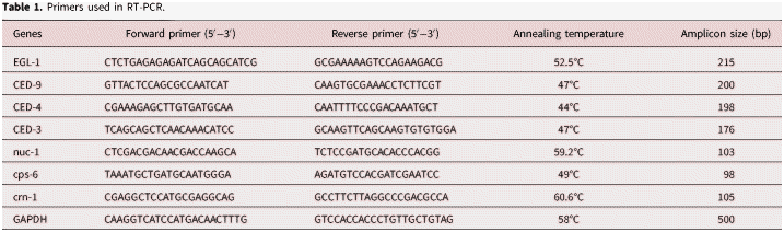
To isolate the RNAs from control and azadirachtin treated adults (n = 2, gravid females for each treatment set) and mf (n = 1 × 10⁵/treatment set), we used the TRI Reagent (Sigma, USA), following the method described earlier (Nayak et al., Reference Nayak2012). The isolated RNA was quantified using a Shimadzu UV-1601 spectrophotometer. Oligo (dT)18 primed reverse transcription (RT) was performed with a First Strand cDNA Synthesis Kit (Fermentas). The RT-PCR was done according to the process described earlier (Nayak et al., Reference Nayak2012). Further, 7 μg of each PCR amplicon was run on a 2% agarose gel, stained in EtBr and analysed under the gel documentation system (Bio-Rad, USA). Primers (apoptotic and housekeeping genes) were designed and confirmed with the aid of the National Center for Biotechnology Information (NCBI) database (http://blast.ncbi.nlm.nih.gov) and the Primer3 Software Input (version 0.4.0). Primer sequences with their annealing temperatures are provided in table 1.
Primers used in RT-PCR.

Immunoblotting of apoptogenic proteins
To detect the changes in the expression of nematode-specific apoptogenic proteins, i.e. EGL-1, CED-9, CED-4 and CED-3, we used immunoblotting (Nayak et al., Reference Nayak2012; Saini et al., Reference Saini2012). Before immunoblotting, the extracted proteins were resolved in 12.5% SDS-PAGE and then silver staining was performed to observe the changes in the expression profile (see supplementary material & method and supplementary fig. S2B). The extracted proteins (60 μg for adult and 120 μg for mf) from the control and treated parasites (adult and mf) were resolved by 12.5% SDS-PAGE and electrotransferred to polyvinylidene difluoride (PVDF) membranes, blocked with 10% skimmed milk, and incubated with different anti-goat primary antibodies viz. EGL-1, CED-9, CED-4 and CED-3 overnight at 4°C. The membranes were thereafter washed in Tris base saline Tween-20 (TBST) solution and incubated with HRP conjugated donkey anti-goat secondary antibody for 120–150 minutes. Alterations in the expression patterns of various apoptogenic proteins were examined by the specific substrate 3,3′,5,5′-tetramethylbenzidine (TMB)-H2O2 (Bangalore GeNei, India). The presence of specific bands was quantified using the Quantity One densitometry software (Bio-Rad, USA).
Determination of toxicity of azadirachtin
In vitro toxicity evaluation
To detect the in vitro toxicity, 5, 10 and 20 μg/ml of azadirachtin was applied to peritoneal macrophages isolated from an adult Wister rat (weight c. 120 ± 20 g) following a previously reported method (Roy et al., Reference Roy2014). In brief, after attainment of specific morphology, the macrophages were incubated with azadirachtin (5, 10 and 20 μg/ml) for 6 h at 37°C with 5% CO2 in a humidified incubator. Control and treated cells were first micrographed, and then harvested by scraping, followed by centrifugation at 5000 rpm for 5 minutes. Cells were washed twice in PBS, and cell death percentages were quantified by MTT assay (Roy et al., Reference Roy2014). The entire experiments were carried out in quadruplicate and repeated three times.
In vivo toxicity evaluation
For in vivo toxicity studies, Wister rats (weight c. 160 ± 20 g) were randomized into control and experimental groups of three individuals per set. Experimental groups were fed azadirachtin orally at 5, 50 and 100 mg/kg body weight once per day, suspended in 0.7% carboxymethyl cellulose for 28 days, in accordance with the Organization for Economic Cooperation and Development (OECD) guidelines (OECD, 1995). The animals had access to food and water ad libitum and were maintained under controlled temperature (27 ± 2°C) and 12 h : 12 h light and dark cycle. The initial body weight of each rat was recorded. At the end of the study, rats were sacrificed, and blood was collected. All the activities, including the corpse clearance, were carried out according to the guidelines provided by the Committee for the Purpose of Control and Supervision of Experiments on Animals (CPCSEA). 1 ml of blood was collected in EDTA for haematological studies, and 2 ml of blood was collected for clinical analysis. Collected blood samples were centrifuged at 1500×g for 15 minutes, and the plasma was collected immediately. All samples were processed within 4–6 h.
Histological preparation
Small portions of the freshly dissected and perfused rat livers were fixed overnight in Bouin's fixative, dehydrated in graded ethanol series (30–100%), and embedded in paraffin. Paraffin sections (5 μm thick) were stained with haematoxylin–eosin following Cardiff et al. (Reference Cardiff, Miller and Munn2014), and Hoechst staining of the liver tissue sections was done following Mukherjee et al. (Reference Mukherjee2015).
Determination of side effects
A study was conducted following the guidelines from the OECD (2009) to determine the serum level toxicity due to different concentrations of azadirachtin exposure. After 28 days of treatment, the rats were sacrificed and the biochemical tests, including the measurement of total serum protein, albumin, globulin, bilirubin (both conjugated and un-conjugated), serum glutamate oxaloacetic transaminase (SGOT), serum glutamate pyruvic transaminase (SGPT) and alkaline phosphatase, were analysed following the conventional methods.
Statistical analysis
All the experiments were repeated at least three times, and data were expressed as the mean ± SD. The results were analysed by one–way analysis of variance (ANOVA) using GraphPad Prism 5.0, and P values <0.001 (**) and <0.0001 (***) were considered to be statistically significant. Densitometry analysis was carried out using the Image J software and expressed as arbitrary units.
Results
Primary evaluation of mortality
Azadirachtin treatment showed dose-dependent enhancement in the mortality of the treated mf (c. 1.0 × 10⁴ of mf/treatment dose). After 24 h of treatment, mortality percentages of mf were 36.6%, 47.0% and 88.2% for 5, 10 and 20 μg/ml, respectively (supplementary fig. S2A). IVM was used as positive control, and the mortality due to azadirachtin exposure was comparable to that of IVM.
Dye exclusion test
The result of the dye exclusion test on the control and treated mf is shown in supplementary fig. S3. Accumulation of Trypan Blue inside the parasite body indicated that the parasite had lost its cellular integrity. However, the control parasites remained unaltered and excluded the dye.
MTT assay and LC50
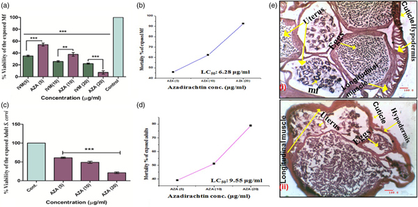
MTT assay confirmed that azadirachtin treatment caused considerable mortality of the exposed mf and adults compared to the control after 24 h of exposure. Mortality percentages of the treated mf were 45.8%, 62.3% and 92.6% for 5, 10 and 20 μg/ml of azadirachtin, respectively (fig. 1a). The LC50 value of azadirachtin against the mf was found to be 6.28 μg/ml, determined by the Origin Pro 8.0 software (fig. 1b). IVM was used as the positive control. The percentages of mortality in adults of S. cervi were noted to be 39.0%, 51.1% and 78.8% for 5, 10 and 50 μg/ml of azadirachtin, respectively, after 24 h (fig. 1c). The LC50 value was found to be 9.55 μg/ml for adult parasites (fig. 1d).
Evaluation of the antifilarial potential of azadirachtin on mf and adults of Setaria cervi. (a) % viability of S. cervi mf (c. 1.0 × 10⁴/treatment set) was evaluated by MTT assay after treatment with 5, 10 and 20 μg/ml of azadirachtin and ivermectin for 24 h. (b) LC50 value of azadirachtin for mf. (c) % viability of adult (2 gravid females/treatment set) S. cervi, evaluated through MTT assay. (d) LC50 value of azadirachtin for adult S. cervi. All the experiments were done in quadruplicate and repeated three times. The bar graph shows mean ± SD values. The following P values were considered for statistical analysis: * P < 0.05, ** P < 0.01, *** P < 0.001, ns = not significant. (e) Morphological alterations of azadiractin-treated S. cervi adults. The control section (i) showed no significant morphological damage (magnification = 20×), whereas (ii) morphological alterations in epithelial lining, muscle layers (longitudinal and hypodermal) and in the cuticular region with disintegrated epicuticle lining were visible in the treated parasites, and are indicated with arrows in the treated parasite section (20 μg/ml). Scale bar (100 μm) was added using Dewinter Biowizard 4.2 software.

Alteration in external morphology
Morphological alterations in the control and treated adult parasites were imaged after conventional haematoxylin–eosin staining under an inverted microscope (Dewinter, Italy). There were visible changes in the epithelial lining, muscle layers (longitudinal and hypodermal) and predominantly at the cuticular region, with disintegrated epicuticle lining in the treated parasites (fig. 1e (ii)) as compared to the control (fig. 1e (i)).
Upregulation of ROS parameters
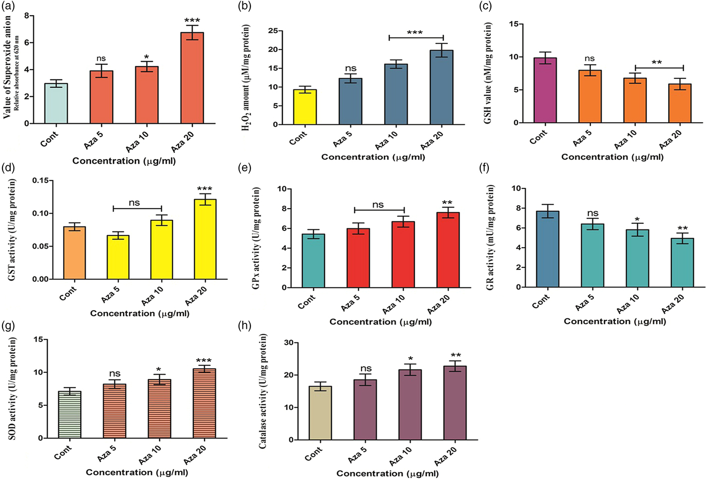
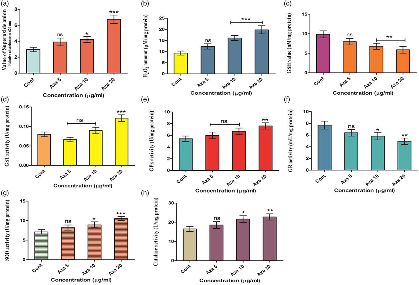
Colorimetric NBT assay showed changes in superoxide anion production in the treated worms. Intriguingly, 31.7%, 42.4% and 127.3% of superoxide anions were found to increase in the treated parasites compared to the control at 5, 10 and 20 μg/ml of azadirachtin, respectively (fig. 2a). Increases of 32.2%, 68.8% and 112.6% in H2O2 level were observed for 5 μg/ml, 10 μg/ml and 20 μg/ml of azadirachtin treatment, respectively (fig. 2b). The GSH level also differed in the treated worms compared to the control, which indicated the misbalance of internal redox milieu. Azadirachtin treatment at 5 μg/ml, 10 μg/ml and 20 μg/ml significantly reduced the GSH level, by 19.0%, 31.2% and 40.2%, respectively (fig. 2c). Azadirachtin at 5 μg/ml caused a decline of GST activity by 16.7% but at the higher doses, i.e. 10 μg/ml and 20 μg/ml, GST activity became upregulated by 12.4% and 52.0%, respectively, compared to the control (fig. 2d). A similar trend of GST activity was observed in immunoblotting (fig. 4a). A dose-dependent elevation in the GPx activity was observed in the treated parasites (fig. 2e). Azadirachtin treatment at 5 μg/ml, 10 μg/ml and 20 μg/ml caused 1.1-fold, 1.23-fold and 1.40-fold increases in the GPx activity, respectively. However, GR activity decreased by 16.9%, 24.4% and 35.6% upon treatment with azadirachtin at doses of 5 μg/ml, 10 μg/ml and 20 μg/ml, respectively (fig. 2f). The superoxide dismutase (SOD) level in the parasite increased 1.15-fold, 1.25-fold and 1.48-fold at 5 μg/ml, 10 μg/ml and 20 μg/ml azadirachtin exposure, respectively (fig. 2g). Catalase activity was recorded to be elevated 1.12-fold, 1.31-fold and 1.38-fold at 5 μg/ml, 10 μg/ml and 20 μg/ml of azadirachtin, respectively, compared to the control (fig. 2h).
Alteration of redox parameters in adult parasites after azadirachtin treatment. (a) An elevation in the generation of superoxide anion was notable with increasing concentrations of azadirachtin. (b) An elevation in the generation of H2O2 was notable. (c) Levels of GSH (nm/mg protein) showed a dose-dependent depletion compared to the control. (d) GST activity (U/mg protein) in treated parasites showed a decrease with the lowest dose (5 μg/ml), whereas there was an increase in the GST activity at higher doses (10 and 20 μg/ml) of azadirachtin. (e) GPx activity (U/mg protein) was also increased in a similar way. (f) Activity of GR (mU/mg protein) showed a dose-dependent depletion. (g) These compounds caused an increased SOD activity (U/mg of protein). (h) A dose-dependent elevation in catalase activity (U/mg protein) was observed in treated parasites. The treated and control S. cervi adults (2 gravid females for each set) were incubated for 24 h. All data represent minimum values of three independent experiments, and the bar graph shows mean ± SD (* P < 0.05, ** P < 0.01, *** P < 0.001, ns = not significant).
Apoptosis detection in azadirachtin-exposed parasites
Determination of DNA fragmentation
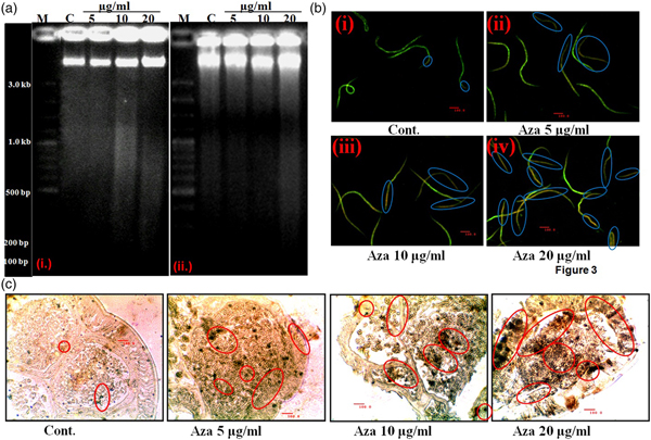
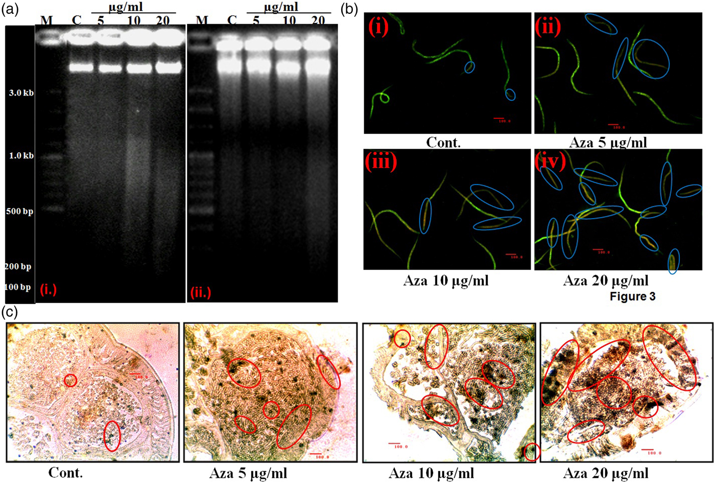
DNA samples obtained from azadirachtin-treated adults and mf showed marked DNA laddering after electrophoresis (fig. 3a). Most DNA breakage was found in the 20 μg/ml treatment dose applied for both mf (fig. 3a(i)) and adults (fig. 3a(ii)). DNA isolated from control parasites showed no significant laddering after electrophoresis.
Induction of apoptosis in azadirachtin-exposed mf (n = 1 × 10⁵) and adults of S. crervi (2 gravid females for each set). (a) Azadirachtin-induced DNA fragmentation in (i) mf and (ii) adults of S. cervi after 24 h exposure. Lane M denotes molecular weight marker (100–3000 bp). DNA fragmentation is observed for treated (i) mf and (ii) adult parasites. (b) AO/EtBr double stained micrographs of azadirachtin-exposed mf. (i) Control mf shows green fluorescence and (ii–iv) show varying degrees of yellow and orange fluorescence in azadirachtin-treated panels, with most orange fluorescence found for the highest dose applied (20 μg/ml). (c) TUNEL positive apoptotic nuclei in treated adult worm sections. Presence of dark brown apoptotic nuclei (marked with red circles) is evident in treated (5, 10 and 20 μg/ml) parasite sections (ii–iv), with very few in the control (i).
Double staining of mf
AO and EtBr double staining can distinguish between live mf and mf in various stages of apoptosis or necrosis. It was observed that the control mf showed green fluorescence (fig. 3b (i)), but varying degrees of yellow and orange fluorescence appeared in azadirachtin-treated mf (fig. 3b (ii–iv)). Intense orange fluorescence was observed in the highest treatment (20 μg/ml) dose (fig. 3b (iv)).
Detection of apoptotic nuclei by in vitro TUNEL assay
The presence of apoptotic nuclei was prominent in the treated adult worm sections (fig. 3c (ii–iv)). The number and intensity of TUNEL positive apoptotic nuclei were most significant at 20 μg/ml. This result confirmed apoptosis as the key event behind the death of the exposed parasites.
RT-PCR of apoptogenic genes
The expression patterns of anti-apoptotic (CED-9) and proapoptotic (EGL-1, CED-4 and CED-3) genes of the execution pathway, and nuc (abnormal nuclease)-1, cps (CED-3 protease suppressor)-6 and crn (cell-death-related nuclease)-1 of the DNA degradation pathway were semi-quantitatively measured by RT-PCR in both the treated and control adults and mf of S. cervi. There was found to be a dose-dependent increase in the expression of proapoptotic EGL-1, CED-4 and CED-3 genes for both the treated mf and adults (see supplementary fig. S4). However, azadirachtin downregulated the expression of anti-apoptotic CED-9 in the treated parasites compared to the control worms (see supplementary fig. S4A, C). Moreover, expression patterns of nuc-1, cps-6 and crn-1 were also found to be upregulated in the treated adults as well as mf (see supplementary fig. S4A, C). Densitometric analyses of the bands were also provided (see supplementary fig. S4B, D). The results suggested that azadirachtin at transcriptional level was able to reduce CED-9 expression and elevate proapoptotic EGL-1, CED-3 and CED-4 genes. Upregulated expressions of different DNA degradation genes were also prominent. The retrieved cDNA sequences of EGL-1 from S. cervi showed significant similarity (>99% with a query coverage of 37% and 42%) with EGL-1 of C. elegans. Setaria cervi CED-9 was 99% homologous to the CED-9 of C. elegans (query coverage of 90%). Accession numbers for partial cDNA sequences of EGL-1 are KC747728 and KC970156 and for CED-9 is KC847047, as provided by NCBI (www.ncbi.nlm.nih.gov).
Changes of apoptogenic protein expression
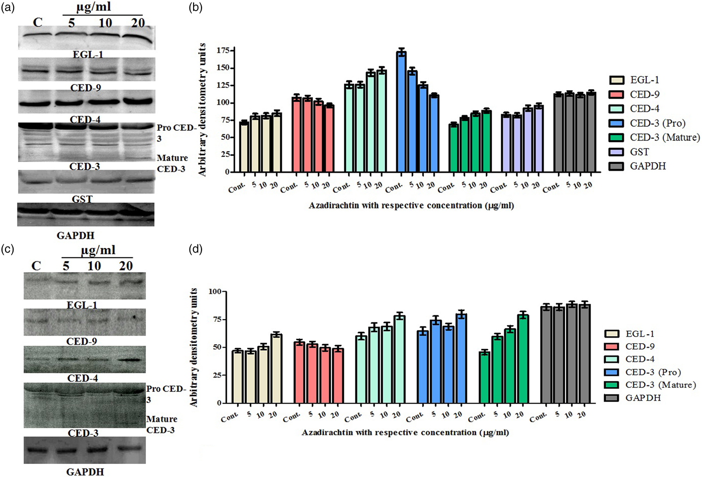
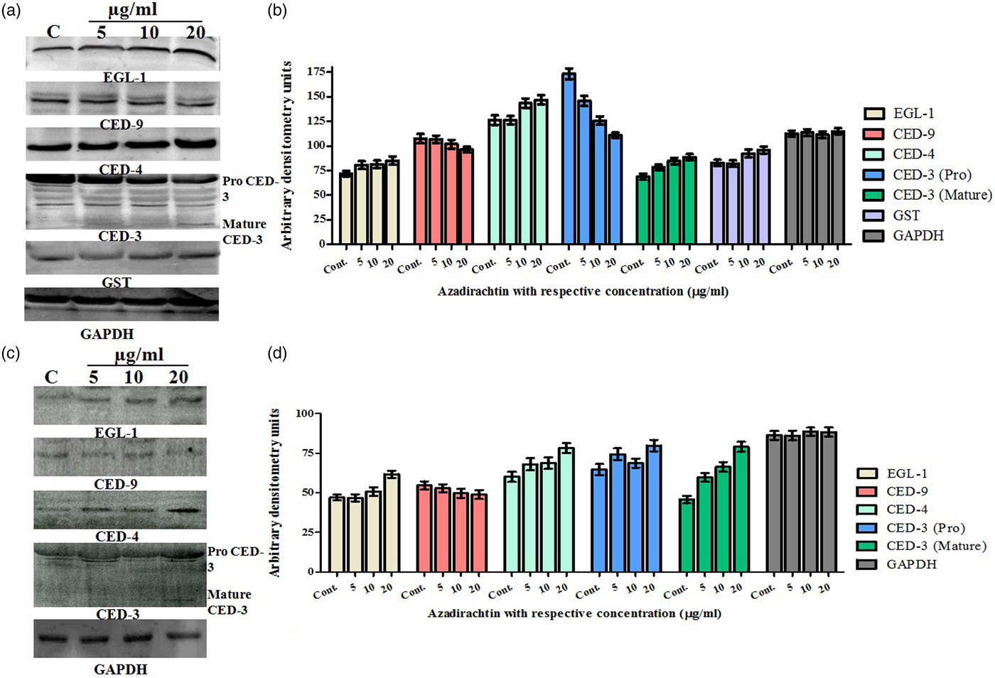
Immunoblotting of nematode-specific apoptogenic proteins EGL-1, CED-9, CED-4 and CED-3 was performed to evaluate apoptosis execution in the exposed parasites (both adults and mf) at the level of translation. Azadirachtin caused downregulation of CED-9 in treated worms (adult and mf). Subsequent elevation in the proapoptotic proteins EGL-1, CED-3 and CED-4 were noted in the treated adults and the mf compared to the control (fig. 4a, c). Densitometric analyses of the blots were also provided (fig. 4b, d). Thus, downregulation of anti-apoptotic protein and simultaneous upregulation of proapoptotic proteins confirmed that azadirachtin exerted its antifilarial efficacy through induction of apoptosis.
Alterations in the expression of anti- and proapoptotic proteins in the treated parasites. (a), (c) showing a dose-dependent elevation of nematode-specific proapoptotic proteins; EGL-1, CED-3 and CED-4 were evident in azadirachtin-treated adults (2 gravid females for each set) and mf of S. cervi (n = 1 × 10⁵). The treatment caused downregulation of anti-apoptotic CED-9 protein expression. However, control worms showed an opposite result in the expression levels of all four proteins. GST activity also showed a similar pattern to that of the enzyme assay, downregulated at the lowest dose (5 μg/ml) and then upregulated. GAPDH served as protein loading control. For adult parasites and mf, 60 μg and 120 μg of proteins were resolved. Images are representative of three independent experiments. Densitometric analyses of the western blot bands of (b) adult parasites and (d) mf are provided.
Determination of toxicity of azadirachtin
In vitro toxicity evaluation
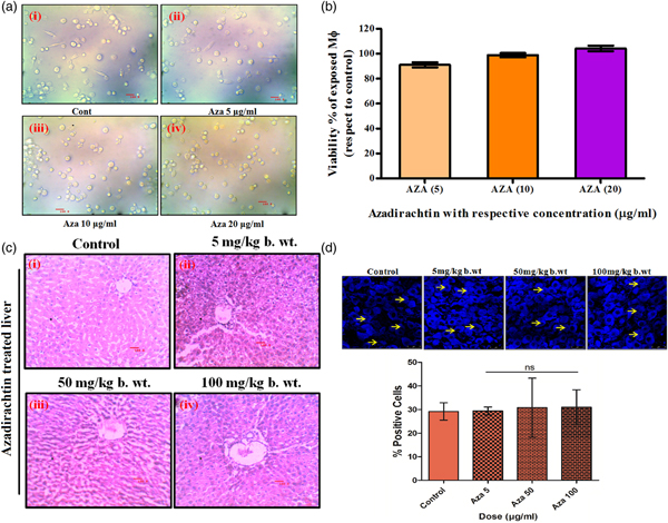
Primary microscopic morphological evaluation of azadirachtin-exposed macrophages did not show any significant death after Trypan Blue staining (fig. 5a (ii–iv)) compared to the control (fig. 5a (i)). MTT assay of azadirachtin-exposed macrophages also confirmed the non-cytotoxic nature of the compound (fig. 5b).
In vitro and in vivo toxicological evaluation of azadirachtin. (a) Trypan Blue stained microscopy images of azadirachtin-exposed rat peritoneal macrophages (ii–iv) at concentrations of 5, 10 and 20 μg/ml, respectively, after 6 h of exposure concerning the control (i). Images are representative of three independent experiments. (b) MTT assay showed no significant death of the exposed macrophages. Results are representative of three independent experiments. (c) Haematoxylin–eosin stained tissue section of liver. Liver tissues after scheduled treatment did not show any observable alterations. Photographs were taken at 400× magnification; scale bar = 100 μm. (d) (i) Hoechst stained liver tissue section (63× magnification; scale bar = 0–25 μm; laser confocal microscope, Leica, Germany); (ii) histogram showing percentage of intact cells in the treated parasite compared to the control. The arrows indicate the Hoechst positive cells with intact nuclear morphology.

In vivo toxicity evaluation
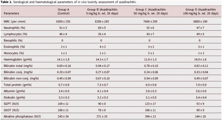
General toxicological observations during the safety assessment included mortality/morbidity observations twice per day, routine clinical observations and weekly body weight measurement. Fur, skin, mucous membrane, eyes, salivation, behavioural pattern, sleep, and food and water consumption of the treated group of rats were found to be normal during the entire treatment period. Lethargy, diarrhoea, tremors and coma were not observed. Body weight gains were normal in all the treated groups compared to the controls. The results of the haematological and serological toxicity parameters of azadirachtin treatment are presented in table 2. It appeared that azadirachtin caused no significant alterations in the haematological and serological parameters. Moreover, the liver function of treated rats was found to be normal. No profound changes were visible in haematoxylin and eosin stained liver sections (fig. 5c). Hoechst staining of the rat liver tissue sections showed no marked alterations, further corroborating the non-toxic nature of the compound (fig. 5d).
Serological and haematological parameters of in vivo toxicity assessment of azadirachtin.

Discussion
Lymphatic filariasis (LF) is known to be a noteworthy cause of long-term or permanent disability in people residing in tropical and subtropical regions. Eight hundred and fifty-six million people in 52 countries across the globe are threatened by lymphatic filariasis and require effective therapeutic measures (WHO, 2017). Lymphatic filariasis mainly affects the poor, and tribal people with poor hygiene practices are the worst affected. Currently available antifilarial chemotherapies include diethylcarbamazine citrate, albendazole and ivermectin. However, their efficacy is limited to the microfilarial stage (Shenoy et al., Reference Shenoy2008).
Natural compounds are advantageous because they are multi-targeted and well tolerated. The toxicity to non-targeted organs is not very significant. However, the available antifilarial agents, such as DEC (diethylcarbamazine citrate) and ivermectin, are associated with undesirable side effects, and there are reports of drug resistance (Lustigman and McCarter, Reference Lustigman and McCarter2007). Therefore, finding new filaricidal components from plants previously reported to have anthelmintic actions remains a feasible research objective. Several crude extracts and phytocompounds were found to be effective against filarial parasites (Mukherjee et al., Reference Mukherjee2014a, Reference Mukherjee2016b). It has been reported that both the aqueous and ethanolic extracts from the leaves of A. indica possess important antifilarial properties (Tripathi et al., Reference Tripathi, Shrivastav and Chaube2012; Mukherjee et al., Reference Mukherjee2014a, Reference Mukherjeeb, Reference Mukherjeec).
A Trypan Blue dye exclusion test confirmed the antifilarial potential of azadirachtin against mf of S. cervi (Gucchait et al., Reference Gucchait2017). Additionally, MTT reduction assay established the antifilarial potential of azadirachtin against both forms at the cellular level (Gucchait et al., Reference Gucchait2017). Significantly, azadirachtin showed promise as a potential filaricidal agent at a considerably low dose, i.e. 20 μg/ml. Morphological alterations were also prominent in the treated mf and adult worms. Therefore experimental evidence confirmed the antifilarial potential of azadirachtin.
Degraded nucleosomal DNA by endogenous endonucleases or DNase, considered to be one of the significant apoptotic markers, was detected in both adults and mf of azadirachtin-exposed S. cervi (Mukherjee et al., Reference Mukherjee2016a). Staining of the control and the azadirachtin-exposed mf with AO/EtBr provided clear indications that the treated mf had shifted towards apoptotic death. The presence of orange/red fluorescence in the mf with increasing doses of azadirachtin is indicative of a dose-dependent apoptotic death (Gucchait et al., Reference Gucchait2017). The appearance of deep brown apoptotic nuclei after TUNEL staining in tissue sections of the azadirachtin-treated adult worms confirmed in situ DNA fragmentation (Mukherjee et al., Reference Mukherjee2016a).
The antioxidant system in nematodes plays a major role in nematode survival and shields the organism from the toxic effects. Experimental data showed that azadirachtin reduced the GSH level in a dose-dependent manner in treated parasites compared to the control. Downregulated GSH in the treated parasites indicates dysregulation in the inherent redox regulation cascade of the exposed parasites, depriving the parasites of their primary defensive molecule against oxidative stress. Upregulation of GST activity during the onset of apoptosis is associated with increased oxidative stress inside the parasite body, and is often regarded as a protective strategy (Syng-Ai et al., Reference Syng-Ai, Kumari and Khar2004). Thus, the reduction of the GSH level and the rise in GST activity in the treated worms are indicative of the ROS generation and oxidative stress. The ability of the antioxidant system of the parasite to combat and neutralize ROS is essential for its survival during altered metabolism (Circu and Aw, Reference Circu and Aw2010), and also to defend itself against the host-generated ROS (Brophy and Pritchard, Reference Brophy and Pritchard1992), mainly in the case of the long-lived parasitic nematodes. The present study showed dose-dependent increases of superoxide anion and H2O2 levels with azadirachtin treatment in adult S. cervi. Changes in the levels of various redox reactive enzymatic and non-enzymatic parameters confirmed that azadirachtin caused an alteration in the redox status of the exposed parasites, which could interact directly with various cellular signalling molecules and consequently trigger apoptosis (Simon et al., Reference Simon, Haj-Yehia and Levi-Schaffer2000).
Upregulation of apoptosis in the azadirachtin-treated adults and mf of S. cervi was prominent, and may be the underlying cause of their death. Furthermore, RT-PCR profiling of various apoptotic and DNA degradation genes in mf and adult stages of S. cervi was estimated and found to be upregulated for the proapoptotic EGL-1, CED-4 and CED-3, and downregulated for the anti-apoptotic CED-9 that marked the onset of apoptosis. Interestingly, cps-6 (C. elegans homologue of mammalian mitochondrial endonuclease G that causes DNA degradation in the dying cell (Lettre and Hengartner, Reference Lettre and Hengartner2006)), crn-1(C. elegans homologue of human flap endonuclease-1 (FEN-1), involved in DNA replication and repair (Parrish et al., Reference Parrish2003)), and nuc-1 were found to be enhanced in adults and mf. The genes nuc-1, cps-6 and crn-1 encode proteins with nuclease activities that participate in the degradation of the dead cell within the engulfing cell (Lettre and Hengartner, Reference Lettre and Hengartner2006). Enhanced CPS-6, CRN-1 and NUC-1 can also minimize TUNEL responses due to further cleavage of DNA into smaller fragments for the downstream activity of CED-3 to produce TUNEL reactive apoptotic nuclei (Yu et al., Reference Yu2015). A study involving expression profiling of various nematode-specific apoptogenic proteins during the execution revealed upregulation of proapoptotic proteins (EGL-1, CED-3 and CED-4) and concomitant downregulation of the anti-apoptotic protein (CED-9). It was previously reported that anti-apoptotic CED-9 and proapoptotic EGL-1 genes of S. cervi have significant homology with their C. elegans counterpart, which suggests the conserved apoptotic pathway of S. cervi and C. elegans (Mukherjee et al., Reference Mukherjee2014b).
The present study also revealed that a subchronic exposure of azadirachtin up to 100 mg/kg body weight in the Wister rat model for 28 days is safe. Furthermore, azadirachtin was found to be safe against peritoneal macrophages in vitro (Roy et al., Reference Roy2014).
It can be concluded from the experimental findings that the azadirachtin-induced increase in ROS has a regulatory role in promoting apoptosis in the treated parasites. The role of ROS in inducing programmed cell death has been reported (Gorman et al., Reference Gorman, McGowan and Cotter1997). Analysing the obtained toxicological data, one can conclude that azadirachtin induces ROS-mediated apoptosis and the compound is relatively safe for use as a potent therapeutic agent against LF. Future studies can be extended for large-scale application of azadirachtin to humans for eliminating LF.
Supplementary material
To view supplementary material for this article, please visit https://doi.org/10.1017/S0022149X18000615
Acknowledgments
NM, NJ and SPS thank the Department of Zoology, Visva-Bharati University, Santiniketan for providing essential laboratory facilities. NJ acknowledges the Department of Science & Technology, Government of India for providing a research fellowship. The manuscript has been checked and corrected critically by Ms Shambhobi Ghosh of Nalanda University, India and the full version of the Grammarly software. Sincere thanks are due to Prof. Amrit Sen, Department of English & Other Modern European Languages, Visva-Bharati University, Santiniketan for critically evaluating the language of the manuscript.
Financial support
Sincere thanks to the Department of Science and Technology (DST), Government of India (Grant No. SR/SO/AS-006/2014) for financial support.
Conflict of interest
None.
Ethical standards
The authors declare that all procedures and protocols for this study comply with the ethical standards of the relevant guides on the care and use of laboratory animals and have been approved by the Institutional Animal Ethical Committee, Visva-Bharati University, Santiniketan, West Bengal, India under the guidelines of Committee for the Purpose of Control and Supervision of Experiments of Animals (CPCSEA); Government of India (Registration No. 1819/GO/Ere/S/15/CPCSEA).