Introduction
Myotonic dystrophy type 1 (DM1) is a slowly progressive muscular dystrophy characterized by multisystemic involvement. It is transmitted in an autosomal dominant manner and is caused by an unstable pathological expansion of (CTG)n repeats (Brook et al. Reference Brook, McCurrach, Harley, Buckler, Church, Aburatani, Hunter, Stanton, Thirion and Hudson1992). Four clinical pictures are distinguished based on the age of onset and the predominant symptoms: a congenital form; a juvenile form; a classical or adult form; and a partial syndrome (Harley et al. Reference Harley, Rundle, MacMillan, Myring, Brook, Crow, Reardon, Fenton, Shaw and Harper1993). Epidemiologically, DM1 is the most frequent neuromuscular disorder with a reported prevalence of 69 to 90 cases per million (Mostacciuolo et al. Reference Mostacciuolo, Barbujani, Armani, Danieli and Angelini1987; Emery, Reference Emery1991). However, the prevalence is significantly higher in Gipuzkoa (North of Spain), reaching 300 cases per million inhabitants (Lopez et al. Reference Lopez de Munain, Blanco, Emparanza, Poza, Marti Masso, Cobo, Martorell, Baiget and Martinez Lage1993).
Previous genotype–phenotype correlation studies described an association between the CTG repeat size and certain clinical features, such as cognitive impairment (Perini et al. Reference Perini, Menegazzo, Ermani, Zara, Gemma, Ferruzza, Gennarelli and Angelini1999; Winblad et al. Reference Winblad, Lindberg and Hansen2006b). Nevertheless, this issue remains controversial (Meola & Sansone, Reference Meola and Sansone2007) and some of these relationships have been questioned on the basis of somatic mosaicism in different tissues (Sergeant et al. Reference Sergeant, Sablonniere, Schraen-Maschke, Ghestem, Maurage, Wattez, Vermersch and Delacourte2001).
Some neuropathological studies of DM1 have described alterations in the central nervous system (CNS) associated with cell loss, atrophy and focal white matter lesions (Ashizawa, Reference Ashizawa1998). More recently, anomalies in tau were also described (Sergeant et al. Reference Sergeant, Sablonniere, Schraen-Maschke, Ghestem, Maurage, Wattez, Vermersch and Delacourte2001), suggesting that cognitive dysfunction in DM1 could be explained by post-transcriptional alterations in this protein (Hernandez-Hernandez et al. Reference Hernandez-Hernandez, Bermudez de Leon, Gomez, Velazquez-Bernardino, Garcia-Sierra and Cisneros2006). Moreover, functional positron emission tomography (PET) studies have identified reduced blood flow affecting the frontal, parietal and temporal lobes (Meola et al. Reference Meola, Sansone, Perani, Scarone, Cappa, Dragoni, Cattaneo, Cotelli, Gobbo, Fazio, Siciliano, Mancuso, Vitelli, Zhang, Krahe and Moxley2003).
Although cognitive impairment in DM1 was reported long ago (Adie & Greenfield, Reference Adie and Greenfield1923), systematic studies to characterize the cognitive profile have only recently been initiated. The available clinical data suggest that patients' difficulties are focused on executive functions (D'Angelo & Bresolin, Reference D'Angelo and Bresolin2003), on visuospatial/constructive abilities (Malloy et al. Reference Malloy, Mishra and Adler1990) and memory (Rubinsztein et al. Reference Rubinsztein, Rubinsztein, McKenna, Goodburn and Holland1997), and even on facial emotion recognition (Winblad et al. Reference Winblad, Hellstrom, Lindberg and Hansen2006a). Although there is still no general consensus about the existence of emotional and personality disorders associated with DM1 (Bungener et al. Reference Bungener, Jouvent and Delaporte1998; Winblad et al. Reference Winblad, Lindberg and Hansen2005), depression and anxiety (Antonini et al. Reference Antonini, Soscia, Giubilei, De Cariolis, Gragnani, Morino, Ruberto and Tatarelli2006), apathy (Rubinsztein et al. Reference Rubinsztein, Rubinsztein, Goodburn and Holland1998) and avoidance (Meola et al. Reference Meola, Sansone, Perani, Scarone, Cappa, Dragoni, Cattaneo, Cotelli, Gobbo, Fazio, Siciliano, Mancuso, Vitelli, Zhang, Krahe and Moxley2003) are features and symptoms most frequently associated with DM1. In addition, more than 50% of patients with classic DM1 are referred because of excessive daytime sleepiness (Rubinsztein et al. Reference Rubinsztein, Rubinsztein, Goodburn and Holland1998; Phillips et al. Reference Phillips, Steer, Soldan, Wiles and Harper1999; Laberge et al. Reference Laberge, Begin, Montplaisir and Mathieu2004).
The aim of this study was to define the cognitive and personality patterns associated with the adult form of DM1, and to analyse both the relationship between such patterns and the association of these clinical patterns with the underlying molecular defect.
Method
Participants
A total of 121 adult DM1 patients (from 68 families) who were consecutively attending the Neuromuscular Unit of the Neurology Service at Donostia Hospital agreed to participate in this study (121 of 126), and they were recruited between October 2005 and March 2007. The inclusion criteria were (1) age between 16 and 70 years and (2) molecular confirmation of the DM1 diagnosis. The exclusion criteria were (1) a history of major psychiatric or somatic illness (according to DSM-IV criteria), acquired brain injury or alcohol or drug abuse, and (2) suffering from a congenital form of DM1. Functional muscle impairment in patients was quantified by the Muscular Impairment Rating Scale (MIRS; Mathieu et al. Reference Mathieu, Boivin, Meunier, Gaudreault and Begin2001) and patients' daytime somnolence was assessed with the Epworth Sleepiness Scale (ESS; Johns, Reference Johns1991).
The data from the patient group were compared with those from a control group of 54 volunteers aged 16 to 70 years. The control subjects were healthy relatives of the DM1 patients (46%), healthy subjects not related to the DM1 patients (30%), and subjects (24%) with limb-girdle muscular dystrophy type 2A, a neuromuscular disease in which CNS involvement has been ruled out (Miladi et al. Reference Miladi, Bourguignon and Hentati1999).
The patients' educational level was registered according to the following classification: primary school (<14 years old), high school (14–18 years old) and university studies (>18 years old).
Neuropsychological assessment
An abbreviated form of the Spanish version of the Wechsler Adult Intelligence Scale-III (WAIS-III; Wechsler, Reference Wechsler2001), composed of Vocabulary, Similarities, Arithmetic, Block Design and Object Assembly subtests, was used to estimate the patients' Intellectual Quotient (IQ) (López et al. Reference López, Rodriguez, Santín and Torrico2003). The neuropsychological assessment also included: WAIS-III digits to assess attention and working memory; the Wisconsin Card Sorting Test (WCST) to assess categorization and cognitive flexibility (Heaton et al. Reference Heaton, Chelune, Talley, Kay and Curtiss2001; the Stroop Color and Word Test to assess automatic response inhibition (Golden, Reference Golden2001); Raven's Progressive Matrices for visual deduction, semantic and phonetic verbal fluency (Raven et al. Reference Raven, Court and Raven2001); the Benton Judgment of Line Orientation Test (BJLOT) as a measure of visuospatial ability (Benton et al. Reference Benton, Sivan, Hamsher, Varney and Spreen1983); Rey's Complex Figure (RCF) for visual-motor organization and planning strategies (Osterrieth, Reference Osterrieth1944); and the California Computerized Assessment Package (CalCAP) for maintained attention and simple and complex reaction time (RT) (Miller, Reference Miller1990).
We used Rey's Auditory Verbal Learning Test (RAVLT) to assess immediate and delayed memory (Rey, Reference Rey1964). An index of learning level was estimated by the following formula:

Where E1 is the number of words learnt in the first RAVLT and E5 the words learnt in the fifth and last assay.
The neuropsychological assessment took approximately 2 h to complete, with a short break to avoid the effects of fatigue. This study was approved by the Donostia Hospital ethical committee.
Personality assessment
Personality traits and psychopathology in our subjects were assessed with the Millon Multiaxial Clinical Inventory II (MCMI-II; Millon, Reference Millon2004). In our opinion, the main advantage of the MCMI is that it has an underlying theory, it is fairly short, and it as valid and reliable as other well-known questionnaires (Choca, Reference Choca2004). This self-report questionnaire contains 175 true–false items that describe 13 defined personality traits according to Axis II of the DSM-IV, nine scales that evaluate clinical symptoms and three validation scales. It has been validated for the Spanish language by Ávila-Espada (1998) (in Millon, Reference Millon2004) and is used to detect possible emotional problems in patients. In clinical practice, the scores are converted directly into base rate (BR) scores, which take into account the prevalence of a particular characteristic in the population. According to Millon's criteria, BR punctuations >75 signify the presence of a trait and BR punctuations >85 are considered very meaningful. All subjects received the questionnaire to complete at home. All the questionnaires completed by the subjects in this study that were labelled as not very reliable or invalid according to the inventory criteria (validity scale=1 or 2) were excluded from the study.
Genetic analysis
DNA was extracted from peripheral blood lymphocytes and the expansion of the CTG repeat in the dystrophia myotonica-protein kinase (DMPK) gene was analysed. The analysis was performed by polymerase chain reaction (PCR) and Southern blotting using the pGB2.6 probe (Hunter et al. Reference Hunter, Tsilfidis, Mettler, Jacob, Mahadevan, Surh and Korneluk1992). The size of the CTG expansion was assessed visually from exposed X-ray films. When X-rays from patients with DM1 showed a smear rather than a distinct band, due to somatic mosaicism, the approximate midpoint of the smear was recorded. Each study subject signed an informed consent.
Statistical analysis
SPSS version 13.0 (SPSS Inc., USA) was used to analyse the data. The variables were described with the most appropriate statistical parameter according to their nature and measurement scale: absolute and relative (percentage) frequencies for categorical variables, and mean and standard deviation (s.d.) for continuous variables. Differences between groups were analysed by the t test or ANOVA in the case of continuous variables and the χ2 test for categorical variables.
To analyse the contribution of different variables on personality traits and cognitive performance, we used multiple linear regression models. This allowed us to define the effect of each individual variable adjusted to the remainders. The coefficient of each individual variable in the model expresses numerically the relationship that exists between this variable and the dependent one. This model takes into account differences in distribution between the patient and the control group to define the intrinsic value of every variable included in the model. A simple linear regression method was used to study the association between two variables.
A significance level was set at 0.05 for all categories.
Results
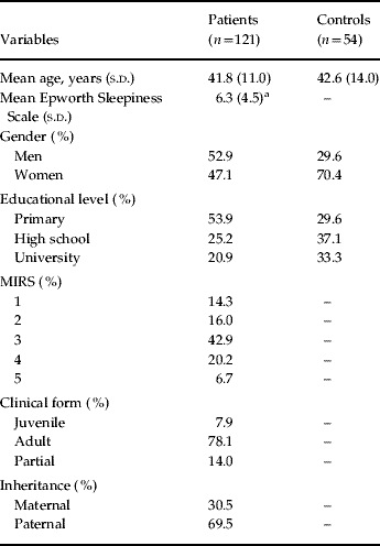
The sociodemographic data describing the subjects analysed in this study are shown in Table 1. There were significant differences between patients and controls in terms of gender and educational level.
Sociodemographic data

MIRS, Muscular Impairment Rating Scale; s.d., standard deviation.
a This value corresponds to 85 patients.
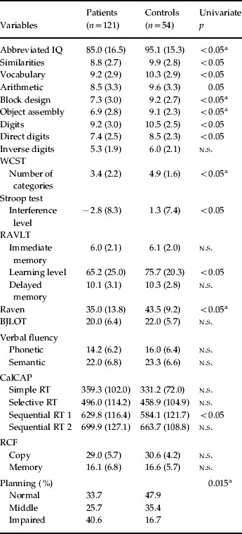
The neuropsychological evaluation identified significant differences between patients and controls in: the abbreviated form of the WAIS-III and all of its subtests, direct digit spam, WCST, interference level of the Stroop test, RAVLT's learning level, Raven, CalCAP's first sequential RT, and planning used to copy RCF (see Table 2). However, the multiple linear regression analysis indicated that it was the actual condition of the patient (i.e. as patient rather than control) and not the effect of other variables, such as the educational level or patients' gender, that was responsible for the poorer performance of the patients compared with the control individuals in the abbreviated WAIS-III and in the Block Design and Object Assembly subtests. The results obtained by the patients were also worse in the Wisconsin categorization test (WCST), in Raven's Progressive Matrices and in the strategy used to copy the RCF. The rest of the variables that were significantly different in the univariate analysis were no longer significant when the subjects' educational level was controlled for. Therefore, it was the fact of having a lower educational level and not the fact of suffering DM1 that was responsible for patients showing lower results in those tests. Including gender as a covariable in the multivariate model did not vary the significant association found between being a DM1 patient and different neuropsychological variables.
Neuropsychological assessment

WCST, Wisconsin Card Sorting Test; RAVLT, Rey's Auditory Verbal Learning Test; BJLOT, Benton Judgment of Line Orientation Test; CalCAP, California Computerized Assessment Package; RT, reaction time; RCF, Rey's Complex Figure; n.s., not significant.
Results presented as mean (standard deviation) or percentage.
a Statistically significant in a multivariate model, including subjects' educational level.
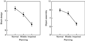
A significant association was found between planning and results in the Block Design and Object Assembly visuoconstructive tasks (see Fig. 1). In both cases, no significant differences were seen in the performance between normal and middle planning groups, although they were evident when the normal and middle groups were compared with the impaired one.
Linear regression analysis: association between planning and results in the Block Design and Object Assembly visuoconstructive tasks (p<0.05).

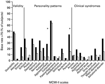
Regarding the results of the personality test, only 68% (n=119) of the participants in this study returned the completed questionnaire to the hospital. The results, after excluding two questionnaires that contained invalid results on the MCMI-II, revealed that there were more patients than controls with BR scores >75 in the following scales: Narcissistic, Antisocial, Aggressive/Sadistic, Paranoid, Thought Psychosis and Sincerity (p<0.05). However, in a multivariate model controlling the subjects' gender and educational level, significant differences were only maintained in the Aggressive/Sadistic and Paranoid scales (see Fig. 2).
Personality assessment using the Millon Clinical Multiaxial Inventory (MCMI)-II. Percentage of subjects with base rate (BR) >75: comparison between patients (▪, n=84) and controls (□, n=33). * Statistically significant in a multivariate model, including subjects' educational level.

When divided according to the expansion size, the patients show statistically significant age and educational-level differences (patients with smaller CTG expansion sizes are older and with a higher educational level than those with large expansion sizes). Moreover, in 85 subjects questioned, a slight (p<0.05, R 2=0.115) but significant correlation was observed between CTG repeat number and daytime somnolence (five out of 89 questioned subjects used nocturnal ventilator assistance and were excluded from this analysis), such that those patients with larger expansion sizes have a greater chance of falling asleep in daily life. In addition, muscular impairment (MIRS) is significantly associated with patients' expansion size, such that patients with larger expansion sizes are physically more disabled.
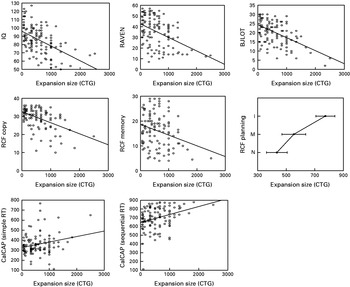
In the genotype–cognitive phenotype correlation, patients with more CTG repeats performed worse than those with smaller expansions in a large number of cognitive tests (see Fig. 3) in a multivariate model that controlled for patients' age, educational level, MIRS and daytime somnolence. However, this was not the case in Digits, WCST, interference level on the Stroop test, immediate and delayed memory and learning level on the RAVLT test and Verbal fluency.
Genotype–cognitive phenotype correlation. Only variables with a statistically significant correlation (after controlling for age and educational level) are shown. I, Impaired; M, middle; N, normal; RAVEN, Raven's Progressive Matrices; BJLOT, Benton Judgment of Line Orientation Test; RCF, Rey's Complex Figure; CalCAP, California Computerized Assessment Package.

The expansion size was also correlated with half of the scales of the MCMI-II: Schizoid, Passive–Aggressive, Autodestructive, Schizotypal, Borderline, Anxiety, Hysteria, Depressive Neurosis, Thought Psychosis and Major Depression.
Discussion
The analysis we have carried out confirms the existence of a cognitive profile associated with adult DM1. Accordingly, patients can be considered as a group with an IQ within the normal range, although considerably lower than that of the control subjects (Portwood et al. Reference Portwood, Wicks, Lieberman and Duveneck1986; Turnpenny et al. Reference Turnpenny, Clark and Kelly1994; Winblad et al. Reference Winblad, Lindberg and Hansen2006b), and with a neuropsychological pattern principally displaying executive function impairment with difficulties in planning and cognitive flexibility, and problems in their visuoconstructive capacity, principally in assembly.
In the cognitive evaluation of genetic disorders it is difficult to separate the influence that some sociodemographic factors affected by the disorder exert over the biological disorder itself. This is particularly relevant for a disorder such as DM1, where socio-economic deprivation has long been noted (Adie & Greenfield, Reference Adie and Greenfield1923). As in previous studies, the educational attainment of the DM1 patients reported here was lower than the controls (Fowler et al. Reference Fowler, Abresch, Koch, Brewer, Bowden and Wanlass1997; Laberge et al. Reference Laberge, Veillette, Mathieu, Auclair and Perron2007). However, the sample size allowed us to control a series of variables, such as age, gender or educational level, with sufficient statistical power so as to be able to detect the real effect of the disease on cognitive performance.
Our findings suggest the existence of alterations that evoke an effect in the prefrontal lobe (Meola et al. Reference Meola, Sansone, Perani, Scarone, Cappa, Dragoni, Cattaneo, Cotelli, Gobbo, Fazio, Siciliano, Mancuso, Vitelli, Zhang, Krahe and Moxley2003; Gaul et al. Reference Gaul, Schmidt, Windisch, Wieser, Muller, Vielhaber, Zierz and Leplow2006), predominantly dorsolateral. Indeed, the dorsolateral cortex is the most specifically centred in executive functions. As described previously, our patients display visuospatial construction but not spatial orientation impairment (Johnson et al. Reference Johnson, Abresch, Carter, Kilmer, Fowler, Sigford and Wanlass1995; Winblad et al. Reference Winblad, Hellstrom, Lindberg and Hansen2006), although the origin of this impairment is not clear. On the one hand, there are functional neuroimaging and structural studies that provide evidence of parietal lobe alterations associated with DM1 (Meola et al. Reference Meola, Sansone, Perani, Scarone, Cappa, Dragoni, Cattaneo, Cotelli, Gobbo, Fazio, Siciliano, Mancuso, Vitelli, Zhang, Krahe and Moxley2003; Antonini et al. Reference Antonini, Mainero, Romano, Giubilei, Ceschin, Gragnani, Morino, Fiorelli, Soscia, Di Pasquale and Caramia2004) that could explain the observed visuoconstructive difficulties. On the other hand, prefrontal cortex impairment gives rise to deficits in strategic planning (Meola & Sansone, Reference Meola and Sansone2007) that could affect a patients' performance in visuoconstructive tasks. In fact, we found a significant association between patients' planning capacity and visuoconstructive ability.
In contrast to other reports (Modoni et al. Reference Modoni, Silvestri, Pomponi, Mangiola, Tonali and Marra2004; Winblad et al. Reference Winblad, Lindberg and Hansen2005), we find similar attention levels in control subjects and patients. In relation to memory, age-related frontal dysfunction and memory impairment have been reported, indicating the possible involvement of the temporal lobe in DM1 (Rubinsztein et al. Reference Rubinsztein, Rubinsztein, McKenna, Goodburn and Holland1997; Modoni et al. Reference Modoni, Silvestri, Pomponi, Mangiola, Tonali and Marra2004). However, in our study the mnesic capacity of the patients was no worse than in the controls, in fact age alone was significantly associated with the variation in memory performance in adult DM1 patients and control subjects. Moreover, irrespective of peripheral factors that could affect speech in DM1, it is accepted that language function is normal. However, although there is evidence of lower phonetic and semantic verbal fluency in adult DM1 patients (Gaul et al. Reference Gaul, Schmidt, Windisch, Wieser, Muller, Vielhaber, Zierz and Leplow2006; Modoni et al. Reference Modoni, Silvestri, Vita, Quaranta, Tonali and Marra2008), those patients have normal fluency.
Genotype–cognitive phenotype correlation is of particular interest because of the ongoing controversy. In contrast to other recent studies (Meola et al. Reference Meola, Sansone, Perani, Scarone, Cappa, Dragoni, Cattaneo, Cotelli, Gobbo, Fazio, Siciliano, Mancuso, Vitelli, Zhang, Krahe and Moxley2003; Modoni et al. Reference Modoni, Silvestri, Pomponi, Mangiola, Tonali and Marra2004), our results confirm a significant negative correlation between many cognitive functions and the molecular defect (Jaspert et al. Reference Jaspert, Fahsold, Grehl and Claus1995; Perini et al. Reference Perini, Menegazzo, Ermani, Zara, Gemma, Ferruzza, Gennarelli and Angelini1999; Marchini et al. Reference Marchini, Lonigro, Verriello, Pellizzari, Bergonzi and Damante2000; Winblad et al. Reference Winblad, Lindberg and Hansen2006b). Hence, we could almost predict the performance of adult DM1 patients' in some cognitive functions according to the number of CTG repeats. However, patients perform worse than controls in cognitive flexibility, where no such correlation was observed. Accordingly, this inflexibility seems to be common to all adult DM1 patients irrespective of the magnitude of their molecular defect.
Nevertheless, the research on this issue has a great limitation, on the accessibility of samples. Brain tissue would be the appropriate source for this analysis because CTG length varies in the different tissues (Fortune et al. Reference Fortune, Vassilopoulos, Coolbaugh, Siciliano and Monckton2000).
Whether the specific patterns of personality or the special attitudes observed in DM1 patients are just the consequence of their motor and cognitive impairment remains to be determined (Bird et al. Reference Bird, Follett and Griep1983; Meola & Sansone, Reference Meola and Sansone2007). With respect to personality, adult DM1 patients in our study demonstrated some paranoid personality traits as described in earlier studies (Delaporte, Reference Delaporte1998), in addition to elevated scores in the Aggressive/Sadistic scale. Nevertheless, it should be noted that an elevated score in a scale on the MCMI-II does not imply a relationship, but rather the clinical symptoms at the time of the examination (Choca, Reference Choca2004). Furthermore, MCMI-II relies on limited empirical information when used as part of a neuropsychological examination.
A jealous and suspicious attitude and also a sense of superiority are the most pronounced characteristics evaluated by the MCMI-II Paranoid scale (Choca, Reference Choca2004). Patients with paranoid traits are characterized by the rigidity of their thoughts and it is often difficult to go against them (Millon, Reference Millon2004). Rash, dogmatic, hostile, competitive, pernicious and explosive are some of the attributes that Millon uses to describe an Aggressive/Sadistic personality. The Aggressive scale is a more pathological variant of the antisocial personality style and it measures a tendency for an individual to be at least aggressive, if not hostile, in his or her interactions with others. An elevation in this scale describes an individual who tends to emphasize the ability to remain independent, competitive, distrusting, excitable and irritable (Choca, Reference Choca2004).
Excessive daytime sleepiness is accepted as one of the most common complaints of patients with DM1 (Ashizawa, Reference Ashizawa1998). Although, to our knowledge, no significant association between daytime sleepiness and CTG repeats has yet been identified (Rubinsztein et al. Reference Rubinsztein, Rubinsztein, Goodburn and Holland1998; Phillips et al. Reference Phillips, Steer, Soldan, Wiles and Harper1999; Laberge et al. Reference Laberge, Begin, Montplaisir and Mathieu2004), we did identify a mild association between daytime sleepiness and the CTG repeat number. Daytime sleepiness does not significantly affect any neuropsychological variable, and it does not significantly alter the association between the CTG repeat number and the different neuropsychological variables. Hence, although this sleep disturbance probably represents a direct effect of the CNS lesions, cognitive impairment cannot be attributed to this symptom in patients with adult DM1 (Broughton et al. Reference Broughton, Stuss, Kates, Roberts and Dunham1990).
In summary, dysexecutive and visuoconstructive impairment in adult DM1 suggest a predominantly fronto-parietal influence. Awareness of these deficits in adults with DM1 should be considered when adjusting the scholastic burden and employment to patients' capacities. Moreover, the predictive value of CTG repeats on the cognitive and personality phenotype should be taken into account in the clinical management of adult DM1.
Acknowledgements
A. Sistiaga was supported by a predoctoral grant from the Spanish Ministry of Education and Science (MEC). We thank the patients (FENEUME), relatives and voluntary subjects who participated in the study. We also acknowledge the technical skills of L. Loinaz (Molecular Diagnosis Unit) and we thank M. Sefton for his help in the preparation of the English version of this manuscript.
Declaration of Interest
None.