INTRODUCTION
Trypanosomatids, including the well-known pathogenic Trypanosoma and Leishmania genera, have complex life cycles exhibiting a number of differentiation stages resulting from transformations that involve both structural and physiological changes (McGhee and Cosgrove, Reference McGhee and Cosgrove1980; Vickerman, Reference Vickerman1994). The Herpetomonas genus, which comprises single host (monoxenous) parasites found in the gut of a wide range of insects (Vickerman, Reference Vickerman1994), represents a very interesting model to study cellular differentiation, since it displays 3 developmental stages during its life cycle (promastigote, paramastigote and opisthomastigote), which are characterized by the position of the kinetoplast relative to the nucleus. In this genus, Herpetomonas samuelpessoai is a flagellated trypanosomatid isolated primarily from the predatory insect Zelus leucogramus, and as predictable its growth occurs at 26°C, the invertebrate vector temperature (Angluster et al. Reference Angluster, Bunn and De Souza1977; McGhee and Cosgrove, Reference McGhee and Cosgrove1980). Interestingly, H. samuelpessoai also grows well at 37°C (Roitman et al. Reference Roitman, Roitman and Azevedo1972, Reference Roitman, Brener, Roitman and Kitajima1976), a characteristic of heteroxenous trypanosomatids that possess a mammalian host in their life cycle.
The differentiation mechanism of H. samuelpessoai is triggered by changes in the culture medium composition, incubation conditions and by adding substances to the culture medium that interact with cellular components. In this context, the cellular differentiation of H. samuelpessoai occurs in the stationary phase and when cell growth is inhibited; also, by high incubation temperature (37°C) or after exposition to metabolic inhibitors like sodium butyrate, 2-deoxy-d-glucose, Concanavalin A, lidocaine, cholinergic drugs and dimethylsulfoxide (DMSO) (Angluster et al. Reference Angluster, Bunn and De Souza1977; Souza et al. Reference Souza, Thomas, Esteves, Angluster and De Souza1980; Castellanos et al. Reference Castellanos, Angluster and De Souza1981; Thomas et al. Reference Thomas, Esteves, Angluster, De Souza and Jurkiewicz1981a, Reference Thomas, Souza, Esteves, Angluster and De Souzab; Nakamura and Pinto, Reference Nakamura and Pinto1989). These substances induce changes in the cell surface saccharide composition (Soares et al. Reference Soares, Alviano, Silva-Filho, Esteves, Angluster and De Souza1984; Santos et al. Reference Santos, Rodrigues, Alviano and Soares2002a), in the cell surface charge (Soares et al. Reference Soares, Alviano, Esteves, Angluster, Silva-Filho and De Souza1988), in the protein synthesis (Santos et al. Reference Santos, Batista, Abreu, Alviano, Angluster and Soares2001), in the phosphatase production (Santos et al. Reference Santos, Souto-Padron, Alviano, Lopes, Soares and Meyer-Fernandes2002b) and in the expression of proteolytic enzymes (Santos et al. 2003 a).
H. samuelpessoai synthesizes an intracellular 45 kDa cysteine peptidase whose expression is modulated during the DMSO-induced differentiation (Santos et al. 2003 a). Cysteine peptidases of parasitic protozoa have been implicated in a variety of biological events including differentiation, nutrition, host cell infection, evasion of the host immune response, pathogenesis and virulence, and some have also been validated as drug targets of high promise, especially providing an alternative to traditional therapy against drug-resistant parasites (reviewed by Sajid and McKerrow, Reference Sajid and McKerrow2002; Vermelho et al. Reference Vermelho, Giovanni-de-Simone, d'Avila-Levy, Santos, Nogueira de Melo, Silva-Junior, Bom and Branquinha2007). Recently, our research group has reported that some insect and plant trypanosomatids presented immunological similarities with cruzipain, the major cysteine peptidase produced by the human pathogen Trypanosoma cruzi (d'Avila-Levy et al. Reference d'Avila-Levy, Araújo, Vermelho, Soares, Santos and Branquinha2005; Santos et al. Reference Santos, d'Avila-Levy, Dias, Ribeiro, Pereira, Elias, Souto-Padrón, Lopes, Alviano, Branquinha and Soares2006a, Reference Santos, d'Avila-Levy, Elias, Vermelho and Branquinha2007a). In addition, previous studies showed that H. samuelpessoai displays close antigenic similarities to T. cruzi (Souza et al. Reference Souza, Reis, Silva and Brener1974) and Leishmania spp. (Elias et al. Reference Elias, Pereira, Silva, Alviano, Soares and Santos2006; Santos et al. Reference Santos, Branquinha and d'Avila-Levy2006b).
In order to further explore the role of peptidases from H. samuelpessoai in the differentiation process, in the present work we have assessed the expression of peptidases in parasite cells grown at distinct temperatures and during the DMSO-triggered differentiation from 24 to 72 h. In addition, we have examined the possible immunological cross- reactivity of some parasite proteins with well-known cysteine peptidases produced by human trypanosomatid pathogens, including cruzipain (T. cruzi) and cysteine peptidase b (cpb) from Leishmania spp., as well as with calpain-like molecules, which refers to a family of neutral cytosolic Ca2+-dependent cysteine peptidases ubiquitously expressed in mammals and invertebrates (Mehendale and Limaye, Reference Mehendale and Limaye2005). Finally, we have compared the expression of the reactive proteins in H. samuelpessoai cells treated or not with DMSO, in order to establish a possible connection with the parasite differentiation process.
Materials and Methods
Microorganisms and cultivation
The protozoan Herpetomonas samuelpessoai (CT–IOC–067) was acquired at the Coleção de Tripanossomatídeos, Instituto Oswaldo Cruz, FIOCRUZ, Rio de Janeiro, Brazil. The trypanosomatid was maintained by weekly transfers in brain heart infusion (BHI) medium. For the experiments, cells were grown in 50 ml Erlenmeyer flasks containing 5 ml of autoclaved BHI medium. DMSO (Sigma Chemical Co.) was filter-sterilized and then added to the medium at 4% (Santos et al. Reference Santos, Batista, Abreu, Alviano, Angluster and Soares2001). The inoculum consisted of 0·5 ml of a 48 h culture, containing about 2×10⁶ cells/ml. Cells were cultivated at 26°C for 24 to 72 h, conditions under which DMSO induced the process of cellular differentiation in H. samuelpessoai (Castellanos et al. Reference Castellanos, Angluster and De Souza1981; Santos et al. Reference Santos, Batista, Abreu, Alviano, Angluster and Soares2001). Alternatively, H. samuelpessoai cells were cultured for 48 h at 37°C (Roitman et al. Reference Roitman, Roitman and Azevedo1972, Reference Roitman, Brener, Roitman and Kitajima1976) in order to study the effect of temperature increase on the peptidase production. The flagellates Trypanosoma cruzi (Dm28c strain) and Leishmania amazonensis (MHOM/BR/75 Josefa) were cultured for 72 h at 26°C in BHI medium supplemented with 10% heat-inactivated fetal bovine serum, in order to obtain epimastigote and promastigote forms, respectively. Parasite growth was estimated by determining the cell concentration in a Neubauer chamber. Cellular viability was assessed by motility and exclusion of trypan blue dye from cells. In order to quantify the percentage of differentiation in H. samuelpessoai, promastigote, paramastigote and opisthomastigote forms were counted after Giemsa-staining. At least 500 microorganisms were examined in each preparation (Santos et al. Reference Santos, Batista, Abreu, Alviano, Angluster and Soares2001).
Cellular extracts
Parasites were collected by centrifugation (500 g/5 min/4°C) and washed 3 times with cold phosphate-buffered saline (PBS; 150 mm NaCl, 20 mm phosphate buffer, pH 7·2). Trypanosomatids (1×10⁸ cells) were resuspended in 100 μl of PBS and lysed by the addition of 1% sodium dodecyl sulfate (SDS). The cells were broken in a vortex mixer by alternating 1 min shaking and 2 min cooling intervals (3 cycles), followed by centrifugation (5000 g/15 min/4°C), leaving the parasite cellular extracts in the supernatants (Santos et al. Reference Santos, d'Avila-Levy, Dias, Ribeiro, Pereira, Elias, Souto-Padrón, Lopes, Alviano, Branquinha and Soares2006a). The Drosophila extracts (kindly provided by Dr Alexandre Afranio Peixoto, Instituto Oswaldo Cruz, FIOCRUZ, Rio de Janeiro, Brazil) were prepared in the extraction buffer (25 mm Hepes, pH 7·2, 5 mm EDTA, 5 mm EGTA, 10 mm β-mercaptoethanol and 250 mm sucrose). About 10 frozen flies were crushed, suspended with 200 μl of cold extraction buffer and homogenized in a Teflon/glass homogenizer with 10–12 strokes at 4°C. After filtration, pure glycerol was added to 20% final concentration. The homogenate was carefully mixed and centrifuged at 100 000 g for 1 h at 4°C. The supernatant was aliquoted and the fractions stored at −80°C. Protein concentration was determined by the method described by Lowry et al. (Reference Lowry, Rosebrough, Farr and Randall1951), using bovine serum albumin as standard.
Gelatin-SDS–PAGE
The proteolytic activities were assayed by 10% SDS–PAGE with 0·1% gelatin incorporated into the gel as protein substrate. The gels were loaded with 40 μg of parasite extracts that were added to SDS–PAGE sample buffer (125 mm Tris, pH 6·8, 4% SDS, 20% glycerol and 2% bromophenol blue). Electrophoresis was performed at a constant current of 60 mA at 4°C for 2 h. After electrophoresis, SDS was removed by incubation with 10 volumes of 1% Triton X-100 for 1 h. Subsequently, the gels were incubated in 50 mm sodium phosphate buffer, pH 5·5, supplemented or not with 2 mm dithiothreitol (DTT) at 37°C, in the absence or presence of the following cysteine proteolytic inhibitors (Sigma) at 1 μm: trans-epoxysuccinyl l-leucylamido-(4-guanidino) butane (E-64) and leupeptin. After incubation for 40 h, the gels were washed twice with distilled water, stained for 2 h with 0·2% Coomassie brilliant blue R-250 in methanol-acetic acid-water (50:10:40), and destained overnight in a solution containing methanol-acetic acid-water (5:10:85), to intensify the proteolytic halos. The gels were dried, scanned and the density profiles digitally processed (Santos et al. Reference Santos, d'Avila-Levy, Dias, Ribeiro, Pereira, Elias, Souto-Padrón, Lopes, Alviano, Branquinha and Soares2006a).
Immunoblotting
Samples containing cellular extracts equivalent to 50 μg of protein were added to SDS–PAGE sample buffer and mixed with 10% β-mercaptoethanol, followed by heating at 100°C for 5 min. Thereafter, protein extracts were separated in 12% SDS–PAGE and the polypeptides electrophoretically transferred at 4°C at 100 V/300 mA for 2 h to a nitrocellulose membrane. The membrane was blocked in 5% low-fat dried milk in PBS containing 0·5% Tween 20 (PBS/Tween) for 1 h at room temperature. Then, membranes were washed 3 times (10 min each) with the blocking solution and incubated for 2 h with the following polyclonal antibodies at 1:500 dilution:anti-cruzipain of T. cruzi (kindly provided by Dr Juan-Jose Cazzulo – Instituto de Investigaciones Biotecnologicas, Universidad Nacional de General San Martin, Buenos Aires, Argentina), anti-cpb raised against Leishmania mexicana cysteine peptidase b (kindly provided by Dr Mary Wilson – Department of Internal Medicine, Biochemistry, Microbiology and Epidemiology, Program in Molecular Biology, University of Iowa, USA), anti-Dm-calpain raised against Drosophila melanogaster calpain (kindly provided by Dr Yasufumi Emori – Department of Biophysics and Biochemistry, Faculty of Sciences, University of Tokyo, Japan), and anti-α-tubulin monoclonal antibody (Sigma). The secondary antibody used was peroxidase-conjugated goat anti-rabbit IgG at 1:2500 followed by chemiluminescence immunodetection after reaction with ECL reagents (d'Avila-Levy et al. Reference d'Avila-Levy, Araújo, Vermelho, Soares, Santos and Branquinha2005). The relative molecular masses of the reactive polypeptides were calculated by comparison with the mobility of GIBCO BRL SDS–PAGE standards (Grand Island, NY, USA).
Densitometric analysis
The densitometric scanning analysis was performed with the use of the Kodak Digital Science EDAS 120 software. In these analyses, bands in each gel were manually selected using the free selection tool provided by the software. Band areas were then determined by repeating this process 3 times, to diminish the probability of errors in these estimations. Values of band area were further integrated with means of grey level in selected bands, generating densitometric values that were used in the comparison between corresponding bands from the different gels. For proteolytic bands analyses, first the images were inverted using the tool provided by the software and then the measurements were performed (Elias et al. Reference Elias, Pereira, Silva, Alviano, Soares and Santos2006).
Fluorescence microscopy and flow cytometry analyses
Parasites (1×10⁷ cells) were collected by centrifugation (500 g/5 min/4°C), washed 3 times with cold PBS and fixed at 4°C in 0·4% paraformaldehyde in PBS for 20 min, followed by extensive washing in the same buffer. After that, the fixed cells were permeabilized with 0·01% Triton X-100 in PBS for 15 min at room temperature and then washed twice in PBS. The fixed and permeabilized cells maintained their morphological integrity, as verified by optical microscopic observation. After this step, the cells were incubated for 1 h at room temperature with a 1:250 dilution of anti-cruzipain, anti-cpb and anti-calpain antibodies and then incubated for an additional hour with a 1:250 dilution of fluorescein isothiocyanate (FITC)-labelled goat anti-rabbit IgG. These cells were washed 3 times in PBS and observed in a Zeiss epifluorescence microscope (Axioplan). For flow cytometry analysis, the cells were examined in a fluorescence-activated cell sorter (FACS) FACSCalibur (BD Bioscience, USA) equipped with a 15 mW argon laser emitting at 488 nm. Untreated cells and those treated with the pre-immune rabbit antiserum and the secondary antibody only were assayed in parallel as controls. The mapped population (n=10 000) was then analysed for log green fluorescence by using a single-parameter histogram.
Results
Effect of temperature on the peptidase expression by H. samuelpessoai
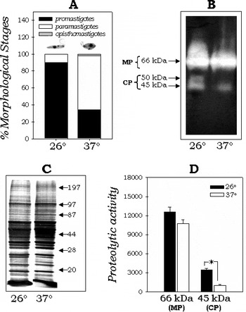
The cellular proteolytic profile of H. samuelpessoai has been previously described and it is composed of a major metallopeptidase of 66 kDa and a minor 45 kDa cysteine peptidase when parasites were grown at 26°C for 48 h (Santos et al. 2003 a; Fig. 1). The comparison of the peptidase expression (by gelatin-SDS-PAGE) in parasites grown for 48 h (exponential phase of growth) at either 26°C or 37°C revealed that the 45 kDa cysteine peptidase had its activity drastically diminished when parasites were cultured at the highest temperature (Fig. 1B and D). In addition, parasites grown at both temperatures revealed similar polypeptide profiles, presenting only minor quantitative differences (Fig. 1C). Curiously, an additional cysteine peptidase of 50 kDa could be observed when parasites were cultivated at 26°C. However, this cysteine peptidase appeared to be very unstable since its presence was transitorily detected in our experiments, in contrast to the major metallopeptidase of 66 kDa and the minor cysteine peptidase of 45 kDa (compare Fig. 1B and Fig. 2B). Besides E-64 (Santos et al. 2003 a), leupeptin, a well-recognized cysteine peptidase inhibitor, was also able to completely block the 45 and 50 kDa hydrolytic activities (data not shown). The modulation of the cysteine peptidase expression is connected to the differentiation process, since the temperature augmentation was able to trigger the promastigote to paramastigote transformation in H. samuelpessoai (Fig. 1A). In contrast, the 66 kDa metallopeptidase produced by H. samuelpessoai (Santos et al. 2003 a) had a similar activity at both temperatures (Fig. 1B). The densitometric analyses corroborated that the metallopeptidase, which shares common epitopes with the leishmanolysin molecule of Leishmania spp. (Elias et al. Reference Elias, Pereira, Silva, Alviano, Soares and Santos2006), had a similar expression at both temperatures (Fig. 1D).
(A) Percentage of each morphological stage observed in Herpetomonas samuelpessoai when cultured in BHI medium for 48 h at 26°C or 37°C. Note in the top of each graphic bar a representative image of the major morphological form observed at 26°C (promastigote form) and at 37°C (paramastigote form). (B) Differential peptidase expression observed in H. samuelpessoai grown at the insect (26°C) and mammalian (37°C) temperatures. The gel strips, containing 40 μg of proteins from parasites after 48 h of growth on both conditions, were incubated at 37°C for 40 h in 50 mm sodium phosphate buffer (pH 5·5) supplemented with 2 mm DTT. Molecular masses of the peptidases, expressed in kilodaltons (kDa), are represented on the left. (C) The protein profiles of both systems in SDS-PAGE were used as a control for sample loading in the gels. Molecular masses of the standard proteins are represented on the right. (D) The densitometric measurements of the digestion halos of the 66 and 45 kDa peptidases are shown in the graphic. The values represent means ± standard deviation of 3 independent measurements. The proteolytic activity was expressed in arbitrary units. The asterisk denotes statistic difference between both groups analysed (P<0·05; Student's t-test). MP, metallopeptidase; CP, cysteine peptidase.

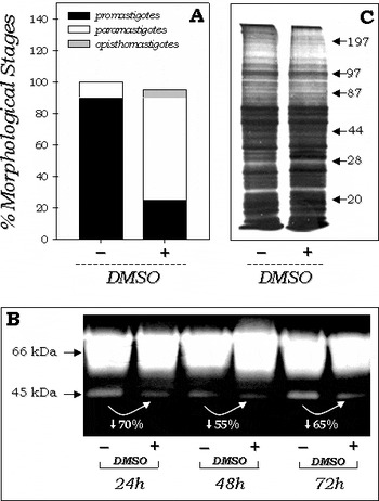
(A) Percentage of each morphological stage observed in Herpetomonas samuelpessoai when cultured in BHI medium for 48 h at 26°C in the absence (−) or in the presence of 4% DMSO (+). (B) Gelatin-SDS-PAGE showing the cell-associated proteolytic activities of H. samuelpessoai grown in BHI medium during 24 to 72 h in the absence (−) or in the presence of DMSO (+). The gel strips were incubated at 37°C for 40 h in 50 mm sodium phosphate buffer, pH 5·5, supplemented with 2 mm DTT. The numbers in white show the reduction of the 45 kDa peptidase activity in DMSO-treated parasites in comparison to the non-treated ones, which were measured by densitometric analysis. (C) SDS-PAGE showing the protein profiles of both untreated (−) and DMSO-treated (+) parasites for 48 h was used as a control for sample loading in the gels. Molecular masses of the standard proteins are represented on the right.

Effect of DMSO and growth phase on the peptidase expression by H. samuelpessoai
Here, we have shown that the 45 kDa proteolytic enzyme had its expression reduced by approximately 70% in the first 24 h (lag growth phase) of parasite growth in the presence of DMSO (Fig. 2B). The 45 kDa band from 48 h (exponential phase) DMSO-treated parasites was then reduced by 55%, and by 65% after 72 h (stationary phase), in relation to the respective controls. This result was validated through the observation that H. samuelpessoai grown in the absence of DMSO presented a preponderance of promastigote forms during 24 to 72 h, and when the parasite was incubated in the presence of DMSO, the number of promastigote forms decreased, while both paramastigote and opisthomastigote forms enhanced (Fig. 2A). Conversely, the 66 kDa metallopeptidase was not significantly altered by the presence of DMSO (Fig. 2B). Since equivalent amounts of proteins were electrophoresed in this set of experiments (Fig. 2C), this result may suggest that the differences in cellular proteolysis observed by gelatin-SDS-PAGE may be a consequence of a quantitative differential expression of the 45 kDa cysteine peptidase in the distinct morphological stages of H. samuelpessoai.
Immunological cross-reactivity of cysteine peptidases expressed by H. samuelpessoai with cpb and calpain
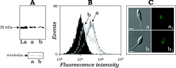
The incorporation of a substrate into SDS-PAGE provides a good method for investigating the overall proteolytic profile of a cell lysate, but a limitation of this technique is that only peptidases of relatively high activity can be investigated easily, as the product of gelatin hydrolysis is soluble and so permeates throughout the gel. In order to reveal whether H. samuelpessoai possesses minor proteins related to the well-known cysteine peptidases from human pathogenic trypanosomatids, cellular protein extracts were probed with the anti-cruzipain of T. cruzi and anti-cpb raised against cysteine peptidase b of L. mexicana. The rabbit polyclonal anti-cruzipain failed to recognize any polypeptide in H. samuelpessoai-derived cellular extract either from parasites grown in the presence or in the absence of DMSO, while it strongly recognized T. cruzi cellular extracts (data not shown). Corroborating this finding, no additional fluorescence was observed in parasites treated with anti-cruzipain antibody when compared with non-treated ones by means of fluorescence microscopy and flow cytometry analyses (data not shown). On the other hand, a single polypeptide band with apparent molecular mass of 35 kDa was recognized by the anti-cpb antibody in both H. samuelpessoai and L. amazonensis lysates (Fig. 3A). The densitometric analysis of this reactive 35 kDa band showed a 25% reduction in the amount of this polypeptide in DMSO-treated cells in comparison to the untreated parasites (data not shown), corroborating the results observed by Western blotting (Fig. 3A) and flow cytometry (Fig. 3B) assays. Fluorescence microscopy showed homogeneous labelling in the whole cellular body (Fig. 3C).
(A) Western blotting showing the cpb-like polypeptide detected in the cellular extract from Herpetomonas samuelpessoai cells grown in the absence (a) or in the presence of DMSO (b) for 72 h at 26°C. The cellular extract of promastigotes from L. amazonensis was used as a positive control (La). Anti-tubulin monoclonal antibody was used as a control for sample loading in the gels. The number on the left indicates the apparent molecular mass of the reactive polypeptide, expressed in kilodaltons (kDa). (B) Flow cytometry and (C) fluorescence microscopy analyses showing the anti-cpb antibody binding in untreated (a) and DMSO-treated (b) H. samuelpessoai permeabilized cells as described in the Materials and Methods section. In the flow cytometry, the black curve represents the autofluorescence of cells. For simplicity, only the autofluorescence of control cells is shown, since the DMSO-treated cells are similar (data not shown). In the same way, parasites treated with secondary antibody or pre-serum presented the same fluorescence intensity depicted in the autofluorescence (data not shown).

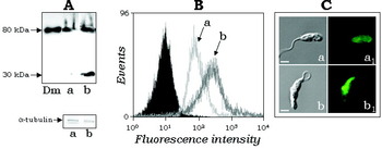
We also detected that H. samuelpessoai expresses a calpain-like molecule of 80 kDa in DMSO-untreated parasites; a similar molecular mass polypeptide was detected in D. melagonaster extract (Fig. 4A). Interestingly, the reactivity of the anti-calpain antibody against the 80 kDa polypeptide was raised by approximately 50% in the extract from differentiated cells, as judged by densitometric analysis, and an additional low-molecular-mass band of 30 kDa was also visualized (Fig. 4A). These results were reinforced by flow cytometry analysis, which demonstrated a significant increase in the fluorescence intensity in DMSO-treated parasites in comparison with non-treated ones (Fig. 4B). The distribution of calpain-like molecules was demonstrated by fluorescence microscopy on both cellular body and flagellum of H. samuelpessoai parasites (Fig. 4C). In addition, an increased fluorescence labelling was observed in the anterior end of DMSO-treated parasites (Fig. 4C).
(A) Western blotting showing the calpain-like polypeptides detected in the whole cellular extract from Herpetomonas samuelpessoai grown in the absence (a) or in the presence of DMSO (b) for 72 h at 26°C. The total cellular extract of Drosophila melanogaster was used as a positive control (Dm). Anti-tubulin monoclonal antibody was used as a control for sample loading in the gels. The numbers on the left indicate the apparent molecular masses of the reactive polypeptides, expressed in kilodaltons (kDa). (B) Flow cytometry and (C) fluorescence microscopy analyses showing the anti-calpain antibody binding in untreated (a) and DMSO-treated (b) H. samuelpessoai permeabilized cells as described in the Materials and Methods section. In the flow cytometry, the black curve represents the autofluorescence of cells. For simplicity, only the autofluorescence of control cells is shown, since the DMSO-treated cells are similar (data not shown). In the same way, parasites treated with secondary antibody or pre-serum presented the same fluorescence intensity depicted in the autofluorescence (data not shown).

DISCUSSION
The life cycle of trypanosomatids is characterized by a complex series of cell type differentiations and variations in metabolism. The differentiation events require stage-specific changes to basic cell biological processes and reflect responses to environmental stimuli and programmed differentiation events. Several factors are able to trigger cellular differentiation in trypanosomatids, including biotic and abiotic stimuli (Parsons and Ruben, Reference Parsons and Ruben2000).
As is well known, growth temperature can modulate the expression of several molecules, including proteolytic enzymes, in several microorganisms. In H. samuelpessoai, high incubation temperature (37°C) and exposure to DMSO induce parasite differentiation from promastigotes (typically found at 26°C with no stimuli) to paramastigote and opisthomastigote forms (Roitman et al. Reference Roitman, Roitman and Azevedo1972, Reference Roitman, Brener, Roitman and Kitajima1976; Angluster et al. Reference Angluster, Bunn and De Souza1977; Castellanos et al. Reference Castellanos, Angluster and De Souza1981; Santos et al. Reference Santos, Batista, Abreu, Alviano, Angluster and Soares2001, 2002 a, b, 2003 a). In order to correlate peptidase expression to the cellular differentiation in H. samuelpessoai, we have assayed these enzymes in parasites grown at 26°C solely, 26°C in the presence of DMSO and at 37°C by gelatin-SDS-PAGE, as well as by Western blotting and flow cytometry analyses employing a panel of antibodies raised against well-known cysteine peptidases: e.g. cruzipain, cpb and calpain.
In our results, cysteine peptidase activities were always significantly diminished when the parasite was grown at 37°C, suggesting a possible influence of growth temperature in the production of cysteine peptidases by H. samuelpessoai cells. The temperature-dependent activity of the 45 kDa cysteine peptidase from H. samuelpessoai may correlate this enzyme to important metabolic processes that occur at 26°C, the invertebrate vector temperature, which must not proceed when the parasite is cultivated at 37°C. Alternatively, it could be associated to parasite differentiation since at 37°C the major morphological stage observed in H. samuelpessoai cultivation was the paramastigote, while at 26°C the promastigote forms predominate (Roitman et al. Reference Roitman, Roitman and Azevedo1972, Reference Roitman, Brener, Roitman and Kitajima1976; Angluster et al. Reference Angluster, Bunn and De Souza1977).
DMSO has also been used as a powerful cell-differentiating agent in numerous biological systems (reviewed by Santos et al. Reference Santos, Figueira-Coelho, Martins-Silva and Saldanha2003b), including the insect trypanosomatid H. samuelpessoai (Castellanos et al. Reference Castellanos, Angluster and De Souza1981). Santos et al. (Reference Santos and Soares2007b) demonstrated that the reduction of parasite proliferation was accompanied by an increase in the differentiation rate in H. samuelpessoai. It has been shown that H. samuelpessoai grown in the absence of DMSO presented a preponderance of promastigote forms during 24 to 72 h (Castellanos et al. Reference Castellanos, Angluster and De Souza1981; Santos et al. Reference Santos, Batista, Abreu, Alviano, Angluster and Soares2001, 2002 a, b, 2003 a). Conversely, when the trypanosomatids were incubated in the presence of DMSO, the number of promastigote forms decreased, while both paramastigote and opisthomastigote forms enhanced by approximately 60–70% during 24 to 72 h (Castellanos et al. Reference Castellanos, Angluster and De Souza1981; Santos et al. Reference Santos, Batista, Abreu, Alviano, Angluster and Soares2001, 2002 a, b, 2003 a). In a preceding work of our group, we also demonstrated that the DMSO-induced differentiation in H. samuelpessoai was dependent on peptidase expression, since cysteine peptidase inhibitors blocked this essential biological process (Santos et al. 2003 a). In this paper, we confirm that DMSO-induced differentiation and the reduction of cysteine peptidase expression in paramastigote cells are inter-connected events. On the other hand, the 66 kDa metallopeptidase expression was not significantly altered by the presence of DMSO, as judged by gelatin-containing gels and Western blotting.
Immunological cross-reactivity between molecules present both in human pathogen trypanosomatids and in lower trypanosomatids, peptidases particularly, seem to be a common feature (d'Avila-Levy et al. Reference d'Avila-Levy, Araújo, Vermelho, Soares, Santos and Branquinha2005; Santos et al. Reference Santos, d'Avila-Levy, Dias, Ribeiro, Pereira, Elias, Souto-Padrón, Lopes, Alviano, Branquinha and Soares2006a, Reference Santos, d'Avila-Levy, Elias, Vermelho and Branquinhab; Reference Santos, d'Avila-Levy, Elias, Vermelho and Branquinha2007a). In our results, we detected the presence of Leishmania cpb-like molecules in H. samuelpessoai cells, with DMSO-untreated flagellates displaying an increased amount of the cysteine peptidase in comparison to the DMSO-treated cells. Nevertheless, no T. cruzi cruzipain homologues were detected by means of the employed methodologies. Since DMSO is able to interfere with several signalling cascades, including some in which Ca2+ participates as second messenger (reviewed by Santos et al. Reference Santos, Figueira-Coelho, Martins-Silva and Saldanha2003b), as well as it causes modification in the cellular architecture during the transformation of promastigote into paramastigote and opisthomastigote observed in H. samuelpessoai (Santos et al. Reference Santos, d'Avila-Levy, Elias, Vermelho and Branquinha2007a), we have investigated the possible participation of calpain-like molecules in the DMSO-stimulated differentiation of H. samuelpessoai. Our results showed for the first time that DMSO-untreated H. samuelpessoai expresses a molecule of 80 kDa that presents homology to D. melanogaster calpain-like protein. In addition, the detection of this polypeptide was raised by approximately 50% in DMSO-induced differentiated cells.
Calpains are Ca2+-regulated cysteine peptidases distributed in a variety of animal cells and localized predominantly in the cytoplasm as heterodimers of an 80 kDa large subunit that contains the active site and a smaller ~28 kDa regulatory subunit. Calpain is involved in a multiplicity of physiological processes that require cellular restructuring, such as proliferation, differentiation and migration (reviewed by Mehendale and Limaye, Reference Mehendale and Limaye2005). Different authors have proposed that several cellular events can increase intracellular calpain activity: a rise in the concentration of cytosolic free Ca2+, translocation of calpains to membranes, dissociation of the calpain subunits, a decrease in the levels of calpastatin, the endogenous inhibitor of calpain, and interaction with calpain activators, such as phospholipids (Sorimachi et al. Reference Sorimachi, Ishiura and Suzuki1997; Goll et al. Reference Goll, Thompson, Li, Wei and Cong2003; Liu et al. Reference Liu, van Vleet and Schnellmann2004). Although dissociation of calpain subunits has been considered as a possible mechanism of calpain activation, recent evidence suggests that calpain can also be activated without dissociation of its subunits (Gil-Parrado et al. Reference Gil-Parrado, Popp, Knoch, Zahler, Bestvater, Felgentrager, Holloschi, Fernandez-Montalvan, Auerswald, Fritz, Fuentes-Prior, Machleidt and Spiess2003).
In trypanosomatids, a calcium-dependent cysteine peptidase was already detected in lysed promastigotes of L. donovani. The enzyme was named caldonopain due to its similarity with the calpain peptidase family (Bhattacharya et al. Reference Bhattacharya, Dey and Datta1993). A calpain-like protein migrating at 80 kDa was also identified by Western blotting assay in promastigotes of L. amazonensis using the anti-calpain antiserum raised against D. melanogaster (d'Avila-Levy et al. Reference d'Avila-Levy, Marinho, Santos, Martins, Santos and Branquinha2006). This calpain-like molecule was identified on the cell surface and in the flagellum of the L. amazonensis by fluorescence microscopy. Additionally, after 48 h of L. amazonensis treatment with the potent calpain inhibitor MDL 28170, the drug exhibited a dose-dependent anti-leishmanial activity, with a 50% lethal dose of 23·3 μm (d'Avila-Levy et al. Reference d'Avila-Levy, Marinho, Santos, Martins, Santos and Branquinha2006). In T. brucei, a cytoskeleton-associated protein (TbCALP1) has similarities restricted only to the catalytic region of calpain-type peptidases. This protein is expressed in procyclics, but not in bloodstream trypanosomes. Finally, d'Avila-Levy et al. (Reference d'Avila-Levy, Souza, Gomes, Vermelho and Branquinha2003) showed by biochemical methods that a released 80 kDa calcium-dependent cysteine peptidase purified from C. deanei belongs to the calpain superfamily. Corroborating these findings, immunoblotting experiments revealed that the calpain-like molecule of C. deanei was distinct from the well-characterized cruzipain from T. cruzi and from mammalian calpains, but some degree of similarity was displayed to invertebrate calpain-related enzymes (d'Avila-Levy et al. Reference d'Avila-Levy, Souza, Gomes, Vermelho and Branquinha2003). Interestingly, in a previous study from our group, we detected a 115 kDa peptidase in H. samuelpessoai that presented proteolytic activity only after phospholipase C hydrolysis, suggesting a close association with the parasite membrane. The enzyme presented some intriguing biochemical features that resemble calpains, such as total inhibition by EDTA, partial inhibition by 1,10-phenanthroline and E-64, and stimulation by Ca+2 (Santos et al. Reference Santos, Abreu, Alviano and Soares2002c).
The biological functions played by peptidases in trypanosomatids are still largely unknown, especially in insect-dwelling parasites. The identification of protein homologues in trypanosomatids with distinct life cycles may help to determine the importance of peptidases in trypanosomatids. The results described above add H. samuelpessoai to the list of parasites whose differentiation seems to be correlated with the expression of different cysteine peptidases, including calpain-like molecules.
This work was supported by grants from Fundação Universitária José Bonifácio (FUJB), Fundação de Amparo à Pesquisa no Estado do Rio de Janeiro (FAPERJ), Conselho Nacional de Desenvolvimento Científico e Tecnológico (CNPq) and Conselho de Ensino e Pesquisa para Graduados da Universidade Federal do Rio de Janeiro (CEPG–UFRJ).