INTRODUCTION
Ciliated protozoans of the family Trichodinidae are one of the primary causes of disease in cultured fish worldwide, which leads to economic losses (Martins et al. Reference Martins, Marchiori, Nunes and Rodrigues2010). Trichodinidae are a diversified group of ciliates with complex structures on their adhesive discs (Basson and Van As, Reference Basson, Van As and Woo2006). These structures are utilized as taxonomic characteristics that can be used for evaluation in silver nitrate-impregnated specimens using the technique described by Klein (Reference Klein1958), whereas the nuclear apparatus is identified by panchromatic staining as proposed by Lom (Reference Lom1958) and Van As and Basson (Reference Van As and Basson1989). Hundreds of trichodinid species are recognized; however, few are as widely distributed as Trichodina heterodentata (Duncan, Reference Duncan1977), Trichodina nigra Lom, 1961, Trichodina mutabilis Kazubski and Migala, 1968, Trichodina acuta Lom, 1961 and Trichodinella epizootica Raabe, 1950, which are commonly found in several countries (Basson and Van As, Reference Basson, Van As and Woo2006). In Brazil, recent studies of the trichodinid fauna of native fish have identified Tripartiella pseudoplatystomae Pinto, Garcia, Figueiredo, Rodrigues and Martins, 2009 in spotted catfish (Pinto et al. Reference Pinto, Garcia, Figueiredo, Rodrigues and Martins2009), Trichodina colisae Asmat and Sultana, 2005 in pacu and its hybrid intergeneric patinga (Jerônimo et al. Reference Jerônimo, Marchiori, Pádua, Dias Neto, Pilarski, Ishikawa and Martins2012) and T. heterodentata in pacu (Pádua et al. 2012). Trichodina fariai Cunha and Pinto, 1928 is not clearly identified because its description was not based on the silver nitrate impregnation method (Lom and Haldar, Reference Lom and Haldar1976).
There are few studies of the relationship between the host and parasite in trichodinid-diseased fish. Among the deleterious effects caused by trichodinids is reduced growth, as observed in Heterobranchus longifilis parasitized by Trichodina maritinkae Basson and Van As, 1991, (Ekanem and Obiekezie, Reference Ekanem and Obiekezie1996) whereas Trichodina sp. rendered Ictalurus punctatus more susceptible to Streptococcus spp. infection (Evans et al. Reference Evans, Klesius, Pasnik and Shoemaker2007). Inflammation and epithelial desquamation of Carassius auratus gills were caused by T. heterodentata (Tang and Zhao, Reference Tang and Zhao2007). Parasitism by T. heterodentata also caused a decrease in the immune response of Nile tilapia vaccinated against Streptococcus iniae (Martins et al. Reference Martins, Shoemaker, Xu and Klesius2011). Few studies have evaluated the effects of trichodinid infestation on fish larvae; however, Basson and Van As (2006) consider the larval stage the most susceptible to trichodinid infestation.
Streaked prochilod Prochilodus lineatus Valenciennes, 1836, is a rheophilic freshwater fish commonly found in South American fish farms. Artificial reproduction of this fish is easy and is performed using specimens maintained in earth ponds (Hainfellner et al. Reference Hainfellner, Souza, Moreira, Nakaghi and Batlouni2012). Streaked prochilod larvae are also used as a live food for South American carnivorous fish, as dourado Salminus brasiliensis (Characidae) and catfish of the genus Pseudoplatystoma (Pimelodidae), commonly known as surubim. Parasitic fauna that affect streaked prochilod in both the wild and captivity are poorly known. Although the occurrence of Trichodina sp. has been reported (Eiras et al. Reference Eiras, Takemoto, Pavanelli and Luque2012), no trichodinid species has been reported. This study shows the occurrence of T. heterodentata that parasitizes the P. lineatus. The histopathological and scanning electron microscopy (SEM) analysis of the fish larvae were also performed and reveals important information about parasite action on the host.
MATERIALS AND METHODS
Study area and fish
Streaked prochilod larvae within 20 days of hatching were utilized for parasitological analysis after observation for larval mortality and abnormal clinical signs. These larvae were utilized as live food for the carnivorous larvae of the cachara catfish Pseudoplatystoma fasciatum that were cultured in earthen ponds (15×10 m) in Jaboticabal city, São Paulo State, Southeast Brazil (22.14′22.7″S; 48°17′33.4″W).
Gross pathology and diagnosis of parasitic infestation
Ten larvae with abnormal behaviour and clinical signs were euthanized by cerebral commotion and examined under a light microscope. When trichodinids were present, the slides were air-dried under dim light, and several slides were fixed with methanol and then stained with Giemsa stain (one drop per 1 mL distilled water) for 120 to 180 min to show the nuclear apparatus. The other slides were impregnated with 2% silver nitrate using Klein's method (Klein, Reference Klein1958) to observe the adhesive disc as described by Lom (Reference Lom1958). Measurements were obtained from 102 specimens impregnated with 2% silver nitrate and 38 specimens stained with Giemsa to observe the nuclear apparatus and obtain a description of the position of the micronucleus (Lom, Reference Lom1958). Specimens are deposited in the National Institute of Amazonian Research (INPA), Manaus, AM, Brazil under the number 008 and 009.
The denticle span was measured from the extremity of the blade to the extremity of the ray as described by Arthur and Lom (Reference Arthur and Lom1984). All measurements were made in micrometres and followed the recommendations of Lom (Reference Lom1958). Measurements were made on photomicrographs obtained with a Nikon E200® photomicroscope equipped with the Moticam 2300® image capture system. The parasite measurements were performed using the ImagePro Plus® 4.1 software. Minimum and maximum values are provided, followed in parentheses by arithmetic mean, standard deviation and number of specimens measured. Schematic drawings of the denticles, as proposed by Van As and Basson (Reference Van As and Basson1989), were produced by means of vectorization using CorelDraw® X5 software.
Histopathological analysis
Five streaked prochilod larvae severely infested with trichodinids were fixed in 10% buffered formalin solution, processed according to usual histopathological techniques, embedded in paraffin, sectioned at 5 μm, and stained with haematoxylin–eosin. The slides were analysed, and photomicrographs were obtained using a Nikon E200® photomicroscope equipped with the Moticam 2300® image capture system.
Scanning electron microscope (SEM)
Five streaked prochilod larvae were fixed in 2·5% glutaraldehyde and post-fixed in 1% osmium tetroxide, both in 0·1 m cacodylate buffer (pH 7·4). Next, the larvae were dehydrated with serial concentrations of ethanol and critical-point dried. The biological material was mounted on aluminium stubs, sputter-coated with gold, and examined using a JEOL JSM-5410 scanning electron microscope at an accelerating voltage of 15 kV.
RESULTS
Parasitic diagnosis
All larvae were severely parasitized by trichodinids (100% prevalence), and no preference of the infestation site was noted. The parasites were observed on the body surface, fins, gills and mouth of the fish. No mixed infestation with other parasite taxa was observed.
Trichodinid description
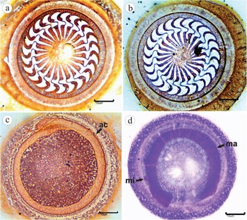
Trichodinids on streaked prochilod P. lineatus larvae showed a disc-shaped body (Fig. 1a and b) diameter of 48·4–65·9 (56·9±3·6; 102), striated border membrane 2·8–5·7 (4·5±0·4; 102) wide, adhesive disc diameter of 39·4–55·3 (47·7±3·6; 102), denticulate ring diameter of 23·0–37·6 (29·4±2·6; 102), and 20·0–26·0 (23·0±0·9; 102) denticles. The denticle length was 5·8–9·3 (7·8±0·7; 102), blade length was 3·8–5·7 (4·6±0·4; 102), central part width was 2·1–4·2 (3·0±0·5; 102), ray length was 6·0–9·9 (7·7±0·8; 102), denticle span was 13·0–17·6 (15·4±1·0; 102) and 6–12 (9·8±1·2; 93) pins per denticle was observed. The adoral ciliature (Fig. 1c) in spiral was 405–430° (418·2±6·7; 34). The horse-shaped macronucleus (Fig. 1d) was 39·8–59·0 (45·8±4·7; 38) in diameter, 10·9–16·1 (8·5±1·4; 38) in thickness and there was a distance between the macronucleus extremities of 23·3–27·5 (16·6±3·9; 38) in length. Occasionally, a rounded micronucleus (Fig. 1d) situated at the +y position was observed; value of +y distance 13·3–25·9 (19·9±4·3; 6); micronucleus length 3·8–4·6 (4·1±0·3; 6) and width 1·8–3·9 (2·9±0·8; 6).
Klein's silver-impregnated specimens of Trichodina heterodentata Duncan, 1977, from Prochilodus lineatus larvae (a–c) showing adoral ciliature (c), and Giemsa stained nuclear apparatus (d) showing the horseshoe-shaped macronucleus (ma) and oval micronucleus (mi). Scale bar = 10 μm.

Denticle description
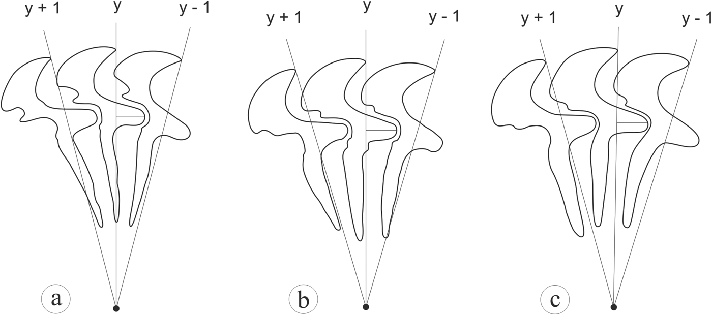
The blade is broad and sickle-shaped and fills the space between the axes y and y+1, in which the apex touches the axis y+1, but in some specimens, crosses the axis y+1. Apophysis of the blade is well-developed and prominent but reduced in a few specimens. The central part of the blade is robust, triangular, with no posterior projection and fills the space between the axes y+1 and y−1. The rays are long, straight and can be projected parallel, either anteriorly or posteriorly in relation to the y axis. It is primarily sharp-pointed and occasionally tapers to a round shape with a short apophysis observed in some specimens (Fig. 2).
Schematic drawing of the denticles of Trichodina heterodentata Duncan, 1977, from Prochilodus lineatus larvae showing morphological variations (a–c).

All morphological characteristics of the parasite on P. lineatus larvae are similar to T. heterodentata.
Clinical signs and gross pathology
The larvae showed lethargy, erratic swimming on the water surface and pond edges, and some larvae presented with irregular whitish lesions on the body surface and head. It was not possible to analyse the cachara P. fasciatum larvae from the same pond as the streaked prochilod P. lineatus larvae because of the small number of animals. It was also not possible to observe the daily mortality of the streaked prochilod P. lineatus larvae because of their small size, although the cachara larvae did not survive, and approximately 50% of the streaked prochilod larvae stocked in the pond reached the fingerling stage.
Histopathological analysis
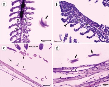
Major histological changes were observed on the gills and were characterized by sub-epithelial oedema with epithelial displacement of the secondary lamellae (Fig. 3a and b). There was also discrete epithelial hyperplasia with focal areas of lamellar fusion and a slight mononuclear inflammatory infiltrate (Fig. 3a and b). Histological changes in the skin of the larvae could not be found. A large number of T. heterodentata was observed on the gills (Fig. 3c), skin (Fig. 3d), inside the mouth and occasionally in the intestine, which presented with rupture signs.
Histological sections of Prochilodus lineatus larvae highly parasitized by Trichodina heterodentata Duncan, 1977. Oedema (asterisk) in the secondary lamella and parasite (arrow) (a); discrete hyperplasia with focal fusion of the secondary lamella (arrow), discrete mononuclear inflammatory infiltrate and oedema (b); trichodinids (arrows) present inside and outside the operculum cavity (oc) showing no altered epithelium (c); parasite (arrow), and pocket scale without alteration (arrow head) (d).

SEM observations
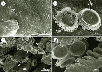
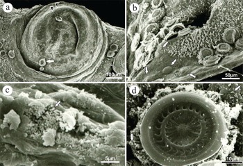
We observed a large number of T. heterodentata on the skin and fins (Fig. 4a), gills (Fig. 4c), cornea and oral cavity of the larvae by SEM. T. heterodentata ciliature and adhesive disc details were observed (Fig. 4b and d). Suction areas in the gill, eye (Fig. 5a) and skin near the nostril (Fig. 5b) caused by attachment of the adhesive disc were observed. Tissue injuries were epithelial desquamation with the formation of ulcers on the skin and fins. On these locations, high numbers of bacteria in the form of cocci associated with the injuries (Fig. 5c). These bacteria were also found among the cilia and on the surface of trichodinids (Fig. 5d).
Scanning electron microscopy analysis of Trichodina heterodentata Duncan, 1977, from Prochilodus lineatus larvae. A large number of parasites (white arrow) on the body surface (a) with detail of the parasite adoral (ADS) and aboral surface (ABS) (b – arrow); trichodinids on gill filaments (c – arrow) with detail of the trichodinid between gill filaments (d).

Scanning electron microscopy analysis of Trichodina heterodentata Duncan, 1977, from Prochilodus lineatus larvae. A large number of parasites on eyes (white arrow) with suction areas (arrow head) (a) and on skin near the nostril with many suction areas (b); ulceration on the dorsal fin with cocci attached (c – arrows) and large number of cocci-shaped bacteria surrounding the ciliate (d – arrows).

DISCUSSION
Trichodina heterodentata is widely distributed in several countries and has significant morphometric variations. The measurements obtained in this study are similar to the descriptions of Basson et al. (Reference Basson, Van As and Paperna1983), Albaladejo and Arthur (Reference Albaladejo and Arthur1989), Al-Rasheid et al. (Reference Al-Rasheid, Ali, Saakran, Baki and Ghaffar2000) and Asmat (Reference Asmat2004). The specimens with the largest-sized dimensions in the majority of taxonomic characteristics were reported in population B of the studies of Duncan (Reference Duncan1977) and Martins et al. (Reference Martins, Marchiori, Nunes and Rodrigues2010), whereas Pádua et al. (Reference Pádua, Martins, Carraschi, Cruz and Ishikawa2012) reported a Brazilian population with a smaller adhesive disc and denticulate ring diameters, denticle span, central part width and adoral ciliature than the present specimens. The variation observed in the schematic drawing of the T. heterodentata isolated from the streaked prochilod are consistent with the description of Van As and Basson (Reference Van As and Basson1989) in which well-developed and robust rays differ from the slender ray described in T. heterodentata isolated from Rhinella pombali tadpoles (Dias et al. Reference Dias, Fernandes, Sartini, Silva-Neto and D'Agosto2009). Ecological and environmental factors could favour the occurrence of a varied shape of this ciliate group (Kazubski, Reference Kazubski1971).
The behavioural alterations and gross pathology observed in the streaked prochilod larvae parasitized by T. heterodentata were not specific, and other Brazilian farmed fish parasitized by Chilodonella hexasticha showed similar changes (Pádua et al. Reference Pádua, Martins, Carrijo-Mauad, Ishikawa, Jerônimo, Dias-Neto and Pilarski2013). In this study, no severe histological alterations were observed that were similar to those observed in Tetraodon fahaka parasitized by Trichodina fahaka Al-Rasheid, Ali, Sakran, Baki and Ghaffar, 2000 (Abdel-Baki et al. Reference Abdel-Baki, Sakran, Fayed and Zayed2011) or Solea aegyptiaca parasitized by Trichodina gobii Raabe, 1959 (Yemmen et al. Reference Yemmen, Quilichini, Ktari, Marchand and Bahri2011a ). In these cases, there were significant proliferative gill disturbances and lamellar fusion, whereas during the inflammatory reaction of Trichodina puytoraci Lom, 1962 in Mugil cephalus, degeneration and gill necrosis were related (Yemmen et al. Reference Yemmen, Ktari and Bahri2011b ). Trichodina heterodentata in streaked prochilod larvae also caused proliferative alterations; however, these alterations were discrete. Tang and Zhao (Reference Tang and Zhao2007) reported the occurrence of inflammation and structural disorders with epithelial desquamation in the gills of C. auratus infested by T. heterodentata. The immune competent cells of the fish immune system are formed during the first days post-fertilization (Zapata et al. Reference Zapata, Diez, Cejalvo, Gutiérrez-de Frías and Cortés2006), therefore, streaked prochilodontid larvae may not have a well-developed immune system necessary for a good inflammatory response.
The pathological alterations observed in this study were acute with the formation of lesions that may result in mortality. Severe proliferative lesions with complete fusion of the gill lamellae during trichodinid parasitism (Abdel-Baki et al. Reference Abdel-Baki, Sakran, Fayed and Zayed2011; Yemmen et al. Reference Yemmen, Quilichini, Ktari, Marchand and Bahri2011a ; Valladão et al. Reference Valladão, Pádua, Gallani, Menezes-Filho, Dias-Neto, Martins and Pilarski2013) are chronic alterations that develop because of prolonged parasite contact. Consistent with these observations, dysfunctions in the respiratory and osmoregulatory capacities cause metabolic disturbances that are lethal to the host. By contrast, the severe infestation of fish larvae observed in this study may cause abrasion that culminates in acute epithelial lesions followed by oedema and displacement of the gill tissue.
From the SEM analysis, suction areas on the cornea, skin and gills were similar to those found on the urinary bladder of Xenopus laevis parasitized by Trichodina xenopodos Fantham, 1924 (Kruger et al. Reference Kruger, Basson and Van As1991). These authors argued that the parasite strategy of attachment to the host prevents the excretion of trichodinids from the bladder, but if trichodinids detach the suction areas disappear with very little if any damage caused. Based on our observations, we agree that this may also be an attachment strategy of T. heterodentata to the external surface of the fish. Particularly, these changes in the cornea may contribute to abnormal clinical signs, as erratic swimming, which may be due to obscured vision and the presence of ciliates on the eyes.
Bacteria associated with trichodinids were also identified by SEM (Khan et al. Reference Khan, Barber and McCann1974), but no information on host lesions was reported. Trichodinid infestation can induce host susceptibility to bacterial infection as shown by Evans et al. (Reference Evans, Klesius, Pasnik and Shoemaker2007) and as demonstrated during infestation by other parasite ciliates (Xu et al. 2012a, b). Our study showed a large number of bacteria associated with the lesions on the host caused by the massive presence of T. heterodentata. The results of this study support the hypothesis that parasitism increases tissue injury that facilitates a secondary bacterial infection, which may favour disease outbreaks.
CONCLUSIONS
This is the first record of T. heterodentata in P. lineatus, a South American freshwater fish. The parasitism caused acute disease associated with an opportunistic bacterial infection, ocular lesions, ulceration on the body surface and fins, as well as compromised respiratory function.
ACKNOWLEDGEMENTS
The authors thank Dr Fernando José Zara for the critical read of the manuscript and some suggestions during the graduation course on Electron Microscopy Applied to Zoology. The SEM procedure was carried on the Electronic Microscopy Laboratory of FCAV (UNESP, Jaboticabal). We thank Claudia Aparecida Rodrigues by the SEM procedure. Thank are also due to Dra. Maria Cecília Rui Luvizotto, Dr Klaus Casaro Saturnino and Camila do Nascimento for histological sections, CNPq (National Council for Scientific and Technological Development) for a grant to Dr M. L. Martins (CNPq 302493/2010-7), FAPESP for scholarships to S. B. Pádua (2010/14490-1) and G. M. R. Valladão (2012/19414-7).