Introduction
Few studies have reported on the management and prognosis of patients with large juvenile nasopharyngeal angiofibroma specific to the eye and optic nerve. Fifty-one patients with juvenile nasopharyngeal angiofibroma were treated in our hospital between 1981 and 2011. Of those, 18 had large juvenile nasopharyngeal angiofibromas that involved the orbit, eye, optic nerve and/or the optic chiasm. This paper introduces the general clinical data for these large juvenile nasopharyngeal angiofibroma cases, and reports three typical patients.
Materials and methods
The study was approved by the hospital institutional review board.
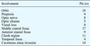
All 18 patients were male, with a mean age of 17.8 years (range, 11–36 years). Table I shows the areas affected by juvenile nasopharyngeal angiofibroma in these patients. It does not, however, indicate tumour involvement of the nasal cavity, paranasal sinus, nasopharynx, pterygopalatine fossa, pterygoid process or infratemporal fossa, all typical sites of juvenile nasopharyngeal angiofibroma involvement.
Juvenile nasopharyngeal angiofibroma involvement

Two patients had esotropia as a result of abducens nerve involvement. Two patients had temporal lobe involvement with epilepsy which resolved following surgery and radiotherapy. Pts = patients
The study used Radkowski's staging system for juvenile nasopharyngeal angiofibroma classification. Of the 18 patients, 7 had Radkowski stage IIIa disease and 11 had stage IIIb disease.Reference Radkowski, McGill, Healy, Ohlms and Jones 1
Different surgical approaches were used to treat the patients: a combined approach via the antrum, infratemporal fossa and nasal cavity, using an extended Caldwell–Luc incision, in 10 cases (in 4 of these, combined intracranial and extracranial approaches were used); a lateral rhinotomy in 3 cases; a Ferguson incision in 2 cases; and a Weber incision, Weber–Ferguson incision and transpalatine incision in 1 case each.
Results
The tumour was completely excised in 12 patients, and subtotal resection was achieved in 6 cases. Eight of the patients experienced visual impairment before surgery. In seven cases, visual acuity improved or remained unchanged post-operatively. One patient, who underwent surgery and radiotherapy, developed blindness in the affected eye.
Three representative cases are described in detail below.
Case one
A 16-year-old boy had suffered from intermittent right epistaxis and nasal obstruction for 2 years, and complained of headache and decreased visual acuity in the right eye in the 2 weeks preceding hospitalisation in April 2005.
Physical examination revealed a huge mass in the right nasal cavity and nasopharynx.
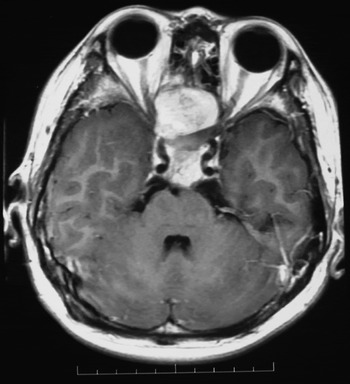
Magnetic resonance imaging (MRI) and computed tomography (CT) revealed extensive damage to the sphenoid bone, sella turcica and clivus, while the pituitary gland, cavernous sinus and internal carotid artery had been pushed away from their original positions. Furthermore, the optic nerve and chiasm were compressed bilaterally (Figure 1).
Axial magnetic resonance imaging scan of case one, showing bilateral involvement of the eyes, optic nerves and chiasms.

Ophthalmological examination revealed a mildly prominent right eye. The patient's visual acuity was at the level of finger counting at 20 cm in the right eye (with no difference for corrected visual acuity), while corrected visual acuity for the left eye was 1.0.
Tumour-feeding vessels from the external carotid artery (ECA) system were embolised.
Three days later, a combined approach via the antrum, infratemporal fossa and nasal cavity, using an extended Caldwell–Luc incision, was utilised to resect the 6 × 7 × 8 cm tumour completely. Intra-operative observation confirmed that the optic nerve and chiasm were partially exposed. A piece of haemostatic gauze soaked in dexamethasone solution was used to protect the optic nerve and chiasm, and iodoform gauze was inserted into the surgical cavity to prevent infection.
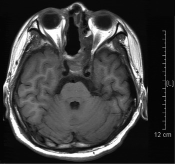
A post-operative MRI scan showed that both eyes and optic nerves were undamaged (Figure 2). Post-operative corrected visual acuity was 1.0 in both the left and right eyes, confirming a successful surgical outcome.
Post-operative, axial magnetic resonance imaging scan of case one, showing the intact eye and optic chiasm. L = left

Six years after the operation, the patient remained healthy.
Case two
A 13-year-old boy had suffered from right-sided, relapsing epistaxis and nasal obstruction for more than 2 years, and had experienced progressive swelling in his cheek and loss of vision during the previous 6 months.
Physical examination revealed proptosis and cheek swelling on the patient's right side, with a mass in the nasopharynx and right nasal cavity.
Ophthalmological examination revealed that the visual acuity was finger counting at 1 m in the right eye (with no difference for corrected visual acuity), while the corrected visual acuity for the left eye was 1.0. The patient's right eye was slightly more prominent than his left eye. The right eye was opened more widely than the left eye, but was not tender.
Bilateral focal electroretinography revealed a normal b-wave amplitude. The pattern visual evoked potential P-wave latency was elongated and the amplitude decreased in the right eye, whereas the P-wave latency and amplitude were normal in the left eye.
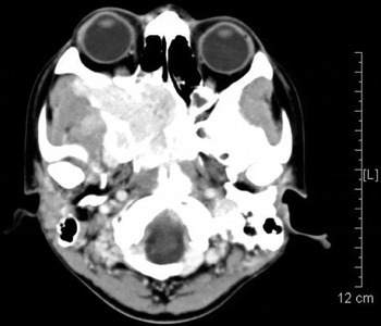
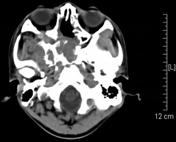
Computed tomography and MRI scans revealed a lobulated mass in the nasopharynx and the right nasal cavity, infratemporal fossa, cheek region and sphenoid sinus. The bones of the right orbit were extensively eroded. The right eye was compressed and displaced forward (Figure 3).
Axial computed tomography scan of case two, showing the tumour compressing the right eye and displacing it anteriorly. L = left

Embolisation of the tumour-feeding vessels from the ECA was performed.
Two days later, on 20 May 2004, we performed a transtemporal-intradural exploration using a combined approach via the antrum, infratemporal fossa and nasal cavity and an extended Caldwell–Luc incision, in order to remove the lobulate tumour.
Post-operative imaging showed tumour residue in the nasal cavity and sphenoid sinus. Thus, a second operation was undertaken to completely remove the tumour (Figure 4).
Axial computed tomography scan of case two taken after the second operation, showing the right eye in the normal position and complete removal of the tumour. L = left

Ophthalmological examination performed on 24 August 2004 showed an improvement in visual acuity, to a corrected visual acuity of 1.0 in the right eye and 1.5 in the left eye.
At seven-year follow up, the patient was in good health but was short-sighted (300° in the left eye and 350° in the right eye).
Case three
A 14-year-old boy had undergone unsuccessful juvenile nasopharyngeal angiofibroma surgery via a transpalatine approach in another hospital 4 years before he was admitted to our hospital in November 2007. His left eye had gradually become prominent, and he had suffered from vision loss over the previous year. Due to the extensive extracranial and intracranial spread of the tumour, the patient had developed obstructive sleep apnoea-hypopnoea syndrome and epilepsy.
Ophthalmological examination revealed 12 mm exophthalmos of the right eye with normal vision and a normal fundus. In the left eye, 14 mm exophthalmos was seen, with temporal bulbar conjunctival injection; examination of the fundus revealed tortuous retinal veins, retinal oedema and macular oedema. Visual acuity was finger counting at 1 m in the left eye.
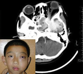
A three-dimensional CT scan revealed juvenile nasopharyngeal angiofibroma specific to the left ethmoid sinus and orbital apex, both sphenoid sinuses, and the left pterygopalatine fossa, infratemporal fossa, temporal fossa and cheek. A tumour lobe spread extensively into the left middle cranial fossa through the superior orbital fissure (Figure 5). The tumour was approximately 8.0 × 8.0 cm in size.
Axial computed tomography scan of case three showing extensive tumour in the region of the left eye, orbital apex and middle cranial fossa. Insert: clinical photograph of the patient; published with the patient's parent's permission.

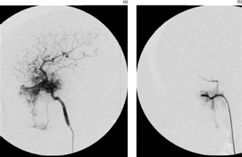
Digital subtraction angiography showed that both ECAs and the left ophthalmic artery supplied branches to the tumour (Figure 6).
Digital subtraction angiography images of case three, (a) pre-embolisation and (b) post-embolisation, showing that the left ophthalmic artery provides feeder branches to the tumour.

After embolisation of the ECA feeding branches, a combined extracranial and intracranial approach was used to remove the tumour.
The patient's obstructive sleep apnoea-hypopnoea syndrome and epilepsy resolved after the operation.
A post-operative CT scan revealed that the tumour lobule that had extended through the superior orbital fissure into the middle cranial fossa had not been completely removed.
However, re-operation was not possible. Thus, in March 2008 local three-dimensional conformal radiation therapy was applied to the left cavernous sinus, orbital apex and the residual intracranial lesion. The radiation dose was divided into 39.6 Gy/22 F.
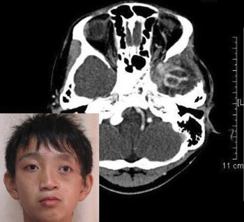
Computed tomography performed in August 2010 showed that the extracranial portion of the tumour had completely resolved (Figure 7), compared with imaging performed 2.5 years earlier (see Figure 5), and the tumour residue in the middle cranial fossa had decreased significantly.
Axial computed tomography scan of case three after surgery and radiological therapy, showing that the tumour has reduced in size in the middle cranial fossa; a reduction in the left exophthalmosis is also seen. Insert: clinical photograph of the patient three years after surgery; published with the patient's parent's permission. L = left

At this stage, the patient's left eye exophthalmos was reduced (see Figure 7), but his left eye dilated pupil, pale optic disc and inability to perceive light remained. Ophthalmological testing showed that visual acuity in the left eye had decreased to finger counting at 1 m; the lack of light perception may have resulted from the radiotherapy. Vision was normal in the right eye.
Discussion
There are few published studies on the treatment and prognosis of patients with juvenile nasopharyngeal angiofibroma involving the eye and optic nerve.
In a study of 218 juvenile nasopharyngeal angiofibroma cases, Stern et al. Reference Stern, Beauchamp and Berlin 2 found exophthalmos in 14 per cent, decreased visual acuity in 5 per cent and partial ophthalmoplegia in 2 per cent. These authors suggested that the observed visual changes were the result of orbital and/or intracranial extension of the tumour.
Cruz et al. Reference Cruz, Atique, Melo-Filho and Elias 3 studied 19 patients with juvenile nasoangiofibroma. They found tumour involvement in the pterygopalatine fossa, nasal cavity, nasopharynx, paranasal sinuses, inferior orbital fissure, orbit and middle cranial fossa, and reported that the inferior orbital fissure was the main route of orbital invasion.
Watnick and TrobeReference Watnick and Trobe 4 reported an 11-year-old boy with a juvenile nasopharyngeal angiofibroma who developed optic disc pallor in one eye and optic disc oedema in the other, a condition known as Foster Kennedy syndrome. The cause was believed to be bilateral, asymmetrical optic nerve compression.
Salcone and PepinReference Salcone and Pepin 5 reported a case of juvenile nasopharyngeal angiofibroma in which the earliest symptom was intermittent esotropia caused by tumour invasion of the superior orbital fissure and subsequent compression of the abducens nerve. In the present study, two of our 18 juvenile nasopharyngeal angiofibroma cases were found to have esotropia caused by abducens nerve compression; this resolved following surgical removal of the tumour.
Because nasopharyngeal angiofibroma is a vascular hamartoma,Reference Liang, Yi and Liang 6 the growing tumour does not infiltrate the surrounding anatomical structures but instead causes erosion of the skull and skull fissures. It may cause exophthalmos and optic nerve and chiasm involvement due to direct compression of the orbital bone and/or spread into the pterygopalatine fossa, infraorbital fissure and superior orbital fissure, and then into the orbital and cranial fossa where it compresses the eye, optic nerve and chiasm. Furthermore, the tumour may displace the pituitary gland, cavernous sinus and internal carotid artery (ICA) away from their original sites. Tumours that do not extend through the oval and round foramina of the skull base are less likely to compress the optic nerve and chiasm.
As some large juvenile nasopharyngeal angiofibromas cause exophthalmos and involve the optic nerve and chiasm, the use of state-of-the-art imaging technology and the in-depth examination of the visual organs are crucial in order to select the best technique of surgical management. This paper presents three typical cases for clinical reference.
Treatment principles
Bilateral vascular supply may be an under-appreciated factor in juvenile nasopharyngeal angiofibroma; hence, thorough pre-operative radiographic investigation involving angiography of both carotid systems should be routinely performed.Reference Wu, Mowry, Vinuela, Abemayor and Wang 7 Prior to tumour resection, all tumour feeder vessels from the ECA should be embolised; special attention should also be paid to feeder vessels from the ICA. Figure 6 shows that, in our third case, the ophthalmic artery branched off from the ICA and supplied small arterial branches to the tumour. This demonstrates the need for tumour feeder branches from the ophthalmic artery to be coagulated delicately, and for the ophthalmic artery to be protected during surgical separation from the tumour.
Surgery is the main treatment option for angiofibroma. Our group has used various surgical approaches for large juvenile nasopharyngeal angiofibromas, such as lateral rhinotomy, Weber incision, Ferguson incision, Weber–Ferguson incision, a transpalatine approach and an endoscopic transnasal approach.Reference Yi, Li, Lin and Ye 8 – Reference Lin, Li, Cheng, Lin, Zhou and Yi 10 However, since 1986 we have preferred a combined approach via the antrum, infratemporal fossa and nasal cavity, using an extended Caldwell–Luc incision (midfacial degloving incision), for these tumours.
This technique consists of two main steps: (1) an endoscopic, transnasal approach to remove tumour involving the eye, optic nerve and sphenoid sinus, as well as removal of tumour spreading into the cranial fossa (in the case of intact dura mater); and (2) a transantral, infratemporal fossa approach to remove tumour in the infratemporal fossa, temporal fossa and cheek region. This is an ideal and minimally invasive surgical technique. It not only preserves the patient's appearance but also allows removal of the tumour under direct visualisation using both hands, thus avoiding the need to use a finger or instrument for blind, rapid removal. Every bleeding point can be seen and treated carefully. In the case of a huge juvenile nasopharyngeal angiofibroma, if the separated portion of the tumour blocks the surgeon's vision and affects management, then it is advisable to resect this portion after tightly suturing the margin of the incision. A wider operative field can then be achieved. If necessary, a combined intracranial and extracranial approach can be used to remove the tumour, as in our third case.
In our study, the symptoms and signs associated with decreased visual acuity and proptosis improved following surgery, as illustrated by cases one and two. A 40 Gy dose of radiation therapy is indicated for cases in which the intracranial residue cannot be removed surgically.Reference Yi, Huang, Lin, Lin, Lin and Li 11 A special effort must be made to protect the eye and optic nerve, as radiation therapy can cause blindness, as illustrated in case three.
-
• Large juvenile nasopharyngeal angiofibromas can compress and displace the optic nerve
-
• Such cases may show visual impairment
-
• Surgery and radiotherapy are the principal treatment options
-
• A combined transantral infratemporal fossa nasal cavity approach with extended Caldwell–Luc incision is described
-
• Restoration of vision and eye position is possible
Follow up is an essential and integral part of the management of juvenile nasopharyngeal angiofibromas.
Conclusion
Patients with juvenile nasopharyngeal angiofibroma may have only mild orbital involvement, with no detectable clinical signs or symptoms. However, when the optic nerve is compressed and displaced, visual impairment may occur.
Surgery and radiotherapy are the principal treatment options for large tumours. We use a combined approach via the antrum, infratemporal fossa and nasal cavity, utilising an extended Caldwell–Luc incision (midfacial degloving), to remove large tumours abutting the eye, optic nerve and optic chiasm. Using this approach, restoration of normal vision and repositioning of the eye to its natural position are possible. In the event of degeneration of the optic nerve and retina, restoration of normal vision may not be possible, but surgery and radiotherapy may remove or decrease the size of the tumour while partially restoring placement of the eye.