INTRODUCTION
The active role of astroglia in neural information processing and storage, as opposed to providing purely metabolic support to neurons, is a relatively novel concept in our understanding of the brain machinery. Rapidly emerging experimental evidence appears to implicate Ca2+-dependent activity of astrocytes in regulation of neural network activity and synaptic transmission including its use-dependent modifications (see Fiacco et al., Reference Fiacco, Agulhon and McCarthy2009; Hamilton and Attwell, Reference Hamilton and Attwell2010; Perea and Araque, Reference Perea and Araque2010 for recent reviews). Because the latter phenomenon is thought to underlie learning and memory formation in the brain, the potential involvement of astroglia attracts intense attention across neuroscience areas. A widely accepted experimental model to explore mechanisms of synaptic memory formation in the brain is long-term potentiation (LTP) of excitatory transmission, in particular its classical forms documented in the hippocampal synaptic circuitry (Bliss and Lomo, Reference Bliss and Lomo1973; Bliss and Collingridge, Reference Bliss and Collingridge1993). Although the basic neuronal mechanisms of LTP induction and expression have largely been established, the picture is far from complete. The most common form of LTP in the hippocampus relies on postsynaptic Ca2+ entry through N-methyl-d-aspartic acid (NMDA) receptors (NMDARs). In addition to glutamate, however, activation of NMDARs depends on the presence of a receptor co-agonist, either glycine or d-serine (Johnson and Ascher, Reference Johnson and Ascher1987), and LTP does require the activation of the NMDAR co-agonist site (Bashir et al., Reference Bashir, Tam and Collingridge1990). Astroglia can synthesise d-serine and release it in a Ca2+-dependent manner (Mothet et al., Reference Mothet, Pollegioni, Ouanounou, Martineau, Fossier and Baux2005). At the same time, excitatory synaptic activity could evoke a variety of Ca2+ responses in astrocytes, most notably through activation of metabotropic glutamate receptors (mGluRs) involving intracellular molecular cascades associated with G-protein coupled receptors (GPCRs) (reviewed in Porter and McCarthy, Reference Porter and McCarthy1997; Haydon, Reference Haydon2001; Volterra and Meldolesi, Reference Volterra and Meldolesi2005). In turn, Ca2+ waves induced inside astrocytes by experimental activation of such cascades, or by direct triggering of the inositol tris-phosphate (IP3)-dependent signalling using photolytic uncaging, have been shown to affect synaptic transmission, by either depressing or enhancing it, in the neighbouring neuropil (Pasti et al., Reference Pasti, Volterra, Pozzan and Carmignoto1997; Fiacco and McCarthy, Reference Fiacco and McCarthy2004; Perea and Araque, Reference Perea and Araque2007; Navarrete and Araque, Reference Navarrete and Araque2008). While the precise physiological circumstances in which such induction may occur in vivo remain to be further investigated, experiments that explored the classical LTP paradigm found a link between astrocytic release of d-serine and use-dependent plasticity of glutamatergic transmission (Yang et al., Reference Yang, Ge, Chen, Zhang, Shen and Wu2003; Panatier et al., Reference Panatier, Theodosis, Mothet, Touquet, Pollegioni and Poulain2006). Indeed, combining two-photon excitation microscopy with patch-clamp electrophysiology in acute hippocampal slices has provided evidence that interfering with intracellular Ca2+ homeostasis in area CA1 astrocytes blocks induction of the classical NMDAR-dependent LTP at nearby excitatory synapses and that the underlying mechanism involves Ca2+-dependent release of d-serine from astrocytes (Henneberger et al., Reference Henneberger, Papouin, Oliet and Rusakov2010). However, the signalling cascade controlling d-serine release remains to be determined: although increased synaptic activity does seem to enhance supply of d-serine by astrocytes, the receptor mechanism which triggers such increases is not known. In addition, the causal connection between mGluR activation and LTP in area CA1 has been a subject of debate in the synaptic literature, with various forms of potentiation involving these receptors depending on the induction protocol used (Bashir et al., Reference Bashir, Bortolotto, Davies, Berretta, Irving and Seal1993; Selig et al., Reference Selig, Lee, Bear and Malenka1995; Lu et al., Reference Lu, Jia, Janus, Henderson, Gerlai and Wojtowicz1997; Manahan-Vaughan, Reference Manahan-Vaughan1997).
GENETIC PROBING OF IP3-DEPENDENT SIGNALLING AND OTHER APPROACHES TO PHYSIOLOGICAL PLAUSIBILITY
Although a major role of mGluRs in generating astrocyte Ca2+ responses in situ has long been demonstrated (Porter and McCarthy, Reference Porter and McCarthy1996), the physiological consequences of astrocyte Ca2+ signalling have been difficult to establish, mainly because the pharmacological action aimed at mGluRs is not normally restricted to astrocytes. However, advances in genetics have enabled researchers to generate genotypes in which astrocytes could be targeted with high specificity (Fiacco et al., Reference Fiacco, Agulhon, Taves, Petravicz, Casper and Dong2007, Reference Fiacco, Agulhon and McCarthy2009). A recent study, which used genetic deletion of the IP3R2 receptor and genetic insertion of MrgA1 receptors in young mice, was unable to associate the astrocyte-specific, Gq GPCR-dependent Ca2+ signalling with excitatory synaptic transmission or its plasticity (Agulhon et al., Reference Agulhon, Fiacco and McCarthy2010). Based on these results, the authors dispute the involvement of astrocytic Ca2+ signalling, in particular Gq/IP3-dependent Ca2+ signalling, in neural function (Agulhon et al., Reference Agulhon, Petravicz, McMullen, Sweger, Minton and Taves2008, Reference Agulhon, Fiacco and McCarthy2010). They argue that the widely reported effects of Ca2+-dependent release of signalling molecules from astroglia on synaptic circuitry are likely to involve either non-physiological manipulations (such as mechanical disturbance of glia or the photolytic uncaging of IP3) or direct physiological action outside astroglia.
Indeed, achieving conditions compatible with brain physiology is an issue of paramount importance in experimental studies in vitro. Ideally, one would aim to identify and activate selectively the molecular trigger(s) of Ca2+-dependent release in astrocytes. One conceptual difficulty is that the current experimental techniques employed to evoke intra-astrocytic Ca2+ signalling (such as uncaging of IP3 or activation of MrgA1) are controlled by detecting Ca2+ elevations throughout the cell (Fiacco and McCarthy, Reference Fiacco and McCarthy2004; Fiacco et al., Reference Fiacco, Agulhon, Taves, Petravicz, Casper and Dong2007). Whether such elevations reproduce the spatiotemporal aspects of physiological Ca2+ signalling is not known. The uncertainty is highlighted by the reports that uncaging of IP3 in astrocytes does affect spontaneous synaptic transmission in the hippocampus (Fiacco and McCarthy, Reference Fiacco and McCarthy2004) whereas activation of MrgA1 does not (Fiacco et al., Reference Fiacco, Agulhon, Taves, Petravicz, Casper and Dong2007; Agulhon et al., Reference Agulhon, Fiacco and McCarthy2010) even though the evoked astrocytic Ca2+ signals appear similar. Furthermore, Ca2+-dependent glutamate release from astrocytes has been associated with an action of endogenous endocannabinoids released by neurons (Navarrete and Araque, Reference Navarrete and Araque2008). This disparity suggests that the origin and propagation of a physiological Ca2+ signal could depend critically on GPCR localisation or, more generally, on the spatial relationships among the intracellular players involved in Ca2+ signalling in astrocytes: GPCRs, IP3Rs, Ca2+ stores and the Ca2+-sensing molecular targets such as the trigger of release. Little is known about the intracellular distribution of such players, either on the scale of the entire astrocytic arbour or within the fine astrocyte processes that approach synaptic structures.
By the same token, a selective alteration of a genome that would boost the expression of a particular receptor, or otherwise would suppress the expression of another receptor, is not expected to habitually occur in physiological circumstances. Although such experimental manipulations provide a powerful tool to address molecular specificity of a particular signalling mechanism, a careful consideration should be given to the potential compensatory or concomitant changes in the function, expression and distribution of signalling molecules that might play part in the effect under study (Hamilton and Attwell, Reference Hamilton and Attwell2010).
Indeed, several recent reports urge caution against the wide extrapolation of the negative results based on the above genetic approach. A study in which the aforementioned genetic model was used has shown that MrgA1 receptor-evoked Ca2+ signals in astrocytes do lead to the enhanced NMDAR activity in nearby neuroblasts migrating to the olfactory bulb (Platel et al., Reference Platel, Dave, Gordon, Lacar, Rubio and Bordey2010). Although these experiments have been carried in a different brain area and focused on a different neural function, they do demonstrate that the Ca2+ activity restricted to astrocytes through a genetic tool could affect activation of NMDARs on other cells. Earlier, conditional astrocyte-selective expression of the SNARE domain of the protein synaptobrevin II (dnSNARE), which suppresses exocytosis in the host cells, was used to associate activity of astrocytes in vivo with modulation of sleep homeostasis, in an ATP-dependent (Halassa et al., Reference Halassa, Florian, Fellin, Munoz, Lee and Abel2009) and NMDAR activation-dependent (Fellin et al., Reference Fellin, Halassa, Terunuma, Succol, Takano and Frank2009) manner. Most recently, Ca2+ signalling in pH-sensitive astrocytes, evoked either endogenously or exogenously, has been found to cause activation of ATP-sensitive neurons in the brainstem in vivo which in turn induced adaptive changes in breathing (Gourine et al., Reference Gourine, Kasymov, Marina, Tang, Figueiredo and Lane2010). These studies have therefore used genetic approaches that are not conceptually dissimilar to that used by Agulhon and co-authors (Agulhon et al., Reference Agulhon, Petravicz, McMullen, Sweger, Minton and Taves2008, Reference Agulhon, Fiacco and McCarthy2010) and yet found what appears to be a causal connection between astrocytic Ca2+-dependent activity and neural function.
An alternative to the genetic targeting of astrocytes, which is designed to facilitate a physiologically plausible exogenous stimulus, is the interference with endogenous signalling from within an individual astrocyte held in whole-cell mode. One common target is Ca2+ signalling cascades which can be modulated by adding combinations of Ca2+ buffers or by the blockade of the Ca2+-sensing effector. Indeed, this type of experimental interference in astroglia has been shown to affect neural function in situ (Jourdain et al., Reference Jourdain, Bergersen, Bhaukaurally, Bezzi, Santello and Domercq2007; Perea and Araque, Reference Perea and Araque2007; Andersson and Hanse, Reference Andersson and Hanse2010; Gomez-Gonzalo et al., Reference Gomez-Gonzalo, Losi, Chiavegato, Zonta, Cammarota and Brondi2010; Henneberger et al., Reference Henneberger, Papouin, Oliet and Rusakov2010; Todd et al., Reference Todd, Darabid and Robitaille2010). One important advantage of these methods is that they allow direct electrophysiological control of an identified astrocyte, thus enabling the monitoring of astrocytic and neuronal responses in real time. Because perturbation of the intracellular medium is inherent to such approaches, tests with control intracellular solutions are normally required to confirm that Ca2+ mechanisms in question are preserved in whole-cell configuration per se.
A common issue in comparative studies of astroglia-neuron signalling is that the effects may depend not only on the maturity of the astrocytes but also on the developmental stage of the neuronal network. Historically, most Ca2+ imaging studies in astrocytes have been performed in tissue obtained from early postnatal to 2–3-week-old rodents, mainly for technical reasons. However, this postnatal period is characterised by the rapid development of the neuronal circuitry: the scope of neuronal plasticity modes and the requirements for their induction vary greatly (Dudek and Bear, Reference Dudek and Bear1993; Bolshakov and Siegelbaum, Reference Bolshakov and Siegelbaum1995; Buchanan and Mellor, Reference Buchanan and Mellor2007), and the astrocyte GPCR signalling itself continues to develop (Cai et al., Reference Cai, Schools and Kimelberg2000). This implies that careful age matching is required to make physiologically relevant comparisons among astrocytic mechanisms under study.
DISTINGUISHING BETWEEN CAUSAL AND CONSEQUENTIAL
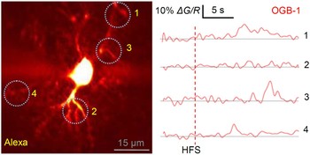
More generally, the observation that the GPCR-dependent Ca2+ rises have no detectable effect on synapses (Fiacco et al., Reference Fiacco, Agulhon, Taves, Petravicz, Casper and Dong2007; Agulhon et al., Reference Agulhon, Fiacco and McCarthy2010) leaves open the possibility that in physiological circumstances such Ca2+ rises could have a common upstream trigger, but not necessarily a causal link, to the Ca2+-dependent astrocytic release machinery. For example, nicotinic acid adenine dinucleotide phosphate (NAADP) elicits relatively small and localised Ca2+ signals that are later amplified many-fold, in a second step prompted by an IP3R-dependent mechanism in mammalian cells (Ruas et al., Reference Ruas, Rietdorf, Arredouani, Davis, Lloyd-Evans and Koegel2010). An initial small Ca2+ transient could easily occur undetected under the current imaging protocols while still providing an efficient molecular signal. Indeed, not only Ca2+ signals in astrocytes seem to have an inherently oscillatory nature and are orders of magnitude slower than evoked Ca2+ entry in neuronal axons or dendrites, such signals often become detectable only seconds after the stimulus (Verkhratsky and Kettenmann, Reference Verkhratsky and Kettenmann1996; Pasti et al., Reference Pasti, Volterra, Pozzan and Carmignoto1997; Haydon and Carmignoto, Reference Haydon and Carmignoto2006; Henneberger et al., Reference Henneberger, Papouin, Oliet and Rusakov2010) (Fig. 1). What happens to small Ca2+ fluctuations during this ‘dormant’ period and whether GPCRs are involved in the underlying machinery is beyond resolution or sensitivity of the currently available imaging approaches.
Processes of a stratum radiatum astrocyte often show delayed Ca2+ responses to a synaptic stimulus, much unlike Ca2+ responses evoked in neuronal axons or dendrites. Left: a CA1 astrocyte held in whole cell mode (a single 2P excitation plane, 40 µM Alexa Fluor 594 ‘red’ channel, λx2p = 800 nm) depicting four regions of interests (ROIs, 1–4). Right: the corresponding Ca2+ responses (ΔG/R, the OGB-1 ‘green’ channel signal increment related to ‘red’ Alexa channel signal to cancel out focus drift (Oertner et al., Reference Oertner, Sabatini, Nimchinsky and Svoboda2002), 200 µM OGB-1); HFS, 100 pulses at 100 Hz applied to Schaffer collateral fibres (Henneberger et al., unpublished observations).

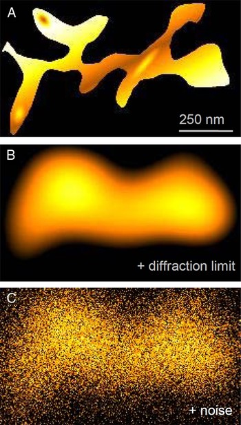
Indeed, non-GPCR sources of Ca2+ signalling provide an additional dimension to the quest for the mechanisms involved in generation of astroglial communication signals. Before GPCRs had taken centre stage in our understanding of how external signalling is translated into intracellular Ca2+ regulation, studies of astroglia documented a variety of other Ca2+ signal transduction mechanisms, including voltage-gated Ca2+ channels, ionotropic receptors or ion exchangers acting in situ (see Porter and McCarthy, Reference Porter and McCarthy1997; Verkhratsky et al., Reference Verkhratsky, Orkand and Kettenmann1998 for a review). In their pioneering study, Porter and McCarthy reported as early as in 1996 that synaptic activity could evoke astrocytic Ca2+ rises when both metabotropic and ionotropic glutamate receptors are effectively blocked (Porter and McCarthy, Reference Porter and McCarthy1996). These Ca2+ mechanisms might well be spatially separated from the GPCR-dependent Ca2+ signalling domains because free Ca2+ inside cells is likely to be highly compartmentalised due to its rapid diffusion and therefore steep concentration gradients arising near local Ca2+ sources and sinks (Fig. 2A). On the other hand, GPCR-independent Ca2+ sources could interact and interfere with store-dependent Ca2+ signalling because the gating of the IP3R itself not only depends on the concentration of IP3 but is also highly sensitive to intracellular Ca2+ levels (see Foskett et al., Reference Foskett, White, Cheung and Mak2007 for a recent review).
Ca2+ imaging does not reveal the nanoscopic landscape of intracellular Ca2+. (A) A hypothetical distribution of nanoscopic Ca2+ sources and sinks in a planar section of an astrocyte process. False colour scale (from dark to bright orange) depicts Ca2+ concentration. (B) Diffraction-limited resolution (~250 nm, Gaussian point-spread function) is imposed on image A. (C) Optics registration noise (~25% of the maximal fluorescence) is added to image B. These transformations represent some typical experimental distortions in two-photon excitation Ca2+ imaging experiments in organised brain tissue. The distortion is likely to be higher with non-confocal registration.

Ca2+ NANODOMAINS
An uneven landscape of free intracellular Ca2+ (Fig. 2A) could potentially be an important factor in considering how Ca2+-dependent signalling is generated inside astrocytes and how the exogenous manipulations might affect it. Indeed, mechanisms that shape Ca2+ activity of astroglia are still poorly understood even at the conceptual level, although important initial data have been obtained in cultured astrocytes addressing the occurrence and basic properties of microscopic Ca2+ domains near vesicle release sites (Marchaland et al., Reference Marchaland, Cali, Voglmaier, Li, Regazzi and Edwards2008) or in thin astrocytic processes (Shigetomi et al., Reference Shigetomi, Kracun, Sofroniew and Khakh2010). One technical limitation is that individual astrocyte processes in situ (organised brain tissue) could be as thin as 20–30 nm (Ventura and Harris, Reference Ventura and Harris1999; Rollenhagen et al., Reference Rollenhagen, Satzler, Rodriguez, Jonas, Frotscher and Lubke2007; Witcher et al., Reference Witcher, Kirov and Harris2007) whereas signals recorded with two-photon excitation in organised brain tissue represent fluorescence volume averaged on an at least an order magnitude coarser scale (see an example in Scott and Rusakov, Reference Scott and Rusakov2006). This averaging will potentially mask a complex distribution of active nanoscopic sources and sinks that might contribute to local Ca2+ homeostasis (Fig. 2B,C), the scenario similar to Ca2+ imaging of thin axons and their synaptic terminals (Koester and Sakmann, Reference Koester and Sakmann2000; Scott et al., Reference Scott, Ruiz, Henneberger, Kullmann and Rusakov2008).
A particularly important aspect of the uncertainty surrounding the interpretation of recorded Ca2+ activity in astrocytes is that, similar to presynaptic terminals, critical release events in these cells could be controlled by hotspots of Ca2+ generated by Ca2+ entry on a 10 nm scale, or Ca2+ nanodomains. If the molecular trigger of release is located strategically in the immediate vicinity of a Ca2+ source, the local concentration of Ca2+ ions entering the cytoplasm is likely to dwarf the numbers of Ca2+ buffer molecules (endogenous or exogenous) stationed in the same locality. This scenario might explain, for instance, why loading astrocytes with EGTA, a high-affinity but relatively slow-binding Ca2+ buffer in whole-cell mode has had little effect on synaptic plasticity at nearby excitatory synapses in the hippocampus (Diamond et al., Reference Diamond, Bergles and Jahr1998; Luscher et al., Reference Luscher, Malenka and Nicoll1998; Ge and Duan, Reference Ge and Duan2007). However, high concentrations of the fast-binding, high-affinity Ca2+ buffer BAPTA could convincingly block Ca2+ and astrocyte-dependent effects on some forms of synaptic activity (see Andersson and Hanse, Reference Andersson and Hanse2010; Todd et al., Reference Todd, Darabid and Robitaille2010 for most recent examples), suggesting that Ca2+ nanodomains occurring near the release trigger machinery could be a preferred mechanism in such cases (Bucurenciu et al., Reference Bucurenciu, Kulik, Schwaller, Frotscher and Jonas2008).
Perhaps surprisingly, high concentrations of intracellular BAPTA did not block LTP to the same extent as Ca2+ clamp, nor did they completely suppress slow changes of intracellular Ca2+ in response to stimulation of afferent fibres in the hippocampal area CA1 (Henneberger et al., unpublished observations). In contrast, an intracellular solution that contains an equilibrated mixture of Ca2+ buffers and Ca2+ ions, a so-called ‘Ca2+ clamp’ approach (Baker et al., Reference Baker, Knight and Umbach1985; Belan et al., Reference Belan, Kostyuk, Snitsarev and Tepikin1993), appears to interfere with this dynamic equilibrium suppressing astrocytic release of the NMDAR co-agonist d-serine and thus inhibiting synaptic potentiation (Henneberger et al., Reference Henneberger, Papouin, Oliet and Rusakov2010). One principal difference between the Ca2+ clamp solution and Ca2+ buffers alone is that the unlimited supply of Ca2+ ions in the former case is likely to prevent free Ca2+ from dropping below the clamped level whereas Ca2+ buffers on their own prevent Ca2+ from going above its equilibrated concentration. This may suggest that the signalling mechanism that eventually leads to d-serine release requires, at some stage, fluctuations of the local Ca2+ concentration rather than monotonic Ca2+ rises. Again, this ambiguity highlights the need for understanding spatial relationships among those cellular systems that (i) trigger the Ca2+ signal, (ii) propagate it on a local scale and (iii) host the molecular target(s) of this signal. An additional complication may arise when individual target systems, such as the Ca2+-dependent release machinery, Ca2+-dependent potassium channels or the sodium–calcium exchanger display different sensitivities to the same local Ca2+ signalling event.
FUTURE DIRECTIONS
It would seem reasonable to conclude that our understanding of Ca2+ signalling in astrocytes is in its infancy. Do the signalling cascades relaying neural code to glial activity occur homogenously across the astrocytic arbour, near synapses or in some specialised areas? What are the origin, spatiotemporal properties and the adaptive purpose of ‘spontaneous’ Ca2+ signals in astrocytes, i.e. cellular Ca2+ elevations that cannot be directly associated with synchronous nerve cell firing or synaptic discharges? How, where and on what time scale do these mechanisms respond to changing patterns of neural activity or its pathological changes? To approach these questions, it would appear critical to improve the sensitivity of Ca2+ measurements and its spatial resolution. The spatial resolution of optical microscopy (including two-photon excitation microscopy) is by definition diffraction limited to 0.3–0.5 µm, which is an order of a magnitude greater than small astrocyte branches (Fig. 2). However, several recently developed super-resolution methodologies may help to overcome this limitation (reviewed in Hell, Reference Hell2007; Wilt et al., Reference Wilt, Burns, Wei Ho, Ghosh, Mukamel and Schnitzer2009). Stimulated emission depletion (STED) microscopy is based on suppressing emission from the outer, doughnut-shaped part of the regular diffraction-limited volume using intense de-excitation light generated by a second laser source. This could potentially enhance resolution by an order of magnitude, and it has successfully been applied to monitor nanoscopic compartments of neuronal dendrites (Nagerl et al., Reference Nagerl, Willig, Hein, Hell and Bonhoeffer2008; Ding et al., Reference Ding, Takasaki and Sabatini2009). The increased resolution of STED microscopy, however, will require increased fluorescence yield and/or excitation light exposure to maintain the acceptable signal-to-noise ratio in turbid media such as organised brain tissue. The latter implies trade-off between achievable resolution, time scale of measurements and potential phototoxic effects. Currently, the light intensity required to obtain STED images seems to be compatible with only a few images per experiment, and using STED in organised brain tissue, may impose additional limitations related to the wavelength- and depth-dependent light scattering. Other advancing imaging methodologies do not rely on intensity measurements but use instead fluorescence parameters independent of fluorophore concentration. Fluorescent lifetime imaging measures the average decay in fluorescence intensity post-excitation on a nanosecond scale. This methodology involves therefore registering photons emitted by fluorophore molecules at different time intervals (1–10 ns) following a very brief (50–200 fs) laser pulse. Because individual signals, or photon counts, recorded during each such duty cycle are very small, thousands of duty cycles are often required to produce the average fluorescence lifetime curve. Fortunately, some Ca2+ indicators, such as the Oregon Green BAPTA series, do have fluorescence lifetimes sensitive to Ca2+ binding (Wilms et al., Reference Wilms, Schmidt and Eilers2006; Gersbach et al., Reference Gersbach, Boiko, Niclass, Petersen and Charbon2009). This should enable direct readout of the free-Ca2+ concentration, albeit on a time scale which has to be sufficiently slow to accommodate multiple duty cycles required to obtain an acceptable signal-to-noise ratio. As for localisation of individual identified molecular targets, much promise is held by photo-activated localisation microscopy (PALM) and stochastic optical reconstruction microscopy (STORM). These two techniques are based on repeated imaging of a small subset of fluorescent molecules (such as protein tags) excited at a low density to avoid overlap between individual point sources of emission. Because each such source could be de-convolved to a relatively precise pair of coordinates, accumulation of data on individual sources in the same preparation generates a combined image with an effective resolution of ~20 nm (Wilt et al., Reference Wilt, Burns, Wei Ho, Ghosh, Mukamel and Schnitzer2009). Again, successful implementation of these two techniques in real-time imaging will depend on the experimental requirements in the time domain: in general, cellular mechanisms under study have to be quasi-stationary during the multiple duty cycles required to construct a complete PALM/STORM image. Accordingly, this imposes a set of constraints on the fluorescent probes, optical system and the acquisition techniques involved, and the entire experiment might thus require a relatively unique combination of all such factors to achieve an optimal outcome. Notwithstanding these potential technical obstacles, such techniques should undoubtedly help to provide important advances in our ability to understand Ca2+-dependent mechanisms underlying functional relationship between neurons and astroglia.
ACKNOWLEDGEMENT
The authors declare no financial interests. The work was supported by Wellcome Trust and MRC (UK).