Arrhythmogenic right ventricular cardiomyopathy is a heritable cardiac condition that is characterised by the replacement of healthy myocardial tissue with fibro-fatty tissue, which can lead to both contractile and electrical abnormalities. In this entity, the right ventricle is classically affected; however, biventricular involvement has also been well documented. Reference Corrado, Link and Calkins1 Mutations in the desmosomal protein required for the cell-to-cell adhesion are the key driver behind the pathogenesis. Clinical manifestations typically occur between the second and fourth decade of life with palpitations or exercise intolerance being the most common presenting symptoms. Reference Corrado, Link and Calkins1 We report an unusual case of arrhythmogenic right ventricular cardiomyopathy in a child who presented with heart failure and was found to have rare de novo mutations.
Case report
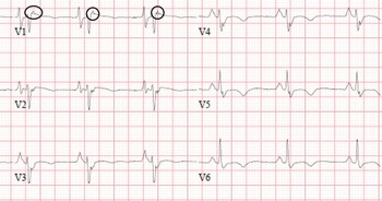
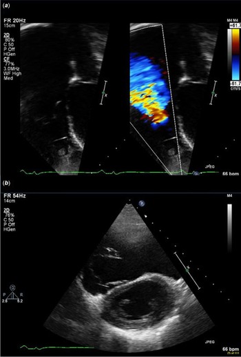
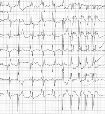
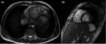
An 11-year-old male presented with signs of right heart failure, including hepatomegaly and recently diminished exercise intolerance. The patient reported symptoms of a gastrointestinal illness a few weeks prior. Physical examination was remarkable for a low frequency, grade III/VI regurgitant murmur at the lower left sternal border, hepatomegaly, and ascites. Abdominal ultrasound demonstrated hepatosplenomegaly, engorgement of the inferior caval vein, and hepatic veins, moderate ascites, and a moderate to large right pleural effusion. Electrocardiogram was remarkable for right atrial enlargement, Epsilon wave in the right-sided precordial leads, and T-wave inversion throughout the precordial leads (Fig 1). Initial echocardiography demonstrated severe right atrial and ventricular dilation with severely depressed right ventricular systolic function. Severe regurgitation due to poor coaptation of the tricuspid valve leaflets was noted. The left ventricular function was also moderately depressed (Fig 2). Brain natriuretic peptide and high sensitivity troponin were elevated at 1000 pg/ml and 84 ng/L, respectively. The patient was initially managed with intravenous diuresis with marked improvement in ascites, hepatomegaly, and mild improvement in both left and right ventricular systolic function. He was then able to be transitioned to oral diuresis. He had multiple runs of non-sustained ventricular tachycardia during which he was hemodynamically stable (Fig 3). Cardiac MRI was performed showing fibro-fatty infiltration within the right ventricle along with areas of focal dyskinesia and aneurysmal formation (Fig 4). His right and left ventricular ejection fractions were estimated at 22% for both. Indexed right and left ventricular end-diastolic volumes were 164 ml/m2 and 68 ml/m2, respectively. Our patient satisfied criteria for diagnosis of arrhythmogenic right ventricular cardiomyopathy as per the modified International Task Force Criteria for the diagnosis of arrhythmogenic right ventricular cardiomyopathy Reference Marcus, Mckenna and Sherrill2 because of the following findings:
Electrocardiogram showing sinus rhythm with right atrial enlargement and T-wave inversion in the lateral precordial leads. Epsilon waves can be seen throughout most of the precordial leads (black circles).

Transthoracic echocardiogram. ( a ) 2D image and colour flow mapping from the apical four-chamber view depicting the severely dilated right atrium with severe tricuspid valve regurgitation. ( b ) Parasternal short-axis view at the level of the papillary muscles demonstrating a severely dilated right ventricle with flattening of the interventricular septum.

Telemetry tracing depicting four beats of wide complex tachycardia (rate approximately 150 bpm).

Cardiac MRI. ( a ) Axial plane with focal areas of dyskinesia and aneurysmal formation (black arrows). ( b ) Sagittal plane with areas of delayed enhancement (white arrows) consistent with fibrosis.

-
Epsilon waves and precordial lead T-wave inversions were present on electrocardiogram,
-
ventricular arrhythmias were documented on telemetry,
-
cardiac MRI demonstrated a dilated right ventricle with an end-diastolic volume >110 ml/m2 along with right ventricular outflow tract dyskinesia and global right ventricular dysfunction (right ventricular ejection fraction <40%).
Additionally, due to lack of MRI findings consistent with myocarditis, chronicity of symptoms, and no clinical improvement over time, an inflammatory process is unlikely. He was deemed an intermediate risk for major arrhythmic events due to non-sustained ventricular tachycardia and moderate biventricular dysfunction. He therefore underwent placement of an implantable cardioverter-defibrillator. Genetic analysis was positive for Desmin gene, c.347A>G variant, and MYH7 gene, c.2146G>T variant mutation. Parents tested negative for the two specific mutations found in the patient. Due to his continued right-sided dilation and depressed systolic function, he is additionally followed by our heart failure specialist. Following transplant evaluation, the decision was made to list him for cardiac transplant. He currently is clinically stable with outpatient medical management.
Discussion
Arrhythmogenic right ventricular cardiomyopathy is transmitted in an autosomal dominant pattern; however, rare autosomal recessive forms have been documented. Reference Bauce, Rampazzo and Basso3 It is a well-established cause of sudden cardiac death in young adults, males being affected more than females. Riele et al in their review of arrhythmogenic right ventricular cardiomyopathy in the paediatric population found that children presented with sudden cardiac death/arrest more commonly than adults did. Reference Te Riele, James and Sawant4 The most common initial manifestations are palpitations or exercise-induced syncope in adults. Reference Corrado, Link and Calkins1 Additional studies looking at paediatric populations show that most young people with arrhythmogenic right ventricular cardiomyopathy present in one of two fashions: asymptomatically when diagnosed as part of a genetic workup of an affected family member or with ventricular arrhythmias and palpitations. Reference Bauce, Rampazzo and Basso3,Reference DeWitt, Chandler and Hylind5,Reference Deshpande, Herman and Quigley6 Few children present as our patient did with heart failure.
Diagnosis of arrhythmogenic right ventricular cardiomyopathy in children can be challenging, keeping in mind that the task force criteria for diagnosis arrhythmogenic right ventricular cardiomyopathy published in 2010 are based on the adult population. Genetic testing has significantly improved the ability to diagnose arrhythmogenic right ventricular cardiomyopathy, especially in familial cases; however, early diagnosis of probands can still be difficult. Reference Bauce, Rampazzo and Basso3,Reference Deshpande, Herman and Quigley6 Electrical abnormalities in children are much less common than in adults. As previously mentioned, our patient uniquely fits many of the electrocardiographic criteria including an Epsilon wave and diffuse T-wave inversion.
Lastly, the pathogenic Desmin gene identified in our patient has been linked to myofibrillar myopathies and cardiomyopathies with a wide spectrum of phenotypes. Reference Ohno7 Clinical manifestations can range from myopathy with cardiac involvement or pure cardiomyopathies. Klauke et al presented a genotype of 22 cohort with arrhythmogenic right ventricular cardiomyopathy, one of which had the same Desmin mutation as ours. Patient initially presented with episodes of syncope at the age of 15 with a complete transplant by 17. Soon after, the patient began to experience skeletal muscle disease. Pathology showed accumulation of desmin and myotilin in the right and left ventricles. Although we do not have confirmed genotype–phenotype in our patient, this reported case provides us a good correlation. Furthermore, Lorenzon et al report a low incidence (2 of 91) of Desmin gene mutations in their sample populations. Reference Klauke, Kossmann and Gaertner8,Reference Lorenzon, Beffagna and Bauce9 These studies suggest that Desmin mutations in Arrhythmogenic Right Ventricular Cardiomyopathy are a rare genetic finding. The sarcomere gene identified, MYH7, although a variant of uncertain significance, we speculate that its presence may contribute to the patient’s severe phenotype as it is also associated with other cardiomyopathies such as autosomal dominant dilated and hypertrophic cardiomyopathy. Both of these were de novo mutations as his parents were not found to have these variants. In a large cohort study, it was reported that patients who were carriers for more than one gene mutation had a five-fold increase in developing left ventricular dysfunction and heart failure. Reference Bhonsale, Groeneweg and James10 With these two positive gene mutations, the biventricular phenotype seen in our patient can be explained.
Conclusion
We report an unusual case of paediatric arrhythmogenic right ventricular cardiomyopathy who presented with heart failure. Our patient fulfilled the 2010 modified criteria for diagnosis of arrhythmogenic right ventricular cardiomyopathy and had uncommon de novo genetic mutations.
Acknowledgements
None.
Financial support
This work received no specific grant from any funding agency, commercial, or not-for-profit sectors.
Conflicts of interest
None.
Ethical standards
This work involved no humans or animal experimentation.