Background
Hypertrophic cardiomyopathy is the most common genetic cardiomyopathy characterised by left ventricular hypertrophy, fibrosis, and myocardial ischaemia.Reference Villa, Sammut and Zarinabad 1 The annual mortality rate of hypertrophic cardiomyopathy ranges from <1% in the general community to 3–6% in tertiary referral centres.Reference Maron 2 Coronary microvascular dysfunction is an important feature of hypertrophy cardiomyopathy, which negatively affects the long-term outcome.Reference Ismail, Hsu and Greve 3 This is illustrated by a reduced coronary flow reserve in the absence of obstructive coronary artery disease.Reference Cecchi, Olivotto, Gistri, Lorenzoni, Chiriatti and Camici 4 Severity of left ventricular hypertrophy and genetic predisposition is implicated in the development of coronary microvascular dysfunction. Abnormal stress perfusion has been associated with the presence of coronary microvascular dysfunction in hypertrophic cardiomyopathy. The presence of left ventricular outflow tract gradient, as well as increased vascular resistance and wall stress resulting from diastolic dysfunction, has also been associated with myocardial perfusion abnormalities in hypertrophic cardiomyopathy.Reference Guclu, Happe and Eren 5 , Reference Hittinger, Mirsky, Shen, Patrick, Bishop and Vatner 6
Although multiple imaging techniques have been utilised to assess myocardial perfusion both at rest and under the effect of a pharmacologic agent, cardiovascular MRI has become increasingly popular in recent years because of it being non-invasive, with no radiation exposure and providing superior spatial resolution when compared with other modalities.Reference Petersen, Jerosch-Herold and Hudsmith 7 Stress perfusion cardiac MRI using a coronary vasodilator has been widely used in adults, but paediatric data are limited;Reference Noel, Krishnamurthy, Moffett and Krishnamurthy 8 the most common experience with perfusion has been with traditional stressors, adenosine and dipyridamole. Moreover, the utility of stress perfusion cardiac MRI to detect microvascular dysfunction in the paediatric setting is unknown. Therefore, the purpose of this study is to report our experience with regadenoson stress perfusion cardiac MRI in a cohort of children with hypertrophic cardiomyopathy examined to assess for coronary microvascular disease.
Methods
The Memorial Healthcare System Institutional research ethics board and institutional review board approved the study. Ethical guidelines and protocol were based on the World Medical Organization Declaration of Helsinki. Written informed consent had been obtained from each patient or parents/guardians in case of minors before testing.
Patient population
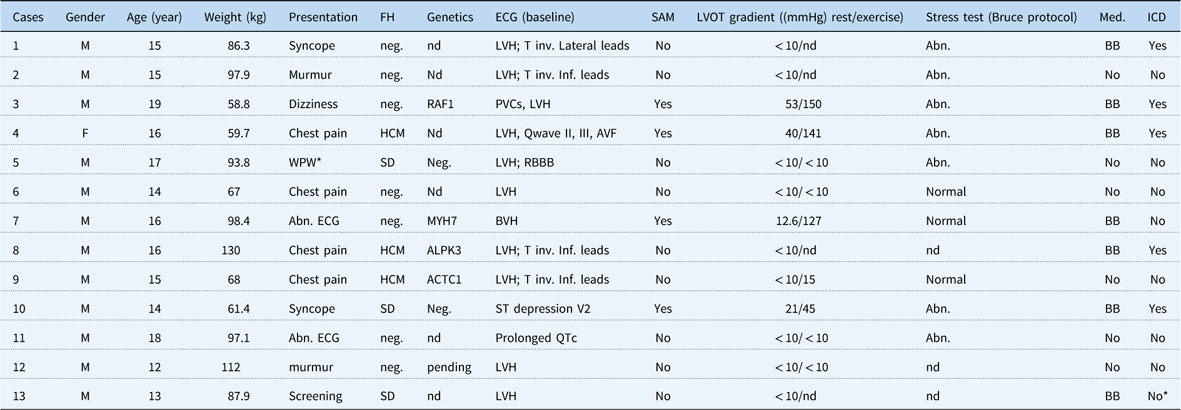
We reviewed our clinical experience using regadenoson stress perfusion cardiac MRI from January, 2016 to January, 2017. In all, 13 subjects, 12 male and one female, were diagnosed with hypertrophic cardiomyopathy based on clinical, echocardiographic, and electrocardiographic data (Table 1). Eight patients (61.5%) had baseline electrocardiogram suggestive of ischaemic (ST-T wave) changes. Nine patients had previously undergone stress echocardiography, and 10 had treadmill exercise testing using the Bruce protocol.
Clinical, electrocardiographic, echocardiographic, and stress test data.

Abn=abnormal; BB=β blockers; BVH=biventricular hypertrophy; ECG=electrocardiogram; FH=family history; f=female; nd=not done; Neg=negative; neg=negative for cardiomyopathy or sudden death; ICD=intracardiac defibrillator; inv=inversion; Inf=inferior; LVH=left ventricular hypertrophy; LVOT=left ventricular outflow tract; Med=medicine; m=male; PVC=premature ventricular contractions; RBBB=right bundle branch block; SAM=systolic anterior motion of the mitral valve; SD=sudden death; WPW=Wolf–Parkinson–White syndrome
* Family refused to proceed with ICD placement
Cardiac MRI protocol
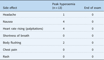
All patients were instructed to avoid caffeinated products for at least 24 hours before the exam. No patient had known contraindication to regadenoson. A cardiac MRI technologist, a paediatric radiology nurse, and a paediatric cardiologist (L.H.) were present in the MRI suite during the entire examination. Heart rate and pulse oximetry were continuously monitored. Patients received 0.4 mg bolus of regadenoson (Lexiscan; Astellas Pharma, Northbrook, Illinois, United States of America) intravenously. Blood pressure was recorded at the beginning of the study, every 2 minutes after the administration, and at the conclusion of testing. Patients were asked for any symptoms during and after administration of regadenoson, and at the end of examination (Table 2). A 50-mg dose of aminophylline (A2A receptor antagonist) was administered intravenously after the stress first-pass perfusion images were acquired to reverse coronary vasodilatory effect. Patients were observed for about 90 minutes following the test for any late adverse events.
Symptoms after the administration of regadenoson.

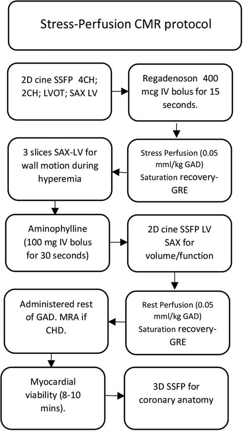
Cardiac MRI images were acquired using a 1.5-Tesla clinical scanner with an 18-element phased array receiver coil (Siemens Magnetom Aera; Siemens AG Healthcare Sector, Erlangen, Germany). The protocol is summarised in Figure 1. A retrospectively gated balanced steady-state free-precession sequence was used to obtain breath-held cine images in three long-axis planes followed by a short-axis stack from the base to the apex for quantification of ventricular size, volume, and ejection fraction. Stress first-pass perfusion sequence was obtained 45 seconds following the administration of regadenoson with 0.05 mmol/kg of gadoteridol (Bayer HealthCare, Whippany, New Jersey, United States of America) injected at a rate of 3.5 ml/second followed by 15 ml of saline at 7 ml/second using a power injector (Medrad Spectris Solaris EP, Bayer HealthCare, Whippany, New Jersey, United States of America). A single-shot, T1-weighted saturation recovery gradient echo was used for first-pass perfusion with typical imaging parameters in a multiple-slice group with three slices in the short axis – basal, mid, and apical – and a single slice in four chambers; the method included 90° saturation preparation pulse for each slice, with repetition time of 5.1 ms, echo time of 1.1 ms, field of view of 360×288 mm, and slice thickness of 8 mm. Three short-axis slices using cine steady-state free-precession of the ventricles were acquired after the first-pass perfusion to assess wall motion in hyperaemic state. Three-dimensional steady-state free-precession electrocardiographic gated and respiratory navigated was obtained for coronary origin and course. Late gadolinium enhancement imaging was performed using two-dimensional phase sensitivity inversion recovery sequences, covering the entire left ventricular myocardium, 10 minutes after gadolinium (gadoteridol) administration in four-chamber, two-chamber, and short axis for myocardial viability.
Stress perfusion protocol. 2D=two dimension; 2CH=two chambers; 4CH=four chambers; CMR=cardiac magnetic resonance; GAD=gadolinium; GRE=gradient echo; IV=intravenous; LV=left ventricle; LVOT=left ventricular outflow tract; mcg=micrograms; SAX=short axis; SSFP=steady-state free-precession image.
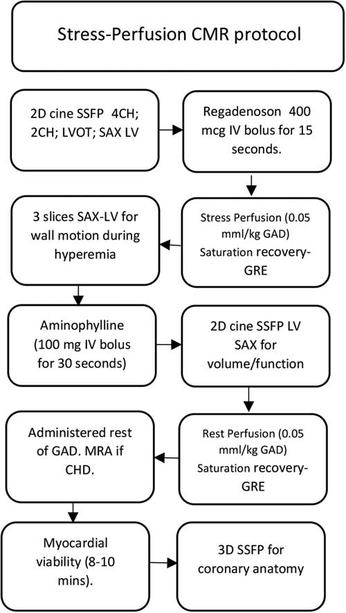
Image analysis
Post-processing analysis of volumes, function, wall thickness, and mass was performed on cvi42 (Circle Cardiovascular Imaging Inc., Alberta, Canada) software. Mass and volumes were indexed to body surface area. Stress first-pass imaging was qualitatively assessed and graded as “clear for diagnosis”, homogeneous myocardium with no significant artefacts; “acceptable for diagnosis”, the presence of dark rim artefact but without other major artefact; and “non-diagnostic”, significant artefact obscuring large amount of the myocardium.Reference Noel, Krishnamurthy, Moffett and Krishnamurthy 8 Simultaneous comparison was performed between the stress and rest first-pass perfusions to identify reversible versus fixed defects. The perfusion defect was considered the result of microvascular disease when no coronary territory pattern was followed.
Statistics
Mean and standard deviation with ranges were used to report the numerical data. Paired t-test was used to compare variables from different haemodynamic conditions. p Values<0.05 were considered to indicate statistical significance. Statistical analyses were performed using MedCalc version 17.2 (Ostend, Belgium).
Results
Clinical, electrocardiographic, echocardiographic, and morphologic assessment
In our hypertrophy cardiomyopathy cohort (Table 1), the mean age was 15.3±1.93 years. The most common presenting symptoms were chest pain, syncope, and abnormal electrocardiogram. Two patients were initially consulted owing to heart murmur. Family history of hypertrophic cardiomyopathy or sudden death was found in six patients. Genetic test was performed in seven patients, of whom four had mutations related to hypertrophic cardiomyopathy. In all, seven patients were on β-blocker therapy at the time of the stress perfusion cardiac MRI. On balanced steady-state free-precession cine imaging, the most common morphologic variant was the hypertrophic cardiomyopathy with sigmoid septal contourReference Noureldin, Liu and Nacif 9 in four patients followed by the symmetric and apical forms with three patients each. Stress echocardiograms were obtained in nine patients. Systolic anterior motion of the mitral valve and left ventricular outflow tract gradient at rest and at peak exercise were present in four patients. Ten patients underwent exercise stress testing with Bruce protocol; this was considered abnormal in seven patients.
Stress perfusion cardiac MRI
All patients completed testing with no interruptions. One patient underwent the exam under general anaesthesia. No major side effects occurred (Table 2). Sensation of rising heart rate and nausea were the most common symptoms in four patients. Body flushing was present in two patients. All symptoms were transient and completely resolved either spontaneously or with aminophylline bolus. The mean heart rate at baseline was 65±11.7 beats per minutes. After the bolus of regadenoson, the mean heart rate increased to 112±21.8 beats per minute (p<0.0001). The mean systolic blood pressure at baseline was 115.4±13.8 mmHg and at peak hyperaemia it was 107±21 mmHg (p=0.06). Neither changes in the atrioventricular conduction nor respiratory symptoms were documented. Patients were discharged as per protocol with normal vital signs and no residual symptomatology. No wall motion abnormalities were identified during the hyperaemic state. The coronary arteries were normal in origin and course in all patients.
Microvascular dysfunction
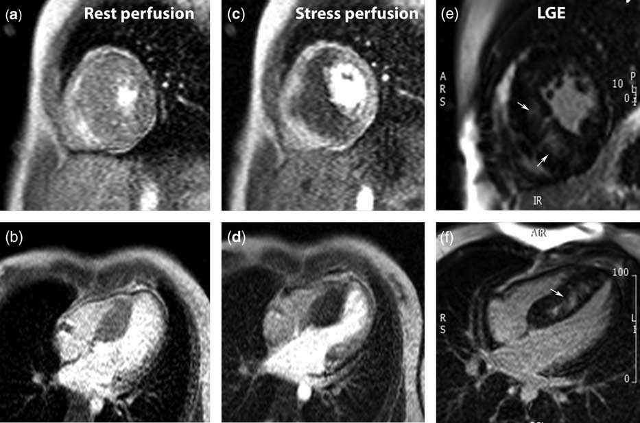
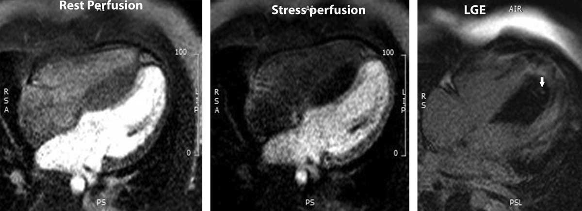
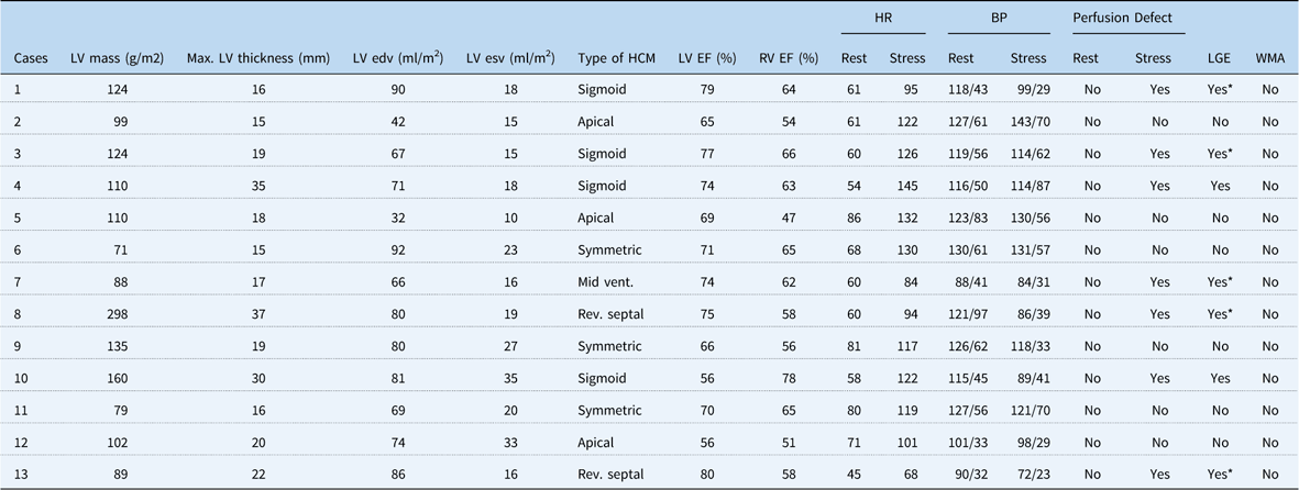
First-pass perfusion sequences at rest and with stress were considered “clear for diagnosis” in the entire cohort. A total of seven patients (53.8%) developed perfusion defect during the hyperaemic state induced by regadenoson (Fig 2). All these patients had normal perfusion at rest (Table 3). Abnormal perfusion was observed in areas of the myocardium unable to be explained by a specific coronary territory. Four patients with abnormal stress perfusion had a maximum thickness at the level of the papillary muscles below 30 mm.Reference Noureldin, Liu and Nacif 9 Late gadolinium enhancement was present in those cases with perfusion abnormalities; however, in five patients there were areas of the myocardium with perfusion defects in which late gadolinium enhancement was not observed (Fig 3). Five patients with positive stress perfusion cardiac MRI have undergone implantable cardioverter defibrillator placement based on current guidelines.Reference Maron 10
First-pass perfusion at rest ( a and b ) and under the effect of regadenoson ( c and d ). Large perfusion defect in the interventricular septum is observed only during the effect of coronary vasodilator (reversible defect). Large mid-wall area of late gadolinium enhancement (arrows) is seen in the interventricular septum ( e and f ). LGE=late gadolinium enhancement.

Patient with a maximum thickness of 22 mm and a large area of perfusion defect in the entire interventricular septum with only a small mid-wall area of delayed enhancement at the apical septum (arrow). LGE=late gadolinium enhancement.

Cardiac MRI data.

BP=blood pressure; edv=end diastolic volume; EF=ejection fraction; esv=end systolic volume; HR=heart rate; LGE=late gadolinium enhancement; LV=left ventricle; Rev=reverse; RV=right ventricle; Vent=Ventricular; WMA=wall motion abnormalities
* Only small areas of LGE extension when compared with larger areas of perfusion defect
Discussion
Feasibility and safety in paediatric hypertrophic cardiomyopathy
The present study demonstrates the feasibility of regadenoson stress perfusion cardiac MRI in children with hypertrophic cardiomyopathy, and its ability to unmask coronary microvascular dysfunction in this cohort of young patients. To the best of our knowledge, this is the first description of the use of regadenoson stress cardiac MRI in paediatric hypertrophic cardiomyopathy. A recent experience with regadenoson stress perfusion cardiac MRI showed safety in the paediatric population,Reference Noel, Krishnamurthy, Moffett and Krishnamurthy 8 and although they included one patient with left ventricular hypertrophy this patient did not have hypertrophic cardiomyopathy. Stress cardiac MRI in hypertrophic cardiomyopathy is commonly experienced by adults, with the majority of studies using adenosine infusion.Reference Ismail, Hsu and Greve 3 , Reference Petersen, Jerosch-Herold and Hudsmith 7
Regadenoson has proven to be safe in children for multiple reasons. Regadenoson is a selective A2A receptor agonist that is administered as an intravenous bolus at a fixed dose with less side effects, unlike an infusion such as adenosine.Reference Al Jaroudi and Iskandrian 11 Its longer half-life in comparison with adenosine allows us to use a single bolus technique. An A2A receptor antagonist such as aminophylline is recommended to reverse the hyperaemia before proceeding with the rest perfusion.Reference Bhave, Freed and Yodwut 12 The safety and effectiveness of regadenoson has been compared with adenosine, a non-selective agonist of multiple adenosine receptors (A1; A2A; A2B; A3), and dipyridamole, an adenosine reuptake inhibitor. In a series on stress perfusion cardiac MRI, Vasu et alReference Vasu, Bandettini and Hsu 13 found a similar vasodilator efficacy of regadenoson and adenosine, much superior than dipyridamole. Theoretical issues regarding post-denervation hypersensitivity to adenosine would also be potentially reduced with regadenoson, which is a more selective agonist. Although other modalities to assess myocardial perfusion have also been used such as positron emission tomography,Reference Timmer and Knaapen 14 , Reference Cianciulli, Saccheri and Masoli 15 cardiac catheterisation using intracoronary Doppler catheter,Reference Litmathe, Stosch, Klues, Boeken, Korbmacher and Gams 16 and also echocardiography,Reference Kutty, Olson and Danford 17 stress perfusion cardiac MRI is considered the test of choice in many centres at present, and it is gaining popularity for use in the paediatric population.
Detection and relevance of coronary microvascular dysfunction in paediatric hypertrophic cardiomyopathy
Coronary microvascular dysfunction is an important feature of hypertrophic cardiomyopathy and has been associated with adverse clinical outcomes.Reference Ismail, Hsu and Greve 3 Although regional perfusion defect has been related to the degree of left ventricular hypertrophy,Reference Jablonowski, Fernlund and Aletras 18 interestingly, four of seven patients (57%) with perfusion defects in our cohort had a maximum wall thickness less than 30 mm. Similar observations were reported by Ismail et alReference Ismail, Hsu and Greve 3 in adult hypertrophic cardiomyopathy where perfusion abnormalities were noted in areas of normal myocardial thickness, suggesting a role for vasomotor dysfunction. Another interesting observation from the present series is that six of the seven (86%) patients with stress perfusion defects had asymmetric septal hypertrophy, of whom four had sigmoidal septal contour, with the highest left ventricular outflow tract gradients and systolic anterior motion of the mitral valve as previously described.Reference Davies and McKenna 19 None of the patients with symmetric concentric hypertrophic cardiomyopathy developed abnormal perfusion. Although the relationship between left ventricular outflow tract gradients and perfusion abnormalities is not precisely known, Guclu et alReference Guclu, Happe and Eren 5 found a significantly lower capillary density in patients with hypertrophic cardiomyopathies and greater left ventricular outflow tract gradients, as compared with normal controls. Clearly the pathophysiology is multifactorial, because in addition to the association of the left ventricular outflow tract obstruction with reduced capillary density,Reference Guclu, Happe and Eren 5 specific genetic mutations have also been implicated in the production of myocardial microvascular abnormalities in hypertrophic cardiomyopathy.Reference Friehs, Moran and Stamm 20 , Reference Maestri, Milia and Salis 21 On the basis of the current guidelines,Reference Maron 10 coronary microvascular dysfunction is not an indication for institution of sudden death prevention measures in patients with hypertrophic cardiomyopathy. Although our cohort had a small sample size, we speculate that coronary microvascular dysfunction may be frequent in children with hypertrophic cardiomyopathy, even in those with only mild or moderate left ventricular hypertrophy. However, further prospective studies are necessary to confirm or refute this.
Implications of abnormal perfusion without fibrosis
Abnormal myocardial perfusion has been considered as a precursor of replacement fibrosis detected by late gadolinium enhancement, which was seen in our cohort in combination with the visual perfusion abnormalities. Although others have shown a relation between late gadolinium enhancement and perfusion abnormalities at rest,Reference Chiribiri, Leuzzi and Conte 22 , Reference Soler, Rodriguez, Monserrat, Mendez and Martinez 23 first-pass perfusion at rest was preserved in our entire cohort (reversible ischaemia), and, as previously reported,Reference Ismail, Hsu and Greve 3 a hyperaemic myocardium was not associated with late gadolinium enhancement. The finding of perfusion defects in areas without late gadolinium enhancement in some of our patients (Fig 3) indicates that gadolinium enhancement by itself could underestimate the true extension of microvascular disease. Therefore, some patients who may not otherwise fulfil criteria for sudden death prevention could be at a higher risk of adverse events. Further investigations are needed to uncover the relationship between cardiac MRI-derived microvascular dysfunction and hard clinical outcomes, and to examine whether microvascular dysfunction would eventually qualify as a solo indicator for sudden death prevention in children with hypertrophic cardiomyopathy. On the basis of these observations, we have increased the frequency of clinical follow-up, surveillance for arrhythmia, and evaluation of sport participation in all patients with perfusion defects.
Limitations
This study had inherent limitations associated with a retrospective cohort study. Findings of this study ought to be confirmed by larger prospective multicentre studies, which will be able to shed more light on the frequency of microvascular dysfunction, and its correlation with ventricular hypertrophy, genetic predisposition, and long-term outcomes.
Conclusions
We demonstrate the feasibility and effectiveness of regadenoson stress perfusion cardiac MRI to unmask myocardial perfusion defects in paediatric patients with hypertrophic cardiomyopathy and microvascular disease.
Acknowledgements
The author would like to thank Dr Jonathan Harvey Soslow and Dr Shelby Kutty for their critical review and edition of this manuscript. The author would also like to recognise our cardiac MRI technicians at Memorial Regional and Joe DiMaggio Children’s Hospitals for their tremendous contribution to this manuscript.
Financial Support
This research received no specific grant from any funding agency or commercial or not-for profit sectors.
Conflicts of Interest
None.
Ethical Standards
Ethical guidelines and protocol were based on the World Medical Organization Declaration of Helsinki of 1975, as revised in 2008, and have been approved by the Memorial Healthcare System Institutional research ethics board and institutional review board.