Introduction
Delayed reconstruction for helical rim and antihelix composite defects typically resulting from traumatic loss like human bites or from oncological resection of skin cancer can be challenging. Previously described techniques often needs multiple stages, produce variable results and need steep learning curve.
Previously published techniques base their indications on the location and size of the defects. Techniques described to deal with defects of less than 2 cm in width, including wedge reReference Tanzersection 1 and Antia–Buch helical chondrocutaneous advancement flaps,Reference Antia and Buch2 produce asymmetric smaller ears and are typically described for mid-third defects.
Wedge excisionReference Tanzer1 can produce a smaller ear, causing punching of the cartilage, which creates obvious asymmetry and unsatisfactory outcome.
The Antia–Buch helical advancement techniqueReference Antia and Buch2 involves mobilising the remaining helical rim skin and cartilage fully to advance the skin and cartilage flaps, is limited to a defect of up to 2 cm in size and can typically address mid-third helical rim defects rather than defects involving the anti-helix. This techniqueReference Antia and Buch2 create a smaller ear, visible scarring extending to the root of the helix, and produce obvious asymmetry in size and shape that may affect the patient’s ability to wear glasses comfortably.
Staged techniques include Converse’s tunnel technique, which is a two-stage procedure involving the use of a prefabricated flap.Reference Converse3 In the first stage, an auricule cartilage graft taken from the contralateral ear is tunnelled under the post-auricular skin close to the helical rim defect. The second stage, performed three weeks later, takes the anteriorly based skin flap along with the cartilage graft and sets it into the helical rim defect.Reference Converse3
An alternative two-stage procedure is Dieffenbach’s flap,Reference Dieffenbach4 which involves harvesting contralateral auricule cartilage and suturing it to the helical rim defect, then elevating post-auricular skin and advancing and covering the cartilage graft. The pedicle requires another stage at three weeks to be divided and inset.
Finally, a few techniques describing the use of staged tubed bipedicle flaps are available in the literature,Reference Dujon and Bowditch5–Reference Steffanoff, Strauch, Vasconez and Hall-Findlay12 typically for delayed reconstruction of the helical rim defects staged over two or three procedures.
All these staged techniques can create confusion for surgeons involved in reconstructing composite helical rim and antihelix defects. These techniques often have a very steep learning curve to master and frequently produce a less satisfactory outcome.
Materials and methods
We describe here a refined, versatile, one-stage technique for delayed reconstruction used to address a variety of size defects of the helical rim and antihelix. Refinements to the originally described technique are introduced.Reference Lin, Lewis and Alrawi13
The refinements extend the use of the technique for larger defects and improve its outcome. This technique was previously used for central helical rim defects, ipsilateral conchal cartilage graft and setting the post-auricular flap in the post-auricularsulcus using bolster alone.
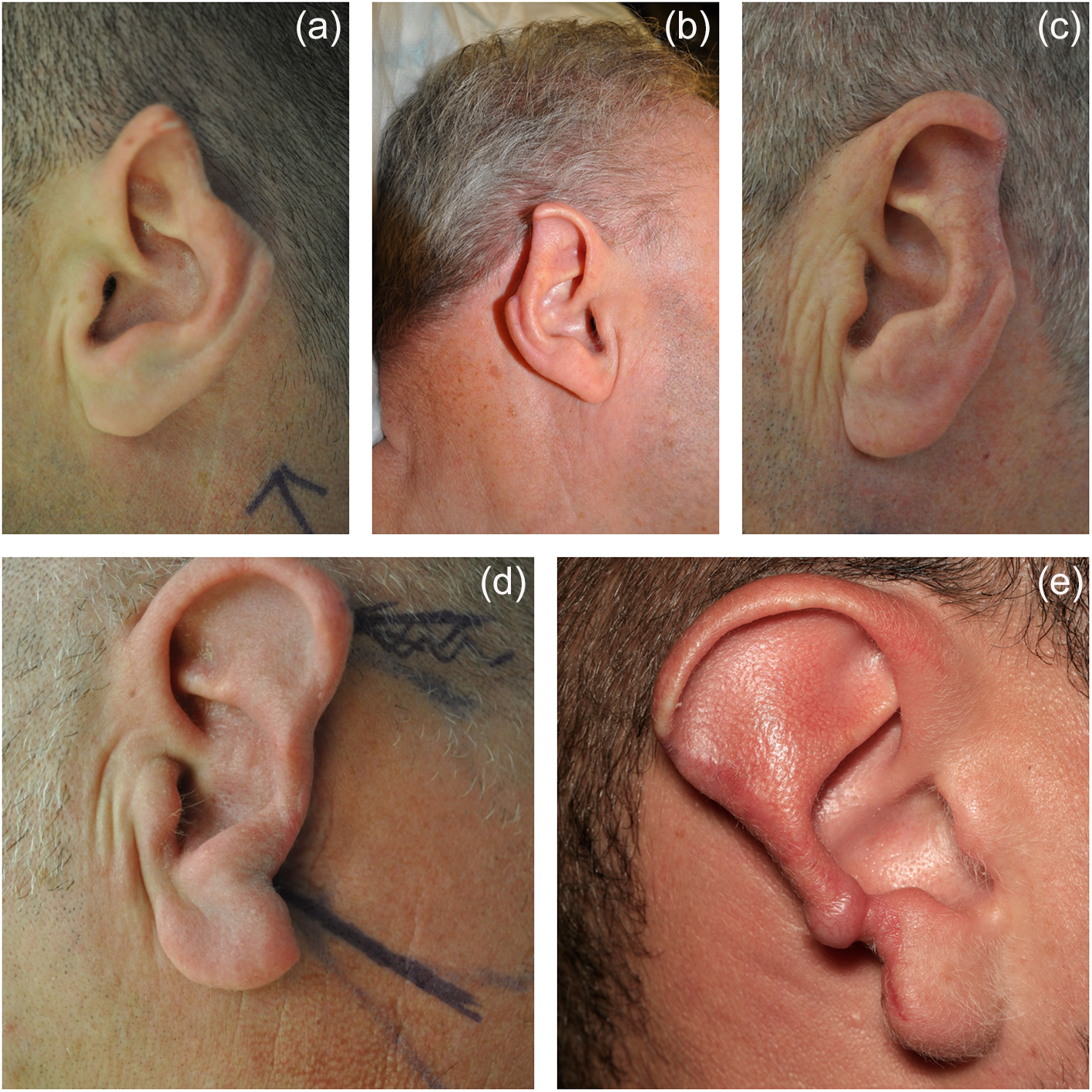
We have extended the use of our technique to reconstruct larger defects than previously described, with use of a conchal cartilage graft allowing reconstruction of defects up to 4 cm. For larger defects (more than 4 cm of the helix and antihelix), we advocate using a carved autologous rib cartilage graft. We have not used a rib allograft, but believe this is potentially an option, if available. Figure 1 shows multiple examples of the types of ear defect prior to reconstruction.
Examples of five different types of partial ear helical rim defects following skin cancer excision (b, c) and traumatic loss as aresults of human bite (a, d, e).

The use of contralateral conchal cartilage grafts is recommended to prevent the risks of weakening the ipsilateral cartilage structure if cartilage graft is to be harvested from the same ear. It also allows the surgeon to set back the contralateral ear to improve ear symmetry in prominence. We also introduced quilting sutures to the post-auricule through the conchal bowl as well as the use of dental roll bolster to transfix and support the post-auricular sulcus and prevent tenting and loss of definition of sulcus.
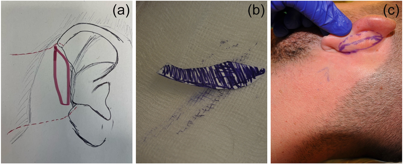
The procedure starts by measuring the defect size and shape, then a template of the cartilage defect is created. The template is used to mark the contralateral ear conchal cartilage (Figure 2).
Schematic representation for marking the post-auricular flap (a), template of cartilage defect (b) and using the template to mark the contralateral conchal cartilage (c).

We found that using an ipsilateral ear cartilage graft causes unwanted weakness of the cartilage structure, resulting in the ear being set back further while wrapping and advancing the skin flap around the cartilage graft. Using a contralateral cartilage graft allows the surgeon to set back the donor ear and create symmetry in prominence.
The procedure is well tolerated under local anaesthetic when using an ear cartilage graft. General anaesthesia is required when rib cartilage is needed. The surgical planning starts by marking the defect and the bipedicle post-auricular flap. We prefer to harvest the cartilage graft from the contralateral ear through the posterior approach, as shown in Figure 2c.
Local anaesthetic infiltration in the post-auricular skin into the subcutaneous plane allows for hydro dissection. Local anaesthetic with adrenaline is infiltrated in the contralateral post-auricle to help harvest the conchal cartilage graft through an incision anterior to the post-auricular sulcus. Once the cartilage graft has been harvested, the donor site can be closed with 3-0 Polydioxanone suture (PDS) concho-mastoid sutures to allow the ear some degree of set back, and the skin can be closed with a 4-0 Monocryl suture in two layers. We discourage harvesting the cartilage graft using the anterior approach as this can produce a scar anteriorly and prevent the surgeon from adjusting the prominence of the ear.
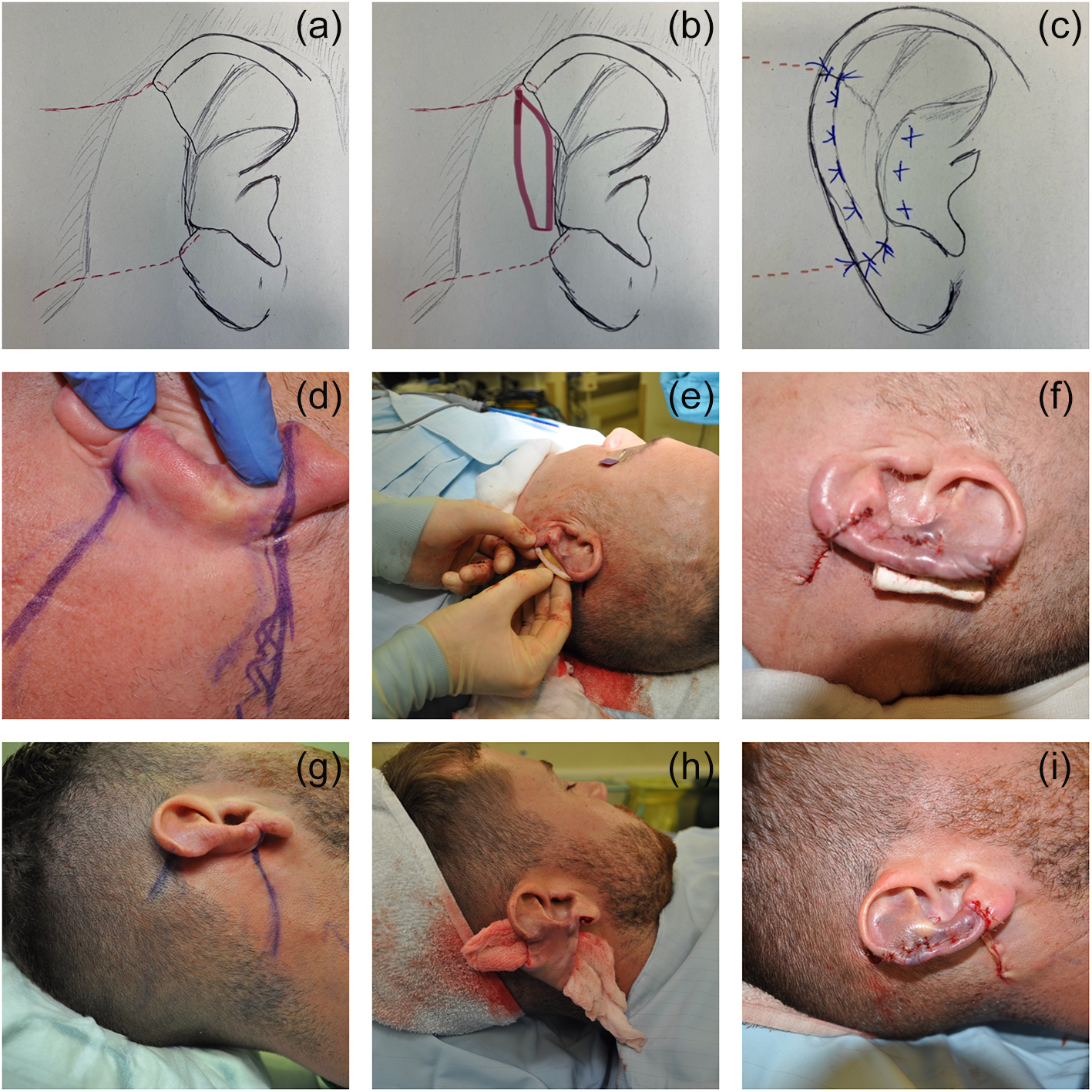
The marking of the posterior bipedicle skin flap starts 2 mm beyond the helical rim defect into the helix in a divergent fashion to include a wider posteriorly based flap and ensure reliable blood supply. The marking can cross the post-auricular hairline if necessary to achieve length and allow safe elevation of the bipedicle flap at the deep subcutaneous plane (Figure 3).
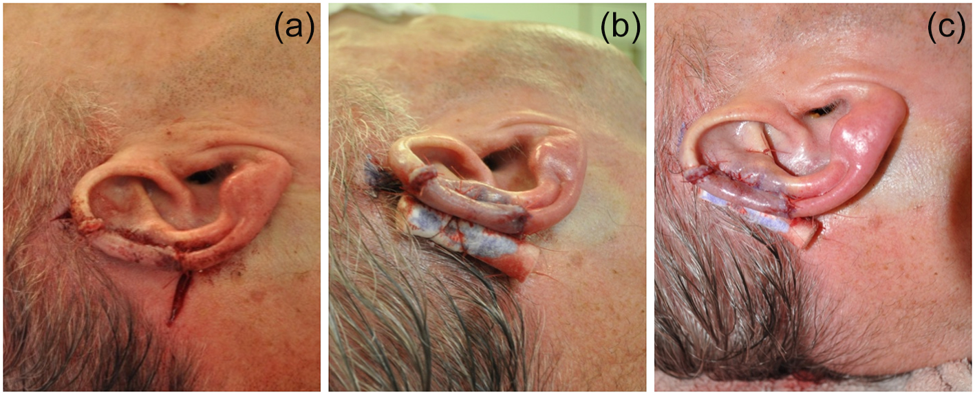
The multiple steps of the procedure. (a) Schematic representation marking, (b) template, (c) reconstruction, (d) marking the post-auricular bipedicle flap, (e) cartilage graft, (f) reconstruction of the defect by re-draping the flap around the cartilage with quilting sutures, (g) marking post-auricular flap, (h) raising the post-auricular bipedicle flap and (i) reconstruction of the defect.

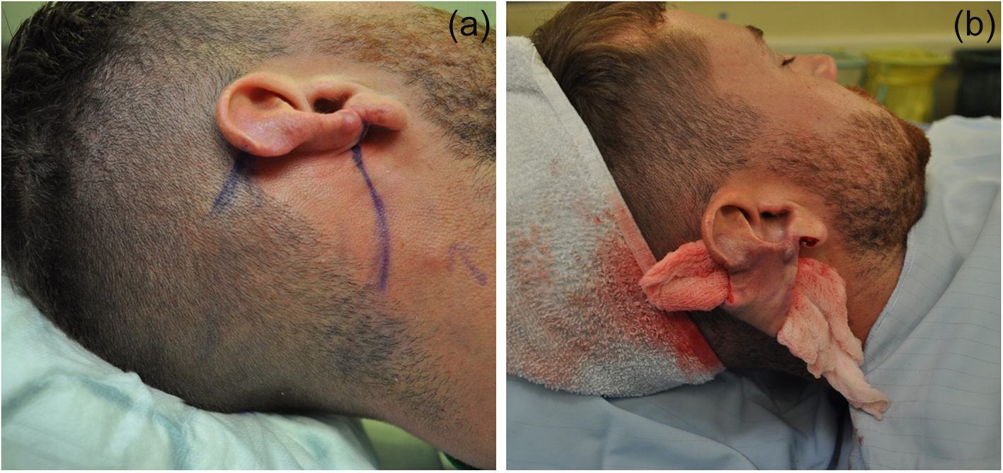
The skin flap on the posterior aspect of the ear and the post-auricular surface is incised and the flap elevated at the subcutaneous plane. The skin overlying the ear cartilage is elevated in a subcutaneous plane, leaving perichondrium behind. The bipedicle flap is fully mobilised and elevated (Figure 4).
Large helical defect. (a) Marking post-auricular flap and (b) full elevation of the post-auricular flap.

If more skin needs to be recruited, the flap incision can be extended to cross the post-auricular hairline and cut as required. The scar along the helical defect is carefully dissected, released and elevated making sure the skin across the scar in the antihelix remain intact while elevating the skin flap. This is important to preserve the skin blood supply across the scar. The bipedicle flap from the helical rim defect to the post-auricular hairline is fully mobilised and released, any scarring on the cartilage defect is carefully removed and the cartilage defect is carefully delineated.
The harvested conchal cartilage graft is secured to the cartilage defect using size 5-0 absorbable sutures (PDS II Polydioxanone; Ethicon by Johnson & Johnson surgical technologies). The bipedicle flap is gradually advanced towards the defect to drape and wrap around the cartilage graft. To ensure the skin drape around the cartilage graft, quilting sutures are used to transfix the skin onto cartilage along the helical rim. Quilting sutures are also used on the post-auricular flap onto the conchal bowl using 5-0 and 4-0 absorbable sutures (Vicryl rapide polyglactin 910; Ethicon) (Figure 5).
(a) Insetting the post-auricular flap, (b) quilting the flap and cartilage graft, and (c) using a dental bolster for the support of the post-auricular sulcus.

Once the defect has been reconstructed the flap is further advanced, secured to the post-auricular sulcus, anchored by 4-0 Monocryl and further secured using 2-3 quilting skin 4-0 Vicryl rapide along the sulcus. Once the defect has been repaired and the post-auricular sulcus secured, the post-auricular flap can be sutured with deep dermal sutures using 4-0 Monocryl suture (Poliglecaprone; by Ethicon) and the skin closed with 5-0 Vicryl rapid suture. We used dental roll bolster sutured into the post-auricular sulcus to provide extra support (Figure 5).
The wounds, including the suture line, are covered with Dermabond skin glue. The ear is covered with non-adherent dressing (MepitelR; Silicone dressing with SafetacR technology by Molnlycke), soft cotton gauze and Gamgee, and a head bandage applied to provide further support.
Both the head bandage and dental roll are removed after three weeks, with the patient being recommended to continue wearing a head band or protective bandage at bedtime for six weeks.
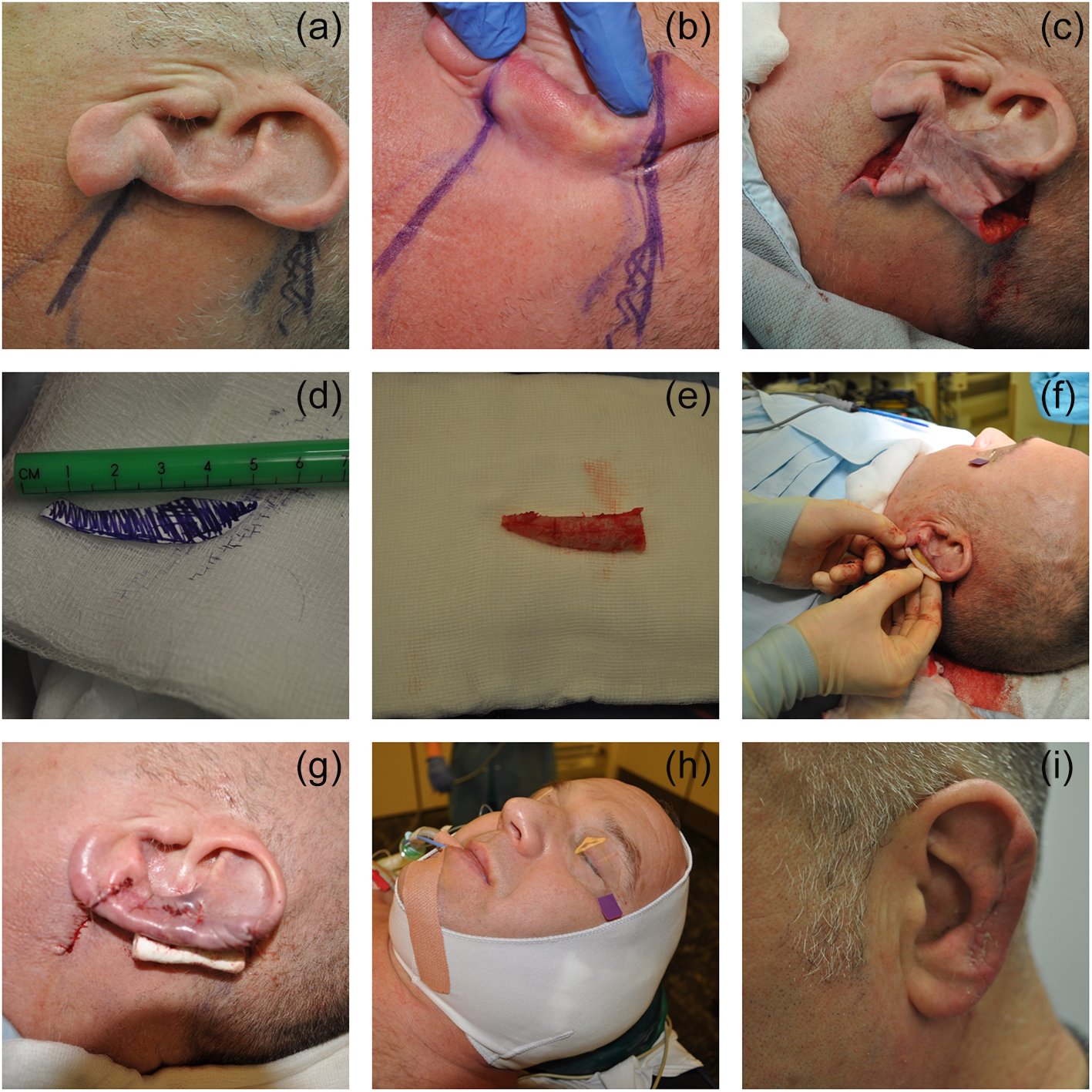
For patients with larger defects involving the helical rim and the anti-helix, a large cartilage graft of 4 cm or more is needed. In our case, the defect was more than 5 cm therefore we used a rib cartilage graft that had been carved according to the template created from the defect, as illustrated in Figure 6.
The multiple steps in the reconstruction of a large (more than 5 cm) ear defect using a carved rib cartilage graft. (a–c) Planning the post-auricular bipedicle flap, (d–f) preparing the rib cartilage graft and (g–i) completing the reconstruction with head dressing and the final results.

We did not use any allograft in our patients, but if this option is available in any unit, it should be considered to minimise donor morbidity. It also avoids the need to perform the procedure under general anaesthetic and hospital admission.
Discussion
There is a lack of consensus around the best technique that can be used to reconstruct variable size defects involving the helical rim and antihelix. Such techniques aim to produce consistent and reliable outcomes. Surgeons are often confused by the considerable number of techniques published and described to reconstruct defects smaller or greater than 2 cm in size.
Since our 2018 paper,Reference Lin, Lewis and Alrawi13 we have further refined the technique to produce stable results. Our unit has now adopted this versatile technique as the standard procedure to deal with such defects. This single-stage technique is clear, versatile and easy to learn. We have shown that using a bipedicle post-auricular flap and contralateral conchal cartilage graft or autologous rib graft for larger defects can reliably deal with variable size defects. Our refined technique is easy to learn and produce reliable and satisfactory outcome preserving ear size and symmetry.
The senior author (MA) describes refinements of the previously described one-stage techniqueReference Lin, Lewis and Alrawi13 to reconstruct a delayed composite defect involving variable size of the helical rim and anti-helix, producing a satisfactory symmetry in size and shape. The refinements strongly emphasise the use of a contralateral ear conchal cartilage graft for defects less than 4 cm. The other refinement is the extended use of this technique to reconstruct larger defects greater than 5 cm of the skin and cartilage using a rib cartilage graft (Figure 6) showing different steps of reconstruction.
We have previously seen that harvesting a conchal cartilage graft from the ipsilateral ear can produce asymmetry in ear prominence and weakness of the remaining cartilage structure of the reconstructed ear. We therefore recommend using a cartilage graft from the contralateral ear through posterior approach because this will allow the surgeon to set back the contralateral ear, providing good symmetry in prominence.
We also advocate the importance of raising a uniformly thick skin flap including the subcutaneous fat, making sure the flap base towards the post-auricular hairline is wide enough to ensure reliable blood supply. Furthermore, the use of quilting sutures to transfix the cartilage graft and the post-auricular sulcus is important, as is keeping the dental roll bolster in place for three weeks until full healing is achieved to prevent tenting of the post-auriclar flap.
Depending on their size and position, composite defects can be reconstructed using several other methods. These previously described techniques, both single and staged, as mentioned earlier for helical rim reconstruction, can create confusion for surgeons, have a steep learning curve, involve multiple staged procedures and multiple hospital admissions, and cost more.
Such procedures produce asymmetryboth smaller in size and different in shape. Antia–Buch helical advancement techniqueReference Antia and Buch2 produces extensive visible scarring extending to the base of the helical root. They also have limited use only address small defects up to 2 cm in size involving the helical rim, the outcome is scars at the helical root with smaller ear which can interfere with the patient’s ability to wear glasses comfortably. Furthermore, helical wedge excisionReference Tanzer1 can produce an asymmetrical smaller ear with punching of the cartilage creating obvious asymmetry.
Techniques involving multiple stages, such as ConverseReference Converse3 and Dieffenbach,Reference Dieffenbach4 can create confusion for surgeons and therefore have a steep learning curve. Furthermore, they can be inconvenient to patients as they involve multiple hospital admissions and can produce variable results.
We believe our technique should be adopted as the first choice to address a wide varieties of complex helical rim defects for patients with delayed presentation (Figure 1). We do not encourage reconstruction to be performed at the time of traumatic loss or oncological excision for obvious reasons with human bites wound tend to be either infected or contaminated , while for excision of skin cancer best to wait to ensure adequate margin of excision. Reconstruction can be discussed with patients and planned in three to six months time.
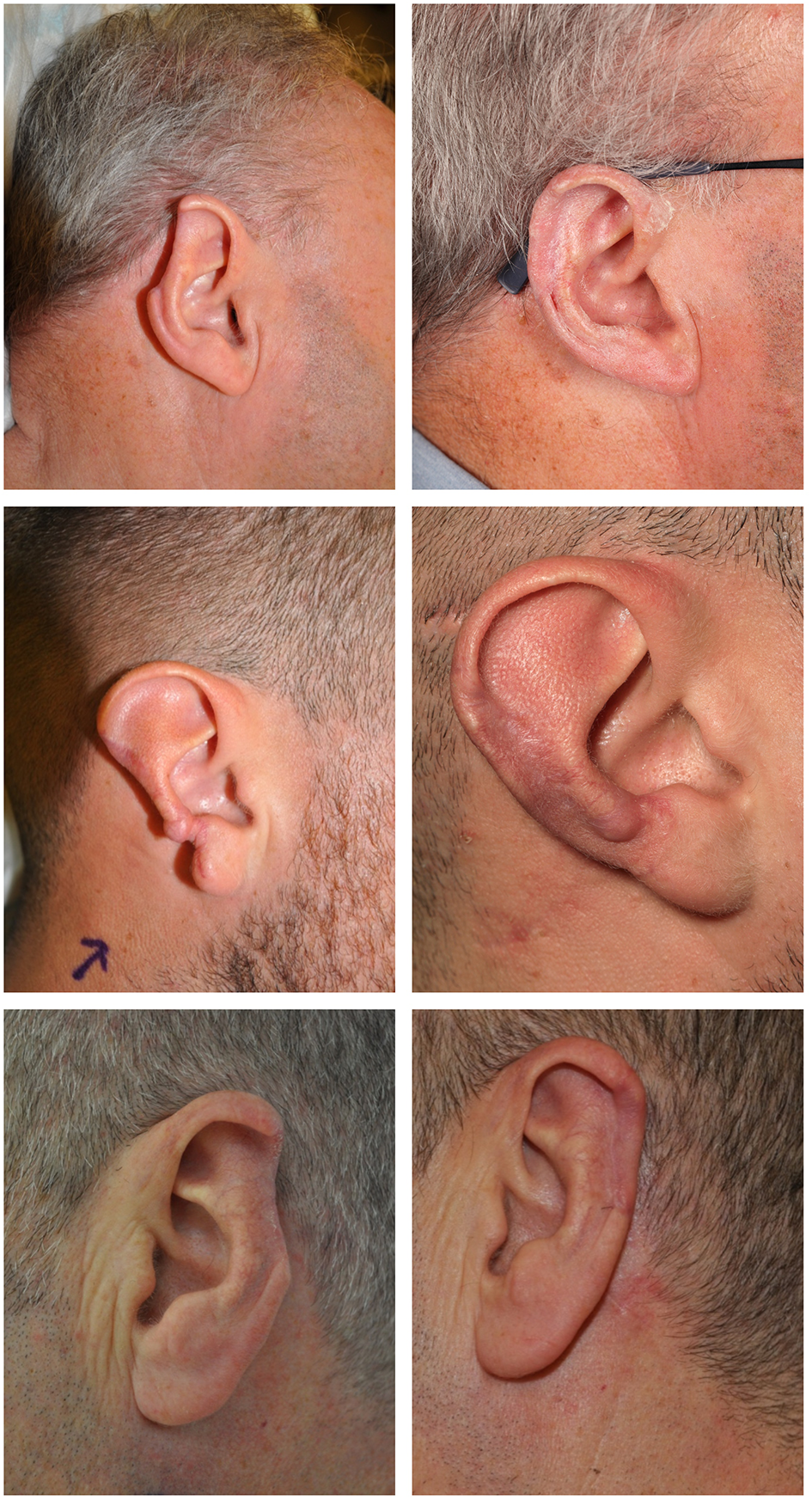
This delayed single-stage bipedicle post-auricular flap and cartilage graft technique is easy to learn and produces satisfactory cosmetic results with stable superior outcomes preserving ear size, shape and contours (Figure 7).
• This paper discusses crucial refinements added to the original novel single-stage helical rim reconstruction using a post-auricular bipedicle flap technique for defects of more than 2 cm
• The refined single-stage helical rim reconstruction technique is versatile, reliable, can be applied to defects larger than 4 cm, and produces satisfactory aesthetic results in terms of size, shape and symmetry
• Published staged techniques for delayed helical rim reconstruction require multiple staged operations, produce inferior outcomes in symmetry of size and shape, and are not easy to learn
• Published single-staged techniques for delay reconstruction of helical rim defects involve ear tissue resection and produce smaller asymmetric ears
• Our refined technique for large partial and subtotal delayed reconstruction is a single stage that uses a post-auricular bipedicle flap and autologous cartilage graft to reliably restore the original ear helix and antihelix
• The refinements discussed here are crucial to standardise the technique and improve outcomes
Three examples (top, middle and bottom) of before and after reconstruction. The size and shape of the ear have been preserved in each case.

Competing interests
None declared.