

INTRODUCTION
The nudibranch genus Thordisa was described by Bergh (Reference Bergh and Semper1877) from the Philippines (family: Discodorididae Bergh, 1891). The key diagnostic characters of the genus are digitiform oral tentacles, a bi-lamiate foot which is anteriorly notched, the dorsum with elongate villous papillae and bi or tri pinnate gills. Radular teeth have hamate inner and middle lateral teeth; these may be smooth or denticulate, and the outer lateral teeth are pectinate. The reproductive system usually has one or two vestibular glands, which may have accessory spines. The vagina and penis are armed or unarmed (Chan & Gosliner, Reference Chan and Gosliner2006, Reference Chan and Gosliner2007).
There are, in all, 30 species belonging to genus Thordisa (family: Discodorididae) that are currently recognized as valid, and the genus is distributed globally (Chan & Gosliner, Reference Chan and Gosliner2007). Of these, only two species were described from India, namely, Thordisa villosa (Alder & Hancock, Reference Alder and Hancock1864) and Thordisa annulata Eliot, Reference Eliot1910. The most recent studies on the genus and its phylogeny were by Chan & Gosliner (Reference Chan and Gosliner2006, Reference Chan and Gosliner2007), who additionally described five new species, including two from the Eastern Pacific.The reports by various authors range from regions such as East Africa and Zanzibar (Eliot, Reference Eliot1903); South Africa (Chan & Gosliner, Reference Chan and Gosliner2007); Andaman (Eliot, Reference Eliot1910); China (Zong-Jie & Lin, Reference Zong-Jie and Lin2005); Indo Pacific Asian region (Coleman, Reference Coleman2008); Panama and Costa Rica (Chan & Gosliner, Reference Chan and Gosliner2007) and the Indo-Pacific region (Gosliner et al., Reference Gosliner, Behrens and Valdés2008). Phylogenetic studies on cryptobranch dorids, including the genus Thordisa, have been carried out by Valdés (Reference Valdés2002).
Thordisa villosa is a large discodorid found in the rocky intertidal areas. Alder & Hancock (Reference Alder and Hancock1864) first described it as Doris villosa based on the collection of Walter Eliot from Waltair, Vizagapatanam, India. The specimen was subsequently re-examined by Sir Charles Eliot (Reference Eliot1906a, Reference Eliotb), who reassigned it to the genus Thordisa. It was later recorded by Satyamurti (Reference Satyamurti1952) from the Gulf of Mannar. Thordisa annulata Eliot, Reference Eliot1910, another species of the genus Thordisa, was described as a new species from the Andaman Islands but has not been recorded from India since then.
MATERIALS AND METHODS
The present specimens were identified on the basis of descriptions provided in the above mentioned literature using radular and external morphological characteristics.
A single specimen of size 40 mm (Bombay Natural History Society (BNHS)-Opistho-341, collected by Vishal Bhave) was found for the first time on 23 February 2010 at Kasheli, Ratnagiri, Maharashtra State (16.741436°N 73.30677°E). A second specimen measuring 40 mm (BNHS-Opistho-741, collected by Amruta Prasade) on 28 January 2012 was found in a shallow tidal pool with corals and sponges at Poshitra, Gujarat State (22.402310°N 69.202669°E) and the third specimen of size 35 mm (BNHS-Opistho-773, collected by Bhavik Patel) was found on 13 March 2012 at Narara reef, Gujarat State (22.477045°N 69.718881°E) (Figure 1).
Study sites of Maharashtra and Gujarat, India: (A) map showing area of study (not to scale, for inset maps scale is provided); (B) Kasheli, Maharashtra—rocky intertidal area; (C) Poshitra, Gujarat—intertidal reef; (D) Narara, Gujarat—intertidal reef.

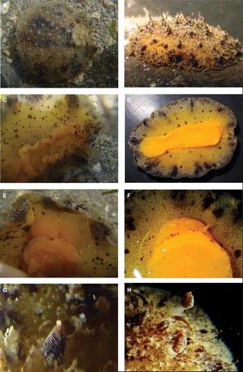
In Gujarat, both the specimens were collected from a rocky intertidal reef. They were hidden under loose rocks, in association with many types of sponges and soft corals (Figure 2A, B). In Maharashtra, however, a single specimen was collected from the underside of a loose rock present at the bottom of a rock pool of the rocky intertidal region. Specimens were sighted while walking along the intertidal area during low tide, manually turning over loose rocks to search for underside fauna. Collected specimens were photographed on the field and in the laboratory for live coloration and morphological details. All the specimens were deposited in the opisthobranchs collection of the BNHS, India.
Thordisa villosa Alder & Hancock, Reference Alder and Hancock1864: (A) from Narara, Gujarat; (B) from Ratnagiri, Maharashtra; (C, D) ventral view of specimen from Gujarat & Maharashtra respectively; (E, F) respective mouth and oral tentacles; (G, H) respective rhinophores.

For the behavioural observations, specimens were kept alive in aerated seawater for a few days before preservation. A brown mucus-like substance was observed coming out from the anal opening, which may have been either due to the irritation, for defence or possibly defecation. The mantle tissue of a specimen (BNHS-Opistho-741) was cut and preserved in 95% ethanol for DNA analysis and the rest of the animal was preserved in 4% formalin. The specimen from Narara, Gujarat (BNHS-Opistho-773) was dissected and the buccal mass was removed for radula isolation.
RESULTS
SYSTEMATICS
EXTERNAL MORPHOLOGY
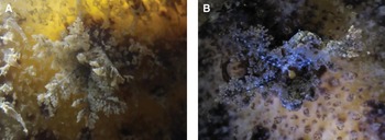
Body is oval, dorsal region with central longitudinal ridge. Dorsal surface is rough and covered with several small tubercles and a few long and slender soft papillae. Colour of animal pale brown on dorsal surface, with dark chocolate brown patches near the middle ridge and at the edge of the mantle (Figure 2A, B). Rhinophores dark brown with distinctive white tip, lamellate with 20–25 lamellae (Figure 2G, H); raised sheath margin tuberculate. Yellowish brown gills, plume-shaped and tripinnate (Figure 3A, B), fully retractile. Villous dorsal structures diagnostic. Hyponotum is yellowish, dark brown patches present at the edge of hyponotum, while few spots are located on the remainder (Figure 2C, D). Foot is bright yellow, which gets pale towards the mantle edge; it bears a few spots around the margin as well as on its upper surface. Brown spots on head and upper lamina with notch and digitiform oral tentacles (Figure 2E, F).
(A) Tripinnate gills of the specimen from Poshitra, Gujarat; (B) tripinnate gills of the specimen from Ratnagiri, Maharashtra.

RADULAR MORPHOLOGY
The dissected specimen was 35 mm (BNHS-Opistho-773). It had a radula formula of 48 × 6.42–44.0.42–44.6 at the 18th row (Figure 4A, B). Rachidian teeth were absent. Middle lateral teeth were hamate (Figure 4C) and of equal size toward the margins. Six outermost lateral teeth were pectinate, out of which the outermost four were smaller in size than the other two (Figure 4D).
Radula of the specimen (BNHS-Opistho-773) of Thordisa villosa from Narara, Gujarat: (A) radula, scale 1000 µm; (B) inner lateral teeth, scale 200 µm; (C) mid-lateral teeth, scale 50 µm; (D) outermost pectinate teeth, scale 200 µm.

DISCUSSION
The Indian discodorid fauna is represented by 14 genera out of the 27 known genera worldwide (Bouchet, Reference Bouchet2012). A total of 33 species of the family Discodorididae were recorded from India till now. The majority of the studies on Indian opisthobranchs were confined to the intertidal reef habitats of the east coast, e.g. the Gulf of Mannar (Rao, Reference Rao1962), Tamil Nadu (Satyamurthi, 1952), Andhra Pradesh (Alder & Hancock, Reference Alder and Hancock1864), Andaman & Nicobar (Ramakrishna et al., Reference Ramakrishna, Sreeraj, Raghunathan, Sivaperuman, Yogesh Kumar, Raghuraman, Immanuel and Rajan2010; Sreeraj et al., Reference Sreeraj, Rajan, Raghuraman, Raghunathan, Rajkumar, Immanuel, Raghunathan and Sivaperuman2010), and on the west coast, e.g. Lakshadweep Islands (Rao et al., Reference Rao, Sivadas and Kumary1974; Apte, Reference Apte2009), Kerala (Rao & Kumary, Reference Rao and Kumary1974; Rao & Rao, Reference Rao and Rao1980), and the Gulf of Kutch, Gujarat (Narayanan, Reference Narayanan1968, Reference Narayanan1969, Reference Narayanan1971; Apte et al., Reference Apte, Bhave and Parasharya2010). Although these areas showed high diversity in discodorid fauna, one needs to look at the many other marine intertidal habitats such as tidal pools and rocky shores of the west coast (Prasade et al., Reference Prasade, Bhave, Apte and Kale2012).
Specimens of Thordisa villosa were present in rocky intertidal pools as well as on sub-tidal reefs, which may be their preferred habitats. The animal is highly cryptic, having a muddy brown colour that resembles the overall substrate.
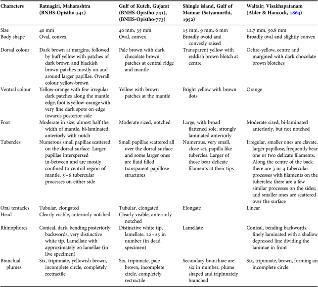
The presence of slender villous papillae is one of the reasons for the naming of the species as Thordisa villosa. The notch in the upper lip of the foot confirms that the species belongs to the Discodorididae; furthermore, two types of processes on the mantle, long soft ones and shorter stiff ones, confirm that it belongs to the genus Thordisa. The present specimens showed a moderately-sized foot (almost half the width of the mantle) that is notched and anteriorly bi-laminated. The characters of our specimens perfectly match those described by Alder & Hancock (Reference Alder and Hancock1864), except for the foot, which is notched in our specimens. No such character of foot was given by Satyamurti (Reference Satyamurti1952).
Chan & Gosliner (Reference Chan and Gosliner2007) described the morphology and anatomy of Thordisa villosa, but the 14 mm specimen described by them had bipinnate gills. The specimens from Ratnagiri and Gujarat as well as those previously described from India, however, have tripinnate gills (Figure 3).
Another key characteristic of the genus Thordisa is the presence of pectinate outermost teeth; in the present specimen of size 35 mm, the six outer lateral teeth are pectinate. Of these, the outermost four are smaller in size than the rest. Barring these dissimilarities may be because of larger specimen size, the rest of the characters were identical to those described by Chan & Gosliner (Reference Chan and Gosliner2007). Table 1 compares all described characters of all Indian specimens of Thordisa villosa.
Comparison of characters of the collected specimens with previous records from India.

ACKNOWLEDGEMENTS
We thank Dr A.R. Rahmani, Director, BNHS for his constant support and motivation. The authors are grateful to the two anonymous referees for their critical comments and suggestions. We thank Richard Willan and Benoît Dayrat, who provided valuable comments and validation of the specimens. The authors also thank team members Pooja Nagale, Vishwas Shinde, Rajendra Pawar and Rajesh Parmar for their invaluable assistance during the fieldwork. We express our thanks to Ms Smita Deodhar for the language editing of the manuscript.