In patients with cardiomyopathy, radiofrequency catheter ablation is performed for ventricular tachycardia, especially for drug-resistant recurrent ventricular tachycardia. An implantable cardioverter-defibrillator is also frequently used to treat ventricular tachycardia. Nearly all patients with a history of sustained monomorphic ventricular tachycardia are candidates for implantable cardioverter-defibrillator insertion for secondary prevention. Here we report the case of a 41-year-old man with cardiomyopathy due to Kawasaki disease in whom radiofrequency catheter ablation was performed to relieve haemodynamically unstable ventricular tachycardia before implantable cardioverter-defibrillator insertion.
Case report
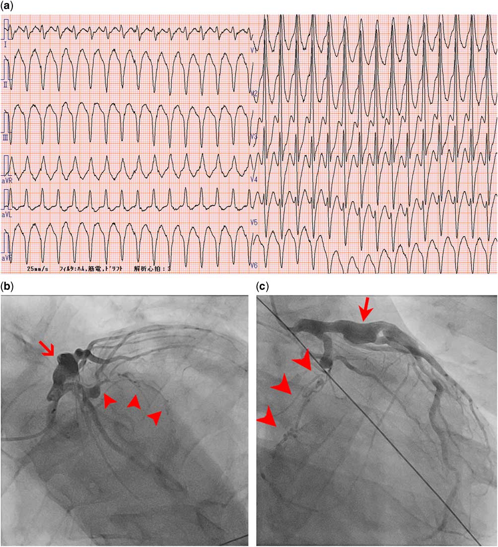
A 41-year-old man who felt a sudden onset of palpitations was taken to our hospital by ambulance. A 12-lead electrocardiogram showed a wide QRS complex with right bundle branch block and superior axis at a heart rate of 188 beats/minute. Atrioventricular dissociation was observed, leading to a diagnosis of ventricular tachycardia (Fig 1a). The ventricular tachycardia subsequently converted to fast ventricular tachycardia with a heart rate of 250 beat/minute, leading to loss of consciousness, but then spontaneously converted to normal sinus rhythm before cardiopulmonary resuscitation was started. Coronary angiography revealed a coronary artery aneurysm in the proximal segment of the left anterior descending artery and chronic total occlusion in the proximal segment of the left circumflex artery (Fig 1b and c). We excluded acute coronary syndrome, as there had been no change in the patient’s coronary arteries since his previous coronary angiography. Two-dimensional echocardiography revealed akinesis of the inferoposterior wall and severe hypokinesis of the lateral wall, with an ejection fraction of 48%. Cardiac MRI showed an epicardial late gadolinium enhancement, and we detected a scar in the posteroinferior wall (Supplementary Figure S1). A Tl-201 single-photon emission CT image taken at rest and under ATP stress (0.12 mg/kg/minute) showed a non-reversible perfusion defect, which indicated an infarcted myocardium without ischaemia.
( a ) A 12-lead electrocardiogram showing sustained ventricular tachycardia (VT) with a rate of 188 beats/minute and a QRS complex compatible with a right bundle branch block and left-axis deviation. ( b and c ) Coronary artery angiography showing the totally occluded left circumflex artery with bridging collateral vessels (arrowhead), as well as a large calcified aneurysm (arrow). The left anterior descending artery has no significant coronary stenosis. aVF=augmented vector foot; aVL=augmented vector left; aVR=augmented vector right.

The patient’s medical records showed that he was diagnosed with Kawasaki disease at the age of 4, and his coronary aneurysm and the old posteroinferior myocardial infarctionReference Tsuda, Matsuo, Naito, Noguchi, Nonogi and Echigo 1 were previously identified. At the age of 26 years, he developed haemodynamically stable sustained ventricular tachycardia, which showed a left bundle branch QRS complex at a heart rate of 167 beats/minute after 5 days. The difference in this bundle branch block from the one 16 years earlier suggests that each ventricular tachycardia has a different re-entry circuit. Radiofrequency catheter ablation was performed. During an electrophysiological study, sustained ventricular tachycardia was induced. A mid-diastolic potential was identified posterior to the mitral valve annulus, where the post-pacing interval was identical to the ventricular tachycardia-cycle length. The interval of the pacing stimulus and QRS was equal to the interval of the mid-diastolic potential and QRS. Radiofrequency catheter ablation was applied to this site during ventricular tachycardia, which resulted in the ventricular tachycardia’s termination. Thereafter, no ventricular tachycardia was induced. Amiodarone was, at the same time, stopped because it was causing elevation of serum transaminases. Thereafter, until his recent event, the patient had been free from any ventricular arrhythmias.
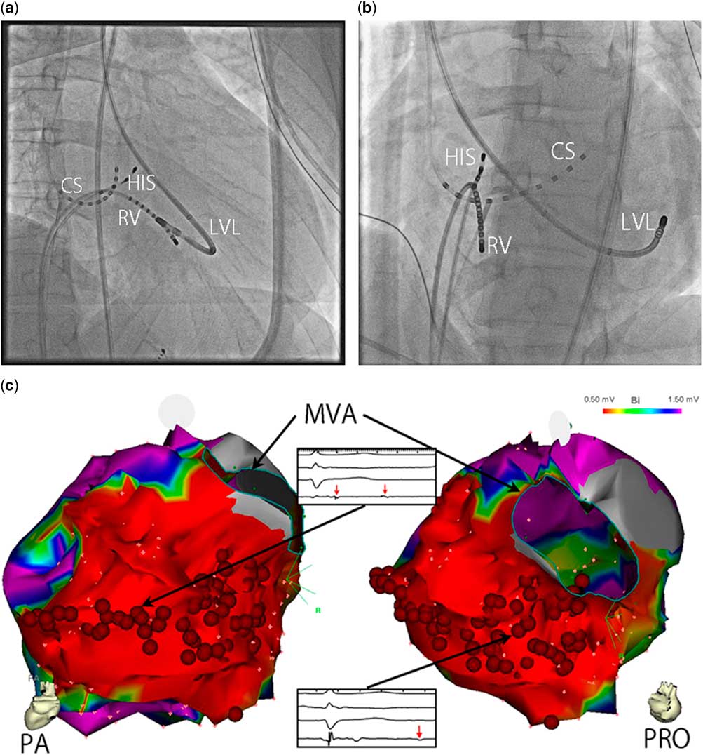
In this case, we treated the patient with radiofrequency substrate catheter ablation for ventricular tachycardia and then implanted an implantable cardioverter-defibrillator. Multi-electrode catheters and an ablation catheter were introduced from the right femoral vein. A three-dimensional mapping system (CARTO-3 mapping system; Biosense Webster, Diamond Bar, California, United States of America) was used for mapping and ablation. A 6-Fr decapolar electrode catheter (IBI inquiry; St. Jude Medical, St. Paul, Minnesota, United States of America) was positioned within the coronary sinus. For mapping, we used a multi-electrode catheter (PentaRay NAV eco; Biosense Webster). In addition, we used an ablation catheter (ThermoCool SmartTouch Catheter; Biosense Webster). Substrate ablation was performed because the induced ventricular tachycardia was haemodynamically unstable. Diastolic fragmented potentials during sinus rhythm, which were recorded in low-voltage areas (<1.5 mV) of the posteroinferior scar, were targeted to be ablated (Fig 2). The end point of this procedure was elimination of the diastolic fragmented potentials. Induction of ventricular tachycardia by programme stimuli was not attempted. After the radiofrequency catheter ablation, an implantable cardioverter-defibrillator (Iperia7 DR-T; Biotronik, se & co., Berlin, Germany) was implanted. We programmed the implantable cardioverter-defibrillator for anti-tachycardia pacing therapy. The patient was followed-up in the outpatient clinic every 6 months and by home monitoring. The onboard memory in the implanted cardioverter-defibrillator showed that no high-rate episodes (>130 bpm) were detected, which suggests that this patient has been free from any ventricular tachycardias for 20 months.
( a ) Deployment of electrode catheters of the left anterior oblique 40°. ( b ) Deployment of electrode catheters of the right anterior oblique 30°. ( c ) The left map is a posteroanterior view (PA), and the right is a right posterior oblique view (RPO). Voltage mapping using a three-dimensional mapping system (CARTO-3) revealing fragmented delayed voltages. The small pink dots are acquired points used to make the maps. The encircling blue line shows the mitral annulus (MVA). Purple colour indicates voltages >1.5 mV and red colour indicates voltages <0.5 mV. A scar area (voltage <0.5 mV) is observed within the posteroinferior lesion. Successful radiofrequency catheter ablation was performed targeting this electrocardiogram (red points). Intracardiac electrograms show the delayed potential recorded from the site. CS indicates catheter in the coronary sinus; HIS=HIS-Bundle; LVL=catheter in the left ventricle lateral wall; RA=right atrial lead; RV=right ventricular lead.

Discussion
In a reported autopsy series, patients with Kawasaki disease exhibited three pathological processes: necrotising arteritis, which is mediated by neutrophils); subacute/chronic vasculitis, which is mediated by lymphocytes, plasma cells, and eosinophils; and luminal myofibroblastic proliferation. There is a complete absence of atherosclerotic changes.Reference Orenstein, Shulman and Fox 2 Because of the difference in the histopathology and natural history from typical adult coronary artery disease, we could not easily extrapolate a treatment strategy from that ailment to the coronary stenosis or secondary ventricular arrhythmia related to Kawasaki disease. We suggest that the old myocardial infarction was probably not owing to atherosclerotic changes but owing to a thrombus or myofibroblastic proliferation with a different aetiology. However, our success with ablation therapy indicates its efficacy against secondary ventricular tachycardia related to Kawasaki disease.
Substrate ablation as a means of treating ventricular arrhythmia is becoming increasingly efficacious.Reference Vergara, Trevisi and Ricco 3 – Reference Zipes, Camm and Borggrefe 8 Substrate ablation targets delayed potentials, including diastolic fragmented potentials. These delayed potentials represent local slow conduction in the surviving myocardium along the scar border zone.Reference de Bakker and Wittkampf 9 These heterogeneous scar border areas are critical lesions for re-entry.Reference Nakahara, Tung and Ramirez 10 Appropriate ablation therapy enables these patients to be free of anti-arrhythmic drugs and reduces the need for an implantable cardioverter-defibrillator.Reference Reddy, Reynolds and Neuzil 11 Further prospective studies may show that it is possible to use ablation as a sole therapy. Indeed, ablation has advantages over aggressive anti-arrhythmic drug therapy for recurrent ventricular tachycardia.Reference Sapp, Wells and Parkash 12 Most notably, ablation frees the young from taking drugs. Cases like this, in which substrate mapping and ablation appear to produce long-term mitigation of clinical ventricular tachycardia, raise the potential for deferring implantable cardioverter-defibrillators. During a follow-up after 20 months, our patient was free of any ventricular arrhythmias. We therefore conclude that substrate ablation could be an initial therapy for unstable ventricular tachycardia in patients with chronic ischaemic cardiomyopathy.
Acknowledgement
None.
Financial Support
This research received no specific grant from any funding agency or from commercial or not-for-profit sectors.
Conflicts of Interest
None.
Ethical Standards
The authors assert that all work reported complies with the ethical standards of the Helsinki convention, and consent for publication has been granted by the patient’s family.
Supplementary material
To view supplementary material for this article, please visit https://doi.org/10.1017/S1047951118000471