Introduction
Vocal fold paresis remains a diagnostic dilemma. It is characterised as a partial motor denervation of the vocal fold, resulting in variable degrees of compromised glottal function.Reference Sulica 1 The difficulties in diagnosing paresis on laryngeal videostroboscopy alone are well known. It is the subtle signs in multiple parameters that make the diagnosis challenging.
Paresis as a clinical entity is well accepted, but its prevalence and clinical importance remain controversial. Simpson et al. indicated that it may not be clinically relevant;Reference Simpson, Cheung and Jackson 2 however, the experience of our voice clinic is that it has a significant impact on quality of life.
The importance of laryngeal electromyography (EMG) in accurately diagnosing paresis is widely accepted in the literature.Reference Sataloff, Praneetvatakul, Heuer, Hawkshaw, Heman-Ackah and Schneider 3 – Reference Syamal and Benninger 8 This paper adds to the growing evidence that now supports its clinical value.
This retrospective study aimed to quantify, for the first time, the impact of paresis utilising the Voice Handicap Index 10. We also compared the diagnostic accuracy of clinical laryngeal videostroboscopy and laryngeal EMG in identifying the paresis and the correct side of vocal fold involved.
Materials and methods
The medical records of patients seen in our tertiary referral voice clinics (St Vincent's Hospital Voice Clinic and Voice Assessment Centre, St Vincent's Clinic, Darlinghurst, Sydney, Australia) between 2012 and 2014 were reviewed. This study included patients with a clinical diagnosis of paresis who had been referred for laryngeal EMG. Their clinical laryngeal videostroboscopy diagnosis was compared to their laryngeal EMG results.
A total of 388 patients presented to our clinics with voice problems in this period. Twenty-nine patients (11 males and 18 females; mean age of 61 years, with an age range of 36–87 years) were diagnosed on laryngeal videostroboscopy with likely vocal fold paresis. Two patients declined laryngeal EMG and were excluded from the study.
Patients presented with a variety of dysphonic symptoms. As discussed by Syamal and Benninger,Reference Syamal and Benninger 8 these symptoms are due to glottal insufficiency and therefore often have elements of hyperfunction. Patients in our study had subtle quality and endurance changes, such as vocal fatigue, poor voice projection, and reduced singing ability with reduced phonation range.
Patients completed the Voice Handicap Index 10.Reference Rosen, Lee, Osborne, Zullo and Murray 9 A chart analysis was also completed to identify occupational voice users. Twenty-seven of the patients went on to have laryngeal EMG.
Laryngeal EMG recordings were obtained in awake patients using concentric bipolar recording needles. The thyroarytenoid muscle and cricothyroid muscle were sampled unilaterally or bilaterally. The posterior cricoarytenoid muscle was not sampled. The thyroarytenoid muscle was sampled 2–3 mm lateral to the midline, at the level of the cricothyroid membrane, whilst the patient was instructed to phonate /i/ in the mid-phonational range. The cricothyroid muscle was sampled lateral to, and slightly above the lateral border of the cricothyroid notch, whilst the patient phonated /i/ in the higher pitch range.
The EMG recordings were made using an analogue Medelec™ Neurostar EMG machine. The EMG signals were amplified, band-pass filtered and displayed at 100 μV per division. Interpretation was performed in real time by 2 senior neurologists who have been performing laryngeal EMG for over 20 years.
Parameters assessed included insertional activity, spontaneous activity, motor unit potential morphology (amplitude and duration (including giant units if present), polyphasia) and recruitment.Reference Heman-Ackah and Barr 10 The level of denervation was graded based on the core findings of reduction in recruitment with or without motor unit potential morphological changes (increased amplitude and duration, polyphasia), fibrillation potentials, and positive sharp waves. Denervation was graded as none, mild, moderate, severe or complete. The diagnosis of vocal fold paresis was confirmed by the presence of denervation on laryngeal EMG, as determined by these criteria.
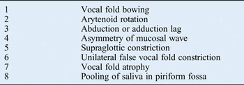
Laryngeal videostroboscopy examinations in our clinics include standard vocal tasks, such as the repetitive phonatory tasks described by Rubin et al.Reference Rubin, Praneetvatakul, Heman-Ackah, Moyer, Mandel and Sataloff 11 The laryngology team (a senior laryngologist and 2 experienced speech pathologists) reviewed the de-identified laryngeal videostroboscopy recordings of the 27 patients included in the study. They were blinded to the laryngeal EMG results and the side of involvement. Consensus on paretic side was determined utilising the clinical indices outlined in Table I.
Laryngeal videostroboscopy clinical indices for vocal fold paresis identification

These visual clues have all been previously described in the literature.Reference Sulica 1 , Reference Simpson, Cheung and Jackson 2 , Reference Koufman, Postma, Cummins and Blalock 7 , Reference Syamal and Benninger 8 , Reference Simpson, May, Green, Eller and Jackson 12 , Reference Sulica and Blitzer 13 Although often associated with vocal fold paresis, they are not conclusive of this diagnosis. Clinical indices included: structural or static signs, such as thinning or atrophy of the vocal folds; arytenoid rotation; and pooling of saliva in the piriform sinus. Other signs seen with vocal tasks included: vocal fold bowing, abduction or adduction lags, generalised supraglottic constriction or unilateral false vocal fold engagement, and mucosal wave asymmetry.
Results
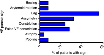
During the period from 2012 to 2014, 388 patients were assessed using laryngeal videostroboscopy. Laryngeal videostroboscopy findings of the 29 patients (7.5 per cent) thought to have paresis showed signs discussed above. In this study, the most prevalent clinical sign was unilateral vocal fold abduction lag, but mucosal wave asymmetry and false vocal fold constriction were also common findings. Patients often presented with multiple clinical signs on laryngeal videostroboscopy. Figure 1 demonstrates the distribution of these findings on clinical laryngeal videostroboscopy.
Vocal fold paresis diagnostic signs. VF = vocal fold

Of the 27 patients who underwent laryngeal EMG, 25 had a confirmed diagnosis of vocal fold paresis. Two patients had normal laryngeal EMG results. One patient had known vocal fold scarring; no cause was found for the second patient's symptoms, which had resolved on subsequent follow up.
Within our cohort, the incidence of paresis was higher in females (64 per cent), with a female:male ratio of 16:9. This is in keeping with the female predominance observed in our voice clinics (60 per cent).
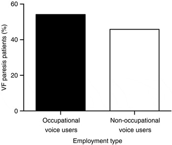
A high percentage of patients in the paresis group (55 per cent) indicated that they were occupational voice users (Figure 2). This may reflect a greater awareness of voice dysfunction in the employment setting and willingness to be referred in order to optimise outcome.
Vocal fold paresis patients’ employment type (occupational vs non-occupational voice users). VF = vocal fold

Laryngeal EMG demonstrated that 9 patients had right vocal fold paresis, whilst 10 had left vocal fold paresis. Six patients (24 per cent) had confirmed bilateral paresis. If we assume that laryngeal EMG is the ‘gold standard’, precision of the initial clinical diagnosis of paresis was 92.6 per cent (25 out of 27 patients). Correct side identification on laryngeal videostroboscopy had a reliability of 78 per cent (21 of 27 patients).
Of the 388 patients seen in the clinics during the study period, 43 (11.1 per cent) were diagnosed with vocal fold paralysis. These patients were comparable to our paresis cohort (20 males and 23 females; mean age of 64 years, with an age range of 22–86 years). The Voice Handicap Index 10 results of patients with diagnosed vocal fold paralysis were compared to the results of patients with laryngeal EMG diagnosed paresis.
The Fisher F-test was utilised to determine if there was a significant difference between the variance of the paralysis group and the paresis group, after which a two-tailed t-test (assuming unequal variances) was conducted to determine statistical differences (p < 0.05) between group means. Data are presented as the mean ± one standard error of the mean.
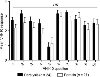
Figure 3 compares total Voice Handicap Index 10 scores between the vocal fold paralysis group and the vocal fold paresis group. There was no significant difference between the groups (p = 0.23; total Voice Handicap Index 10 score was 26.08 ± 0.21 for the vocal fold paralysis group vs 22.93 ± 0.17 for the paresis group).
Voice Handicap Index 10 (VHI-10) results for vocal fold paralysis and paresis groups. Although vocal fold paralysis patients scored higher for the majority of questions, responses were comparable. Ns = non-significant

The scores for the individual questions within the Voice Handicap Index 10 indicate that mean responses were again similar between the groups. Paresis patients scored marginally higher in the functional indicator ‘my voice problem causes me to lose income’ (which may be an indication of a greater proportion of professional voice users within the group) and in the emotional indicator ‘my voice problem upsets me’, but these differences were not significant.
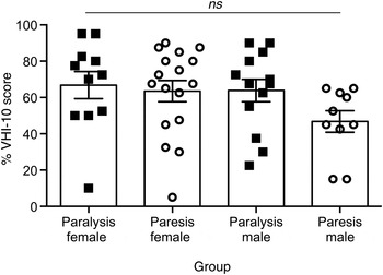
There was no statistical difference in Voice Handicap Index 10 scores between the vocal fold paresis and paralysis patients when grouped by gender (Figure 4).
Voice Handicap Index 10 (VHI-10) results for vocal fold paralysis and paresis groups by gender. Ns = non-significant

Discussion
Vocal fold paresis is a well-established diagnosis, but its implications have not been fully appreciated until now. Rosen et al., in their Voice Handicap Index 10 validation study, grouped paresis and vocal fold atrophy together, producing a high score, though not as high as paralysis patients.Reference Rosen, Lee, Osborne, Zullo and Murray 9 Tested independently, this study confirms that vocal fold paresis has a similar impact on a patient's quality of life as vocal fold paralysis. Patients have difficulties with social interaction and employment, and complain of emotional upset as a consequence of their vocal deficit.
Our concern is that, unlike vocal fold paralysis, vocal fold paresis has subtle endoscopic signs on laryngeal videostroboscopy which can be easily missed, making diagnosis elusive unless there is a high index of suspicion. Consequently, we suspect that vocal fold paresis is a universally underdiagnosed condition. This reduced ability to predict vocal fold paresis on laryngeal videostroboscopy has been reflected by a number of retrospective studies.
Repetitive phonatory tasks have been used to promote vocal fold fatigue, to help identify vocal fold lag and accentuate asymmetry.Reference Rubin, Praneetvatakul, Heman-Ackah, Moyer, Mandel and Sataloff 11 In a cohort of 75 patients, repetitive phonatory tasks correctly predicted unilateral paresis in 82.6 per cent of patients. However, in 30 per cent of these, bilateral paresis was found. This was similar to the 24 per cent of bilateral paresis cases in our study. Overall, individual predictions based on repetitive phonatory tasks alone were correct in only 48 per cent of cases. Rubin et al. ultimately concluded that a more accurate prediction was reached if the entire examination was utilised.Reference Rubin, Praneetvatakul, Heman-Ackah, Moyer, Mandel and Sataloff 11 This increased overall prediction to 76 per cent; however, this remains suboptimal in comparison to laryngeal EMG.
Heman-Ackah and Barr completed a cohort study of patients with mild vocal hypomobility who presented to their tertiary laryngology referral clinic over a 13-month period.Reference Heman-Ackah and Barr 10 Of these patients, 86.4 per cent had neuropathy on laryngeal EMG. Hypomobility was described as a mobile vocal fold with mild ‘sluggishness’ in adduction, abduction and/or longitudinal tension. Forty-six per cent of 134 patients presenting with vocal complaints had hypomobility. Importantly, this finding was undiagnosed by their referring otolaryngologist on initial examination, and was identified during tertiary referral assessment. We have found that asking the patient to phonate at a modal or low fundamental frequency, but at a high intensity during laryngeal videostroboscopy, exaggerates asymmetry and optimises clinical interpretation.
This study, like others before, highlights the difficulty in accurately determining the side involved. Simpson et al. reported that 19 of 23 patients (83 per cent) with mucosal wave asymmetry on laryngeal videostroboscopy had laryngeal EMG evidence of vocal fold paresis, but prediction of vocal fold side involvement was poor, being correct in only 35 per cent of cases.Reference Simpson, May, Green, Eller and Jackson 12 It is important to note, however, that they investigated only those patients with isolated vibratory asymmetry, and intentionally did not include patients who had other signs such as abduction lag. With all the additional clinical signs, our predictive rate was 92.6 per cent, but our ability to accurately predict side was similarly reduced.
The treatment of paresis remains difficult, largely because of the heterogeneity of the underlying aetiologies and the often unknown prognosis. Syamal and Benninger concluded that ‘LEMG [laryngeal EMG], when used judiciously as an adjunct, can provide useful prognostic information’.Reference Syamal and Benninger 8 Treatment options are based largely on anecdotal evidence,Reference Sulica and Blitzer 13 but do include both augmentation of the vocal fold (either unilateral or bilateral) and framework surgery. There is still debate as to whether unilateral or bilateral augmentation is best, and, if the former is preferred, laryngeal EMG is again important to correctly identify which side is affected.
Distinguishing presbylarynges from vocal fold paresis in dysphonic patients of advanced age also remains difficult. Stager and Bielamowicz reviewed laryngeal videostroboscopy findings for 52 patients aged over 64 years who presented with hoarseness, and compared those with normal versus abnormal laryngeal EMG findings.Reference Stager and Bielamowicz 14 Analysis of laryngeal videostroboscopy findings showed that the most sensitive index was impairment of arytenoid movement. The study concluded that laryngeal EMG was especially useful in distinguishing between bilateral paresis and presbylarynges.
Laryngeal EMG is a useful investigation for patients with subtle voice changes who have grossly normal vocal fold movement. Although not essential in making the initial diagnosis, laryngeal EMG can help differentiate vocal fold paresis from other differential diagnoses and aid prognosis, and may be of use if unilateral treatment options are considered. It is, however, not always available in a general otolaryngology setting.
Using a validated quality-of-life measurement, we have for the first time been able to show the impact that vocal fold paresis has on a patient's life. It is comparable to the impact of full vocal fold paralysis, yet making the diagnosis is substantially harder, with only subtle changes on laryngeal videostroboscopy that are often missed. For the patient, confirming a diagnosis of vocal fold paresis by laryngeal EMG provides insight and reassurance for patients persistently suffering from debilitating dysphonia. The diagnosis enables patients to be streamed to an appropriate treatment plan, which will ultimately result in more efficient voice use and optimised voice management.
-
• Vocal fold paresis is incomplete paralysis of the vocal folds
-
• Clinical diagnosis can be easily missed because of the subtle clinical signs
-
• Patients’ quality of life is significantly affected, comparable to the impact of vocal fold paralysis
-
• Making the diagnosis is important for the patients’ wellbeing and ensures appropriate treatment options are explored
Given the significant morbidity associated with vocal fold paresis and the difficulties of diagnosing it on laryngeal videostroboscopy, it is important that clinicians have a high index of suspicion when performing their examinations. If dysphonia patients with normal clinical examination findings on nasendoscopy do not improve with targeted and effective speech rehabilitation, they should be referred to a specialised voice assessment clinic for laryngeal videostroboscopy and consideration of laryngeal EMG.
Conclusion
Vocal fold paresis is a debilitating and underdiagnosed condition. Its acceptance as an entity and awareness of its potential will help to validate the patient's clinical presentation and facilitate appropriate treatment. Laryngeal EMG should be added to the repertoire of diagnostic tests for patients with persistent dysphonia and apparently normal vocal fold movement, to aid in the diagnosis of vocal fold paresis.