INTRODUCTION
Bivalves constitute a prominent commodity in aquaculture but are also crucial to preserve the complexity and function of ecosystems (Zannella et al., Reference Zannella, Mosca, Mariani, Franci, Folliero, Galdiero, Tiscar and Galdiero2017). Mussels are important suspension filter feeders and thus represent an essential component in the ecology of coastal waters. Mussels are favoured commercially because many species can tolerate and accumulate xenobiotics in their tissues at levels higher than those of the aquatic environment (Torre et al., Reference Torre, Trischitta and Faggio2013b; Faggio et al., Reference Faggio, Pagano, Alampi, Vazzana and Felice2016; Pagano et al., Reference Pagano, Capillo, Sanfilippo, Palato, Trischitta, Manganaro and Faggio2016, Reference Pagano, Porcino, Briglia, Fiorino, Vazzana, Silvestro and Faggio2017; Capillo et al., Reference Capillo, Silvestro, Sanfilippo, Fiorino, Giangrosso, Ferrantelli, Vazzana and Faggio2018). Moreover, since mussels are edible, nutritious, and sessile species, they have been harvested and cultured worldwide for human consumption. Bivalve species of the genus Mytilus are dominant mussels living in the intertidal and subtidal zones, withstanding either more impacted or pristine sites (Roginsky & Lissi, Reference Roginsky and Lissi2005; Hamer et al., Reference Hamer, Korlević, Durmiši, Nerlović and Bierne2012). The Mediterranean mussel Mytilus galloprovincialis Lamarck, 1819 is an important commercial species in Slovenia and Croatia, in the northern Adriatic Sea, and thus it has been extensively maricultured (Gombač, Reference Gombač2010; Pavičić-Hamer et al., Reference Pavičić-Hamer, Kovačić, Koščica and Hamer2016).
The study of parasites and diseases affecting molluscs with ecological and economic interests is critical for the management of natural stocks and aquaculture (Boehs et al., Reference Boehs, Villalba, Ceuta and Luz2010). The most severe mussel diseases are caused by viruses, bacteria and protistans, which can be responsible for mortality outbreaks, thus posing a substantial commercial impact (Tuntiwaranuruk et al., Reference Tuntiwaranuruk, Chalermwat, Pongsakchat, Meepool, Upatham and Kruatrachue2008; Ceuta & Boehs, Reference Ceuta and Boehs2012; Mladineo et al., Reference Mladineo, Petrić, Hrabar, Bočina and Peharda2012). To a lesser extent, other diseases are caused by ciliates, turbellarians (Cova et al., Reference Cova, Serafim Júnior, Boehs and Souza2015), trematodes (Özer & Güneydağ, Reference Özer and Güneydağ2015) and fungi (Santos et al., Reference Santos, Hauser-Davis, Santos and De Simone2017). The spread of fungal strains may be facilitated by a combination of environmental conditions, such as wind or human-conveyed propagules (Sallenave-Namont et al., Reference Sallenave-Namont, Pouchus, Robiou du Pont, Lassus and Verbist2000). Fungi can also be present in the marine environment in latent forms on host organisms or potential symptomless carriers (Ein-Gil et al., Reference Ein-Gil, Ilan, Carmeli, Smith, Pawlik and Yarden2009).
According to the World Organisation for Animal Health (OIE), notifiable diseases are those with socioeconomic and/or public health importance within countries or that are significant for the international trade of aquatic animals and their products (Aranguren & Figueras, Reference Aranguren and Figueras2016). The current list of notifiable diseases of bivalves and the causative pathogens includes infection with Marteilia refringens, Bonamia sp., Xenohaliotis californiensis and Perkinsus sp. (Carnegie et al., Reference Carnegie, Arzul and Bushek2016).
The increased use of coastal areas and the sea due to expansion in urbanization, population growth and tourism associated with the intense traffic of tourist vessels and maritime transport have detrimental effects on the health status of bivalves (Burgos-Aceves and Faggio, Reference Burgos-Aceves and Faggio2017; Savorelli et al., Reference Savorelli, Manfra, Croppo, Tornambè, Palazzi, Canepa, Trentini, Cicero and Faggio2017; Burgos-Aceves et al., Reference Burgos-Aceves, Cohen, Smith and Faggio2018; Faggio et al., Reference Faggio, Tsarpali and Dailianis2018). Studies have shown that environmental factors including poor water quality and the presence of parasites can impact the health status of mussel stocks in the UK (Bignell et al., Reference Bignell, Dodge, Feist, Lyons, Martin, Taylor, Stone, Travalent and Stentiford2008; Lynch et al., Reference Lynch, Morgan, Carlsson, Mackenzie, Wooton, Rowley, Malham and Culloty2014). A decrease in water quality can affect the immunological response in aquatic organisms thus making them more susceptible to parasitic infection and increasing parasite prevalence (Khan, Reference Khan1991). It becomes evident that particular attention coupled with protective measures are needed both for native and cultured mussel stocks.
The study of pathogens from different mussel populations in response to regional differences is a subject of major interest, particularly in aquaculture, where mussels originate from various sources (Bratoš et al., Reference Bratoš, Glamuzina and Benović2004; Pavičić-Hamer et al., Reference Pavičić-Hamer, Kovačić, Koščica and Hamer2016). The objectives of this study are (a) to investigate the health status of mussels on the northern Adriatic coast (Croatia and Slovenia) concerning effects of on-site water quality; (b) to identify the range of parasites and fungi present in aquaculture and wild areas and the possible route of exposure; and (c) to evaluate the health status of mussels and their likely high infection risks due to detrimental effects of pollutants.
MATERIALS AND METHODS
Collection of mussels and description of sampling sites
Mussels were collected from six sampling stations (N = 10 per station) along the coast of the northern Adriatic, in Slovenia and Croatia (Figure 1), during February and April 2014. Strunjan Bay (45°16′24″N 13°34'57″'E) and Piran Bay (45°07′50″N 13°44′10″E) harbour the most important Slovenian aquaculture sites, which are located 11.4 km and 20.6 km away from Koper, respectively. The mussel farm in Strunjan Bay (site code 0024) is inside a natural reserve (Krajinski park Strunjan), and the mussel farm near to Seča (site code 0035) is located in the inner part of Piran Bay. The third sampling site was close to the Marina Koper in Slovenia (site code 00TM), where mussels were collected from a natural bed in the vicinity. The two other impacted sites are Adriatic Croatia International Marinas (ACI), the ACI Marina Pula (44°87′55″N 13°84'67″E) and ACI Marina Rovinj (45°04′32″N 13°38′08″E). They are located near a sewage outflow and boat processing area characterized by elevated pollutant concentration in biota and sediment (Center for Marine Research, 2014). St Catherine Island in Croatia (45°07′67″N 13°62′96″E) is located ~5 km away from the urban and sewage outflow area and is considered a non-impacted environment (Bihari et al., Reference Bihari, Mičić, Fafanđel, Hamer, Jakšić and Batel2004; Center for Marine Research, 2014). Environmental parameters were measured with an MSS 90 multiparameter probe (Sea & Sun Technology), at 2 m depth at the time of sampling using standard procedures (ARSO, 2014; Center for Marine Research, 2014) (Table 1).
Geographic location of the sampling areas and source of mussels in the northern Adriatic Sea (Slovenia and Croatia).

Environmental parameters estimated for the sampling sites during February and April in 2014, measured at 2 m depth.

Histopathology
Ten mussels from each station per month were sampled and immediately transported to the laboratory where digestive glands were removed, fixed with n-hexane, and cooled in liquid nitrogen. The digestive glands were placed in a straight row on aluminium cryostat chucks (five per chuck). The chucks were stored at −80°C until analysis. Before cryosectioning, the samples were embedded in O.C.T.™ compound (Microm Inc. GmbH, Germany) and cut into 10 µm sections with a cryostat (Zeiss Hyrax C 50, Microm GmbH, Germany). Sections were stained with haematoxylin and eosin (Sigma-Aldrich, USA). Parasite morphology was studied using a light microscope (Nikon, UK). Authorities for parasite and endobionts identification included Vázquez & Cremonte (Reference Vázquez and Cremonte2017) for prokaryotic and basophilic inclusion bodies; Francisco et al. (Reference Francisco, Hermida and Santos2010), Cova et al. (Reference Cova, Serafim Júnior, Boehs and Souza2015) and Tuntiwaranuruk et al. (Reference Tuntiwaranuruk, Chalermwat, Pongsakchat, Meepool, Upatham and Kruatrachue2008) for Nematopsis sp., Ancistrocoma-like ciliates, and turbellarian Urastoma cyprinae von Graff, 1882; Bignell et al. (Reference Bignell, Dodge, Feist, Lyons, Martin, Taylor, Stone, Travalent and Stentiford2008) for haplosporidian-like plasmodia; and Zhang et al. (Reference Zhang, Mu, Feng, Kang, Zhang, Gu, Wang, Ma and Zhu2009) for fungi. All micrographs were captured using an Ikegami ICD-803P digital video camera and the Lim Screen Measurement™ Lucia G image capture system (Nikon, UK).
Following Saffo (Reference Saffo1992), we used the term ‘infection’ in referring to all organisms parasitically and endobiotically associated with their hosts. The prevalence and intensity of infection of each parasite were calculated according to Bush et al. (Reference Bush, Lafferty, Lotz and Shostak1997), as follows: the prevalence is the number of individuals infected divided by the total number of individuals in a sample, expressed as a percentage; the intensity is the number of parasites found in an infected mussel.
Diagnostic detection of M. refringens by PCR
Infection by M. refringens was surveyed by polymerase chain reaction (PCR). For this, a small piece of the mussel digestive gland of each collected individual was preserved in 95% ethanol. DNA was extracted using the DNeasy Blood & Tissue Kit (QIAGEN GmbH, Hilden, Germany) according to the manufacturer's instructions. After isolation, DNA samples were amplified in a SureCycler 8800 Thermal Cycler (Agilent Technologies, Santa Clara, California, USA) using standard primers for the M. refringens DNA, M2A (5′- CCGCACACGTTCTTCACTCC-3′); M3AS (5′-CTCGAGTTTCGACAGACG-3′), according to the protocol by Le Roux et al. (Reference Le Roux, Lorenzo, Peyret, Audemard, Figueras, Vivarès, Gouy and Berthe2001). The presence of PCR products was verified by agarose gel (1.5% w/v) electrophoresis.
Statistical analysis
The relationship between prevalence and sampling site and time was evaluated by a Chi-square test. The relationship between the intensity of infection and sampling site and time was assessed by a Kruskal–Wallis test. All tests were performed with Statistica 9.0. software at significance P < 0.05.
RESULTS
Along the coast of the northern Adriatic Sea, mussels were sampled from six sites in Croatia and Slovenia that were previously evaluated and classified as aquaculture sites (Strunjan Bay and Piran Bay, Slovenia), non-impacted sites (St Catherine, Croatia), and impacted sites (Marina Koper, Slovenia; ACI Pula and ACI Rovinj, Croatia).
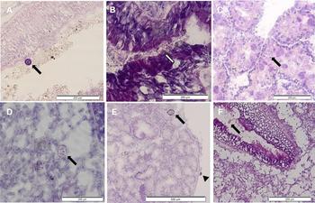
No significant infections, i.e. parasites that cause significant mortalities or heavy infestations, were observed in situ during the study. Histological and molecular analyses did not show any evidence of the presence of M. refringens, an OIE-listed pathogen. The most common pathogens found in mussels from the impacted and non-impacted sites were haplosporidian-like organisms, prokaryotic inclusion bodies, Ancistrocoma-like ciliates, Nematopsis sp. and the turbellarian U. cyprinae (Figure 2). Filamentous fungi of Ulocladium sp., Psylocibe sp. and Alternaria sp. were detected in mussels from St Catherine, ACI Pula, ACI Rovinj (Croatia) and Marina Koper (Slovenia), but not from aquaculture sites (Figure 3). The number of parasite groups per site was higher in impacted sites (N = 9) compared with non-impacted (N = 1) and cultivation (N = 0) sites.
Light microscopy of transverse cryosections through the mussel digestive gland of Mytilus galloprovincialis containing: (a) basophilic prokaryote-like organisms (arrow) in the lumen of the digestive gland; (b) eosinophilic prokaryote-like organisms (arrow) in the lumen of the digestive gland; (c) plasmodia (arrow) of a haplosporidian-like organism in the digestive tubule epithelium; (d) host phagocyte containing three Nematopsis sp. oocysts in the connective tissue between the digestive tubules; (e) Urastoma cyprinae (arrow) and Ancistrocoma-like ciliate (arrowhead) at the edge of the digestive gland and digestive tubules; (f) Ancistrocoma-like ciliate (arrow) in the lumen of digestive tubule.

Light microscopy of the transverse cryosections through the mussel digestive gland of Mytilus galloprovincialis containing spores of (a) Psilocybe sp.; (b) Ulocladium sp.; and (c) Alternaria sp. attached to the digestive tubules.

Specimens of a haplosporidian-like organism were detected in mussels from Koper (10% prevalence of infection). Plasmodia of haplosporidian-like organisms were spread into the epithelium of digestive tubules with more than 100 plasmodia per section (at 100× magnification).
Prokaryotic inclusion bodies that were spherical to oval shaped with basophilic structures 5–10 µm in size were found in the digestive tubules and between them (Figure 2a). In some cases, spherical inclusions were eosinophilic stained (Figure 2b). Their prevalence ranged from 10% (ACI Pula, February 2014) to 40% (ACI Pula, April 2014). A 30% prevalence was detected in all the other infected mussels (Table 2). The intensity of infection ranged between one and 12 prokaryotic inclusion bodies per section (Table 2). The site influenced both prevalence (χ2 = 6.66, df = 2, P < 0.05) and intensity of infection (Kruskal–Wallis test, H = 6.32, P < 0.05, Table 4).
Prevalence (P) and intensity of infection (I) with the range in parentheses of prokaryotic inclusion bodies, Nematopsis sp., and Ancistrocoma-like ciliates from Mytilus galloprovincialis mussels collected in the northern Adriatic Sea.

Prevalence (P) and intensity of infection (I) with the range in parentheses of the fungi Psilocybe sp. and Ulocladium sp. from Mytilus galloprovincialis mussels collected in the northern Adriatic Sea.

Effect of sampling time and site on the prevalence and level of infection of parasites and fungi in mussels from the northern Adriatic Sea.

Significant difference (P) of χ2 test for the prevalence and Kruskal–Wallis test (H) for the intensity of infection.
Specimens of Nematopsis sp. with ungrouped and grouped oocysts were detected in the connective tissue between digestive tubules of mussels from non-impacted and impacted sites (Figure 2c). A single thickened membrane was bounding oocysts with a basophilic vermiform sporozoite inside. In infected mussels sampled in April 2014, the prevalence ranged from 20% (St Catherine and ACI Pula) to 80% (ACI Rovinj) (Table 2). In February 2014, ACI Rovinj had the highest prevalence of infection with Nematopsis sp. (80% prevalence). The intensity of infection was variable, with < 15 oocysts per section in most of the cases (maximum 47 oocysts/mussel). Prevalence (χ2 = 6.00, df = 2, P < 0.05) and intensity of infection (Kruskal–Wallis test, H = 9.18, P < 0.05) were significantly different between sampling sites (Table 4).
Specimens of the turbellarian U. cyprinae (10% prevalence) were observed at the edge of the digestive gland from mussels sampled in ACI Pula (Figure 2e).
In mussels from the same sampling site, oval-shaped Ancistrocoma-like ciliates, characterized by a ciliary fringe and a basophilic horse-shoe shaped nucleus, were observed (Figure 2f). Prevalence of Ancistrocoma-like ciliates in wild mussels ranged from 10% (Marina Koper, ACI Pula, ACI Rovinj, February 2014) to 20% (ACI Pula, April 2014) (Table 1), with an intensity of infection from 1 to 2. Site influenced both prevalence (χ2 = 8.57, df = 2, P < 0.05) and intensity of infection (Kruskal–Wallis test, H = 7.50, P < 0.05).
The fungi were observed in digestive glands of wild mussels. Alternaria sp. individuals (10% prevalence) were found within the connective tissue in mussel digestive gland from mussels sampled in Marina Koper (Figure 3a). Spores of Psilocybe sp. were recorded in the epithelial cells of the digestive tubules in the mussel digestive gland from mussels of impacted sites. Prevalence ranged from 10% (ACI Rovinj, April 2014) to 30% (Marina Koper, February 2014). We observed specimens of Ulocladium sp. (10% prevalence) inside the digestive tubules of mussels from impacted (Marina Koper and ACI Rovinj in February) and non-impacted (St Catherine) sites in both months. The intensity of infection was low (up to five spores per section). No significant difference in the prevalence of fungi (P > 0.05) was observed between cultured and wild mussels.
DISCUSSION
A diversity of endobionts and parasites was observed in cryosections of wild mussels from the northern Adriatic Sea, including Ancistroma sp., Nematopsis sp., U. cyprinae, and fungi Ulocladium sp., Psylocibe sp. and Alternaria sp. One of the most devastating pathologies in mussels, infection with M. refringens, an OIE-listed disease, was not detected by diagnostic PCR in mussels from the northern Adriatic during this survey. There was a significant difference between sites (impacted, non-impacted and aquaculture sites) concerning parasite infection. Histological examination of paraffin-embedded tissue sections is considered to be the standard screening diagnostic method. We identified parasites and endobionts in cryosections since this method preserves the tissue as close as possible to its natural state (Kovačić & Pustijanac, Reference Kovačić and Pustijanac2017). Nowadays, for detection of parasites causing OIE-listed diseases, more specific and sensitive molecular diagnostic techniques have been used (Aranguren & Figueras, Reference Aranguren and Figueras2016). For a presumptive diagnosis of a disease, the standard method is histopathology, but for confirmatory diagnosis, the standard methods are PCR and DNA sequencing. As the result of adopting European legislation for monitoring diseases according to OIE on shellfish farms in Slovenia, analyses of M. refringens in Mediterranean mussels was established from 2004 (Gombač, Reference Gombač2010). Marteilia refringens ciliates were detected in mussels from Slovenian aquaculture sites in recent years, at intensities that would cause significant pathologies (Gombač et al., Reference Gombač, Kušar, Ocepek, Pogačnik, Arzul, Couraleau and Jenčič2014). At the time of our study, cultured Mediterranean mussels were free from M. refringens. This protozoan is widespread from the Atlantic Ocean to the Persian Gulf (Bower et al., Reference Bower, McGladdery and Price1994) and has also been detected in the northern Adriatic Sea (Zrnčić et al., Reference Zrnčić, Le Roux, Oraić, Sostarić and Berthe2001; Gombač et al., Reference Gombač, Kušar, Ocepek, Pogačnik, Arzul, Couraleau and Jenčič2014). A haplosporidian-like organism was found in M. galloprovincialis from only one site (ACI Pula, Croatia). Multinucleated plasmodia were observed in Mytilus edulis, M. galloprovincialis and their hybrids from Southampton Water, Hampshire, and the Exe River, Devon in the UK (Bignell et al., Reference Bignell, Dodge, Feist, Lyons, Martin, Taylor, Stone, Travalent and Stentiford2008). Besides, a haplosporidian infection in Mytilus sp. from the Exe River estuary and Southampton Water was described in 2004 and 2005 (Bignell et al., Reference Bignell, Dodge, Feist, Lyons, Martin, Taylor, Stone, Travalent and Stentiford2008). However, in the northern Adriatic Sea, this is the first record of a haplosporidian-like organism in mussels.
In our study, prokaryotic inclusion bodies were found infecting epithelial cells or the lumen of the digestive tubule in mussels from the Northern Adriatic, similarly to their detection in several bivalve species (Cova et al., Reference Cova, Serafim Júnior, Boehs and Souza2015; Vázquez & Cremonte, Reference Vázquez and Cremonte2017). They occurred as small rounded intracellular basophilic colonies, evoking the lysis of infected cells of the digestive tubule. In some cases, colonies caused the lysis of the infected cells and were released from the lumen of the tubule into organs of bivalves (Vázquez & Cremonte, Reference Vázquez and Cremonte2017), as we detected in this study. In the host tissue, the parasites can be associated with an entry mechanism via food particle capture (Queiroga et al., Reference Queiroga, Vianna, Vieira, Farias and Da Silva2015). In these cases, granulocytes can cross the digestive epithelium reaching the lumen of the gut where they capture and digest food particles, and cross back transporting nutrients to bivalve tissues (Torre et al., Reference Torre, Trischitta, Corsaro, Mallamace and Faggio2013a, Reference Torre, Trischitta and Faggiob; Matozzo et al., Reference Matozzo, Pagano, Spinelli, Caicci and Faggio2016; Sehonova et al., Reference Sehonova, Svobodova, Dolezelova, Vosmerova and Faggio2018). Moreover, some inclusions detected in our study were coated by an eosinophilic fibrous cover, which is interpreted as an encapsulation response by the host (Gulka & Chang, Reference Gulka and Chang1985). Despite their intracellularity being unclear, these inclusions were reported as Rickettsia-like (RLOs) and Chlamydia-like organisms (Vázquez & Cremonte, Reference Vázquez and Cremonte2017).
In our study, Ancistrocoma-like ciliates occurred on the surface of the digestive gland and in the lumen of the tubule. Ancistrocoma-like ciliates are presumed to be ubiquitous in many species of bivalves (Adlard et al., Reference Adlard and Chollet2003; Rayyan et al., Reference Rayyan, Damianidis, Antoniadou and Chintiroglou2006). Most of them are extracellular and found in the lumen of the digestive gland tubules in the intestine. Some ciliates may be attached to or located near the gills, mantle and labial palps (Sphenophrya-like, Trichodina sp., Ancistrum-like). Although ciliates are mostly harmless and commensals, the intracellular ciliates that are Sphenophrya-like and Rhynchodid-like can disrupt the epithelia of the digestive tubule of M. edulis (Adlard et al., Reference Adlard and Chollet2003).
Our histological examination revealed Nematopsis sp. in the form of a single or numerous dense oocysts. Mladineo (Reference Mladineo2008) also found a high prevalence of Nematopsis sp. in the horse-bearded mussel Modiolus barbatus Linnaeus, 1758 in Mali Ston (Adriatic Sea, Croatia). Urastoma cyprinae turbellarians were found in low prevalence in our study, as was similarly observed in M. galloprovincialis from Baja California, north-west Mexico (Caceres-Martinez et al., Reference Caceres-Martinez, Vasquez-Yeomans and Sluys1998) and from the Black Sea coast at Sinop, Turkey (Özer & Güneydağ, Reference Özer and Güneydağ2015). Moreover, low prevalence of U. cyprinae was found in the mangrove oyster Crassostrea rhizophorae (Guilding, 1828) in the estuary of the Graciosa River in Taperoá, Bahia State, north-east Brazil (Cova et al., Reference Cova, Serafim Júnior, Boehs and Souza2015). Specimens of U. cyprinae are usually found among the gill filaments. However, we found them at the edge of the mussel digestive gland, like in a previous study (Kovačić & Pustijanac, Reference Kovačić and Pustijanac2017).
The results obtained in our study include the first record of fungal spores of Ulocladium sp., Psylocibe sp. and Alternaria sp. in M. galloprovincialis in the northern Adriatic Sea (Croatia). Epibiotic and endobiotic fungi live on the surface and in the inner tissues of many invertebrates and algae (Zhang et al., Reference Zhang, Mu, Feng, Kang, Zhang, Gu, Wang, Ma and Zhu2009). Filamentous fungi were found to be associated with several wild and farmed bivalves and are potentially toxic (Zvereva et al., Reference Zvereva, Zvyagintsev and Ivin2012; Borzykh & Zvereva, Reference Borzykh and Zvereva2015; Santos et al., Reference Santos, Hauser-Davis, Santos and De Simone2017). Besides, fungal spores were abundant in salts from the Atlantic and Pacific Oceans (Biango-Daniels & Hodge, Reference Biango-Daniels and Hodge2018).
In this study, the sampling site influenced the diversity and abundance of parasites present in mussels. Mussels from impacted sites (marinas and port) were more infected in comparison to mussels from the non-impacted and aquaculture sites. An association of parasite diversity and their abundance with impacted polluted sites has been observed in many studies (Aarab et al., Reference Aarab, Pampanin, Nævdal, Øysæd, Gastaldi and Bechmann2008; Bignell et al., Reference Bignell, Dodge, Feist, Lyons, Martin, Taylor, Stone, Travalent and Stentiford2008; Morley, Reference Morley2010; Bignell et al., Reference Bignell, Stentiford, Taylor and Lyons2011), because mussels are under physiological stress and thus are susceptible to infection. Several pathogens affect cell morphology and tissue architecture, leading to altered physiological functions especially during massive infections. This is especially pronounced in the mussel digestive gland, where infection with parasites leads to considerable displacement of the digestive epithelium with individual parasites occupying much of the cytoplasm of the digestive cells, thus increasing sensitivity to the impact of pollutants (Bignell et al., Reference Bignell, Dodge, Feist, Lyons, Martin, Taylor, Stone, Travalent and Stentiford2008). Moreover, pollution is often associated with a decrease in dissolved oxygen, which creates a favourable environment for bacteria and viruses, while inert suspended solids can damage the tissue epithelium and make individuals more susceptible to infection with fungi (Svobodová et al., Reference Svobodová, Richard, Jana and Blanka1993; Lynch et al., Reference Lynch, Morgan, Carlsson, Mackenzie, Wooton, Rowley, Malham and Culloty2014). The dissolved oxygen together with the pollution could be the parameters that most influenced parasite and fungi diversity at sampling sites in Croatia. All the sampling sites (two aquaculture and four wild) along the Slovenian and Croatian coastlines were included in biomonitoring programmes in previous years. The site Marina Koper is near to Port Koper and is characterized by elevated pollutant concentrations in biota and sediment compared with mussel farms (Ramšak et al., Reference Ramšak, Ščančar and Horvat2012; Tsangaris et al., Reference Tsangaris, Moschino, Strogyloudi, Coatu, Ramšak, Abu, Rana Carvalho, Kosyan, Lazarou, Hatzianestis, Oros and Tiganus2016). Likewise, ACI Pula and ACI Rovinj were characterized by elevated pollutant concentration in biota and sediment compared with mussel farms in Croatia (Kanduč et al., Reference Kanduč, Šlejkovec, Falnoga, Mori, Budič, Kovačić, Pavičić-Hamer and Hamer2018). Seasonal changes in metal concentration (Se, Cu, Pb, Cd, As and Zn) in mussels and metallothionein content did not reveal significant differences between sites in Slovenia (Ramšak et al., Reference Ramšak, Ščančar and Horvat2012; Tsangaris et al., Reference Tsangaris, Moschino, Strogyloudi, Coatu, Ramšak, Abu, Rana Carvalho, Kosyan, Lazarou, Hatzianestis, Oros and Tiganus2016). On the other hand, significant differences in metal concentrations (Mn, Co, Ni, Cu, Zn, Se, Cd and Pb) were found in mussels from polluted sites along the Croatian coastline (Kanduč et al., Reference Kanduč, Šlejkovec, Falnoga, Mori, Budič, Kovačić, Pavičić-Hamer and Hamer2018). Values of bioconcentration factors (BCF) for metals were below the maximum values recommended by the International Atomic Energy Agency (IAEA, 2004), and no differences were found when investigating the genotoxicity of seawater by micronuclei frequency on Slovenian sites (Kristan et al., Reference Kristan, Kanduč, Osterc, Šlejkovec, Ramšak and Stibilj2014). No signs of organic pollution from sewage were confirmed in the mussels from investigated sites in Slovenia (Kristan et al., Reference Kristan, Kanduč, Osterc, Šlejkovec, Ramšak and Stibilj2014) and Croatia (Kanduč et al., Reference Kanduč, Šlejkovec, Falnoga, Mori, Budič, Kovačić, Pavičić-Hamer and Hamer2018), measured as isotopic composition of carbon (δ13C) vs nitrogen (δ15N) in mussel tissue. Moreover, biochemical biomarkers (catalase, GST activity and AChE) confirmed stress in mussels from sampled sites in Slovenia (Tsangaris et al., Reference Tsangaris, Moschino, Strogyloudi, Coatu, Ramšak, Abu, Rana Carvalho, Kosyan, Lazarou, Hatzianestis, Oros and Tiganus2016). In contrast to the wild sites, no significant parasites and/or pathogens were found in mussels from Slovenian aquaculture sites in the northern Adriatic, in this study. Natural characteristics of the northern Adriatic Sea in addition to aquaculture activities such as collecting mussel seeds from surrounding wild areas and/or Italian areas could pose risks of spreading parasites in such small, protected coastal regions as the Strunjan Bay and Piran Bay (Gombač et al., Reference Gombač, Kušar, Ocepek, Pogačnik, Arzul, Couraleau and Jenčič2014). Mussel seeds are collected from native beds or collector ropes and transferred to aquaculture areas for on-growing (FAO, 2015). Occasionally, small quantities of mussel seeds are bought in Italy (Gombač, Reference Gombač2010). Moreover, because water currents generally flow from the south along with the Istrian coast and turn west along the Slovenian coast (Bricelj & Rejec Brancelj, Reference Bricelj and Rejec Brancelj2009), they can transport parasites preferentially northwards (Kovačić et al., Reference Kovačić, Pavičić-Hamer, Pfannkuchen and Usich2016) to the aquaculture areas in the northern Adriatic, particularly during winter and early spring. Moreover, the tides in the Gulf of Trieste are very high for the Mediterranean Sea and the difference between low and high tides can reach more than 180 centimetres, representing the largest tidal range in the Adriatic Sea (Bricelj & Rejec Brancelj, Reference Bricelj and Rejec Brancelj2009). Tides could transfer the parasites with indirect life cycles from hosts living at the bottom to their intermediate hosts, such as bivalves (Boehs et al., Reference Boehs, Villalba, Ceuta and Luz2010). Since the transmission route of parasite infection can vary (Cable et al., Reference Cable, Barber, Boag, Ellison, Morgan, Murray, Pascoe, Sait, Wilson and Booth2017), we decided to perform a study in which farmed and wild Mediterranean mussels were collected in late winter and early spring.
During this study, farmed mussels from two protected areas in Slovenia were free of endobionts and parasites. This may indicate that aquaculture areas are somehow protected against infection, at the same time that they are protected from transfer of endobionts and parasites by natural water currents or translocation of mussels, at least for now. Although wild mussels from Croatian and Slovenian coasts were infected with a high diversity of parasites and fungi, they did not present infection with M. refringens, an OIE-listed disease pathogen.
Finally, a variety of stressors in these coastal communities coupled with different modes of species translocation (e.g. ballast water, wind dispersion) and climate change may further impact parasite dynamics. These relationships need to be further evaluated, particularly regarding the OIE-listed diseases.
ACKNOWLEDGEMENTS
The authors thank Assist. Prof. Vedrana Nerlović and Dr Lorena Perić from the Ruđer Bošković Institut, Center for Marine Research, Croatia, for mussel sampling.
FINANCIAL SUPPORT
The Croatian Ministry of Science and Education partly funded this study through project No. 098-0982705-2725 (grant to Dr Nevenka Bihari). The work of Andreja Ramšak was financed by the P1-0203 Coastal Sea Research program, Slovenian Research Agency (ARRS), Slovenia.