Introduction
Gestational diabetes mellitus (GDM) is one of the most common metabolic disorders of pregnancy, defined as varying degrees of glucose intolerance with first recognition during gestation. It affects an estimated 4%–6% of pregnancies worldwide, with prevalence steadily rising due to increasing obesity rates, sedentary lifestyles, and delayed conception. Reference Choudhury and Devi Rajeswari1,Reference Quintanilla Rodriguez, Vadakekut and Mahdy2 GDM most often develops in the third trimester, coinciding with a natural increase in insulin resistance that facilitates fetal nutrient supply. While glucose tolerance frequently returns to normal after delivery, women with prior GDM have more than sevenfold increased risk of developing type 2 diabetes mellitus (T2DM), and up to half may progress within a decade. Reference Damm, Houshmand-Oeregaard, Kelstrup, Lauenborg, Mathiesen and Clausen3 The impact of GDM extends beyond the mother. Offspring exposed to intrauterine hyperglycemia face a two-to eightfold greater risk of obesity, metabolic syndrome, impaired insulin sensitivity, and T2DM compared with children of normoglycemic pregnancies. Reference Damm, Houshmand-Oeregaard, Kelstrup, Lauenborg, Mathiesen and Clausen3 Risk factors for GDM include maternal overweight or obesity, a family history of diabetes, polycystic ovary syndrome (PCOS), dyslipidemia, particularly low HDL cholesterol and elevated triglycerides, and impaired glucose tolerance. Reference Quintanilla Rodriguez, Vadakekut and Mahdy2 The maternal microbiome is yet another important factor that may contribute to or prevent GDM.
The maternal microbiome, often termed the “second genome,” is increasingly recognized as another key player in GDM pathogenesis. It influences host metabolism, immune regulation, and nutrient signaling. Reference Brown, Al-Memar and Marchesi4 During pregnancy, natural shifts in gut microbial composition occur, with reduced diversity and a rise in pro-inflammatory taxa in late gestation. In GDM, these changes are amplified, favoring bacterial profiles that promote low-grade inflammation, insulin resistance, and lipid metabolism disturbances. Reference Balleza-Alejandri, Peña-Durán and Beltrán-Ramírez5,Reference Dualib, Taddei and Fernandes6 Beyond metabolic effects, the microbiome can modulate the epigenome through metabolites such as short-chain fatty acids (SCFAs), which serve as cofactors for histone acetylation and DNA methylation enzymes. Reference Miro-Blanch and Yanes7 Butyrate-producing bacteria, for instance, have been linked to improved glycemic control and beneficial methylation of metabolic genes, whereas dysbiosis may drive epigenetic changes associated with β-cell dysfunction. Reference Qin, Wang, Wang, Ning, Huai and Yang8
Emerging evidence points to epigenetic programming as a crucial mechanism linking maternal hyperglycemia to long-term metabolic risk in offspring. Intrauterine exposure can induce persistent modifications in DNA methylation, histone modifications (acetylation, methylation, phosphorylation), and non-coding RNA expression, particularly in genes involved in insulin signaling, β-cell function, adipogenesis, and inflammation. Reference Priviero9,Reference Talpur, Bano and Abdullah10 Altered methylation patterns in LEP (leptin) and ADIPOQ (adiponectin) have been associated with dysregulated adipokine signaling, while imprinting changes in IGF2 (insulin-like growth factor 2) may influence fetal growth. Reference Shamsad, Gautam, Singh and Banerjee11 Importantly, such epigenetic marks are potentially reversible, offering opportunities for targeted early-life interventions. Taken together, GDM represents more than a transient complication; it acts as a metabolic “stress test” that reveals latent vulnerabilities in maternal and fetal physiology. Its origins lie in a complex interplay of genetic predisposition, epigenetic modifications, microbiome composition, and environmental factors. Reference Mittal, Prasad, Lemos, Arevalo and Hirani12 Unraveling these interconnected pathways may pave the way for precision strategies, including microbiome-targeted approaches and epigenetic-based interventions, to reduce the intergenerational transmission of metabolic disease risk.
Diagnostic criteria of gestational diabetes
The diagnosis of GDM is challenging and contentious due to variability and a lack of consensus in screening and diagnostic criteria. A meta-analysis compared the criteria of the Diabetes in Pregnancy Study Group of India (DIPSI) and the World Health Organization (WHO) 1999 and concluded that DIPSI criteria showed higher specificity. Reference Rawat, Zangmo and Chowdhury13 Additionally, a study established diagnostic criteria for the 75-g 2-h glucose tolerance test, defining GDM as fasting plasma glucose ≥5.4 mmol/L, 1-h plasma glucose ≥10.5 mmol/L, and 2-h plasma glucose ≥8.4 mmol/L. Reference Sfameni, Wein and Sfameni14 Comparing diagnostic criteria, the International Association of Diabetes and Pregnancy Study Groups (IADPSG) criteria diagnosed more cases of GDM than the National Institute for Health and Care Excellence (NICE) criteria, with varying impacts on predicting adverse pregnancy outcomes across different racial/ethnic groups. Reference He, Ching Wan Ma and McIntyre15 However, international studies may not provide conclusive diagnoses for the Indian population, as exemplified by the Hyperglycemia and Adverse Pregnancy Outcome (HAPO) study, which lacked adequate representation of Indian participants in its sample. Thus, the Diabetes in Pregnancy Study Group of India presently recommends comprehensive screening using a single 75 g oral glucose tolerance test (OGTT) without fasting, where a 2-hour blood glucose level exceeding 140 mg/dL is considered indicative of GDM. Reference Mantri, Goel and Patel16 These criteria are crucial in identifying and managing GDM to improve maternal and fetal health outcomes.
Maternal and fetal complications associated with GDM
GDM poses significant risks to both mothers and fetuses, leading to various complications. Studies have shown that women with GDM have higher rates of adverse pregnancy outcomes, including an increased likelihood of cesarean sections, macrosomia, neonatal hypoglycemia, and respiratory distress. Reference Khare, Kumar and Sinha17,Reference Oros Ruiz, Perejón López, Serna Arnaiz, Siscart Viladegut, Àngel Baldó and Sol18 Additionally, GDM is associated with a higher incidence of gestational hypertension, preeclampsia, and operative deliveries, leading to potential morbidities for both mothers and babies. Reference Shaikh, Stanley, Singla and Arora19 Furthermore, inflammatory biomarkers in women with GDM have been linked to alterations in offspring anthropometry during the first year of life, suggesting a potential impact on the developing offspring’s weight and BMI. Reference Antoniou, Quansah and Gilbert20 Effective management strategies, including stringent glucose monitoring, personalized treatment plans, and timely interventions, are crucial to mitigate these maternal and fetal complications associated with GDM.
Regulatory role of the maternal microbiome in metabolism and epigenetics
During pregnancy, the maternal microbiome undergoes marked compositional and functional shifts that significantly influence maternal metabolism, insulin sensitivity, and fetal growth. This interaction occurs through multiple mechanisms, including the production of microbial metabolites, modulation of immune responses, and regulation of host gene expression. Reference Miko, Csaszar, Bodis and Kovacs21–Reference Ziętek, Celewicz and Szczuko25 Microbial metabolites, specifically SCFAs like propionate, acetate, and butyrate, serve as energy sources for intestinal epithelial cells and control the energy balance of the body by binding to G-protein receptors (GPR), Reference Ziętek, Celewicz and Szczuko25 thereby regulating processes like gluconeogenesis and lipogenesis. Reference Miko, Csaszar, Bodis and Kovacs21,Reference Pessa-Morikawa, Husso and Kärkkäinen22 SCFAs also enhance insulin sensitivity by modulating adipokine secretion and inflammatory pathways, thereby improving maternal glucose handling Reference Pessa-Morikawa, Husso and Kärkkäinen22 while modulating the expression of angiopoietin-like protein 4 (ANGPTL4, fasting-induced adipose factor), thereby influencing lipid storage and gestational insulin resistance. Reference Ziętek, Celewicz and Szczuko25,Reference Gao, Chen and Li26 Beyond these metabolic effects, SCFAs serve as potent epigenetic regulators. Butyrate acts as an endogenous histone deacetylase (HDAC) inhibitor, increasing histone acetylation and promoting transcription of genes involved in insulin signaling and lipid metabolism. Reference Liu, Wang and He27,Reference Stein and Riber28 Propionate and acetate also contribute to one-carbon metabolism and methyl donor availability, influencing DNA methylation at insulin-responsive loci such as PPARγ (peroxisome proliferator-activated receptor gamma), ADIPOQ, IRS1/2 (insulin receptor substrate 1/2), and SLC2A4 (solute carrier family 2, member 4 (GLUT4)). Reference Anderson, Sant and Dolinoy29–Reference Rohde, Klös and Hopp31 These epigenetic changes alter gene expression in maternal liver and adipose tissue, contributing to long-term regulation of glucose uptake, adipogenesis, and insulin sensitivity.
In addition, SCFAs modulate transcription factor activity by activating AMP-activated protein kinase (AMPK), a key energy sensor, and peroxisome proliferator-activated receptors (PPARs), which govern lipid and glucose metabolism. Reference Gao, Chen and Li26 Together, these findings demonstrate that the maternal microbiome exerts its effects not only through short-term metabolic signaling but also through transcriptional and epigenetic mechanisms, thereby shaping maternal and fetal metabolic trajectories with potential long-term health implications. However, certain bacterial profiles are associated with healthier metabolic outcomes. Reference Miko, Csaszar, Bodis and Kovacs21,Reference Dreisbach, Morgan, Cochran, Gyamfi, Henderson and Prescott32 The maternal diet plays a crucial role in shaping gut microbiota composition and metabolic activities. High-fat diets can alter microbial diversity and function, potentially impacting maternal weight gain and metabolic health during pregnancy. Reference Dreisbach, Morgan, Cochran, Gyamfi, Henderson and Prescott32 Moreover, probiotic dietary interventions have shown potential in enhancing glucose metabolism and lowering risks of complications like gestational diabetes. Reference Edwards, Cunningham, Dunlop and Corwin23,Reference Dreisbach, Morgan, Cochran, Gyamfi, Henderson and Prescott32 Although there is growing evidence that the maternal microbiome is linked to metabolic and epigenetic regulation, most of the research out there remains largely associative. The majority of human studies utilize cross-sectional or observational designs, which limit the ability to infer causality. Reference Li33 Though mechanistic insights from animal models indicate that microbial metabolites, such as SCFAs, can directly affect host epigenetic mechanisms, applying these findings to human pregnancy is difficult due to individual variability, dietary factors, and environmental confounders. Reference Kopczyńska and Kowalczyk34 Moreover, discrepancies in reported microbial signatures across studies underscore the necessity for standardized methodologies in microbiome profiling. Consequently, although the microbiome–epigenome axis constitutes a persuasive mechanistic framework, prudence is required in interpreting these associations as causal in the absence of substantial longitudinal and interventional evidence. Reference Rubas, Torres and Maunakea35
Maternal microbiome and epigenetic alterations in GDM
Pregnancy is associated with significant changes in the maternal microbiome that extend beyond digestive functions, influencing immune regulation, metabolic adaptations, and ultimately fetal development. Reference Damm, Houshmand-Oeregaard, Kelstrup, Lauenborg, Mathiesen and Clausen3 Although these microbial alterations are widely reported, their interpretation remains complex. Differences in microbial composition across studies, particularly in taxa such as Firmicutes and Actinobacteria, suggest that these changes are unlikely to serve as universal biomarkers of GDM. Reference Liu, Sun, Wang, Ma, Zhang and Lin36 Instead, they appear to reflect context-specific variations shaped by factors such as diet, ethnicity, geographic location, and study design. Similarly, while an increased Firmicutes/Bacteroidetes ratio is often linked to metabolic dysfunction, its reliability as an independent marker of GDM remains uncertain. Reference Liu, Sun, Wang, Ma, Zhang and Lin36 These variations highlight the need to carefully distinguish consistent microbial trends from findings that may be specific to certain populations.
In early pregnancy, the maternal gut microbiome generally resembles that of healthy non-pregnant individuals. However, as pregnancy progresses, particularly into the third trimester, it undergoes notable restructuring, characterized by reduced microbial diversity and a relative increase in pro-inflammatory taxa. Reference Edwards, Cunningham, Dunlop and Corwin23,Reference Sajdel-Sulkowska37 In women with GDM, these changes appear more pronounced and skewed toward dysbiosis. For example, increases in Phascolarctobacterium, Lachnospiraceae, and Christensenellaceae have been reported, alongside variability in the abundance of Firmicutes and Actinobacteria across different cohorts. Reference Cortez, Taddei and Sparvoli38 The Firmicutes/Bacteroidetes ratio, often considered an indicator of metabolic imbalance, is frequently elevated in GDM, suggesting a shift toward microbial communities that promote insulin resistance and impaired glucose tolerance. Reference Wu, Zhou and Mo39
The vaginal microbiome also shows distinct alterations in GDM. Protective genera such as Lactobacillus and Atopobium are relatively depleted, while opportunistic taxa, including Enterococcus, Klebsiella, Veillonella, and Escherichia, become more abundant. Reference Cortez, Taddei and Sparvoli38 These communities are linked with local inflammation and altered barrier function, processes that may indirectly exacerbate maternal metabolic stress. These observations highlight that the influence of microbiota in GDM is not confined to the gut but extends to multiple maternal niches. Reference Wang, Li and Yang40,Reference Yi, Wang, Xu and Zhang41 An emerging body of evidence connects these microbial shifts to epigenetic regulation. Microbial metabolites such as SCFAs are key mediators of this relationship. Butyrate, for instance, functions as a natural histone deacetylase inhibitor, enhancing histone acetylation at promoters of metabolic genes and thereby modulating insulin sensitivity. Reference Liu, Wang and He27,Reference Garcia-Gutierrez, O’Mahony, Dos Santos, Marroquí and Cotter42 Propionate and acetate contribute to one-carbon metabolism and methyl donor availability, linking gut dysbiosis to altered DNA methylation patterns. Such mechanisms are particularly relevant in GDM, where differential methylation has been observed in genes regulating insulin signaling and adipokine secretion. Reference Wang, Li and Yang40,Reference Sharma, Li, Stoll and Tollefsbol43
Beyond maternal tissues, epigenetic alterations extend to the placenta and developing fetus. Aberrant methylation of imprinted genes such as maternally expressed gene 3 (MEG3) and IGF2 has been reported in GDM pregnancies, with potential consequences for fetal growth trajectories and long-term metabolic health. Reference Yi, Wang, Xu and Zhang41,Reference Lizárraga and García-Gasca44 These findings provide a mechanistic basis for the well-documented observation that children born to GDM mothers carry an increased risk of obesity, insulin resistance, and type 2 diabetes later in life. Overall, the interplay between maternal microbiome composition and epigenetic reprogramming appears central to the pathophysiology of GDM. Reference Kweon, Mun, Choi, Kim and Ahn45 Rather than acting as isolated processes, microbial dysbiosis and epigenetic modifications converge to shape maternal insulin resistance and fetal metabolic programming.
Maternal microbiome transmission to offspring
The maternal microbiome plays a crucial role in the development of the infant microbiome. It has been demonstrated that several body sites are possible sources for microbial transmission from mother to child. Reference Kweon, Mun, Choi, Kim and Ahn45,Reference Browne, Shao and Lawley46 The key body sites include the gut, vagina, oral cavity, skin, and breast milk as major locations of the maternal microbiome. Each site presents unique microbial communities with the capability to be transferred to the infant during and after birth Reference Kweon, Mun, Choi, Kim and Ahn45 (Figure 1). The mode of transmission of microbiota from mother to infant has been observed to take place by several mechanisms:
Factors influencing neonatal gut microbiota composition and colonisation pathways across different stages of offspring (Upward arrow: increased abundance; Downward arrow: decreased abundance).

Figure 1. Long description
A diagram illustrating the factors influencing the maternal microbiome and its impact on the offspring’s microbiome during and after birth. The diagram is divided into several sections, each representing different factors and their associated microorganisms. The central circle contains an image of a baby and various microorganisms, symbolizing the offspring’s microbiome. Surrounding this central image are sections labeled Gestational Age, Delivery Mode, Feeding, Post-Natal Contact, Environmental, and Maternal. Each section lists specific microorganisms influenced by these factors. For instance, the Gestational Age section includes Full Term and Preterm, each with their own set of microorganisms. The Delivery Mode section includes Normal and Cesarean, again with specific microorganisms listed for each. The Feeding section includes Breastfeeding and Formula, with corresponding microorganisms. The Post-Natal Contact section highlights the interaction between the mother and the baby. The Environmental section lists microorganisms influenced by the environment. The Maternal section includes factors related to the mother’s health and microbiome.
At birth
Mode of delivery significantly impacts on the initial microbial colonization in infants. In most circumstances, it has been estimated that a vaginally delivered infant may pick up microbes from vagina and gut flora, while that from the cesarean section, though not properly documented in different case studies, would appear much different, almost resembling those derived from skin. Reference Browne, Shao and Lawley46,Reference Xiao and Zhao47 For example, the microbiota of vaginally delivered infants resembles the vaginal microbiome of the mother that is dominated by Lactobacillus, Escherichia, and Bacteroides. On the contrary, c-section babies had a similar microbiome to mother’s skin microbiome and depended on the environment of the operating theater. Their microbiome consists of Staphylococcus, Enterococcus, and Klebsiella. Reference Xiao and Zhao47,Reference Zhou, Qiu and Wang48
Postnatal contact
Beyond birth, infants obtain microorganisms through direct contact, breastfeeding with their mothers, or environmental exposure. The enteromammary transfer hypothesis has been a supposition wherein maternal microbiota are conveyed through a breast milk rich with beneficial bacteria such as Bifidobacterium species. This continuous interaction further refines the infant’s microbiome during this period, making its diversity more increased. Reference Browne, Shao and Lawley46,Reference Xiao and Zhao47,Reference Tian, Li and Zheng49
Geographical and environmental factors
The maternal microbiome would be influenced by factors that include geographical location, habits of diet, and way of life which in turn impacts the variability in the microbiome in offspring. For instance, different populations may show unique microbial patterns passed to the infants. Reference Tian, Li and Zheng49,Reference Hartz, Bradshaw and Brandon50
Impact of GDM on fetal microbiome development and epigenetics
The infant microbiome is seeded primarily from the mother and continues to develop through delivery and early postnatal exposures. Vertical transfer during childbirth, along with breastfeeding and skin contact, plays a central role in establishing early microbial communities. Breast milk contributes both beneficial bacteria and prebiotic compounds that promote the growth of protective taxa such as Bifidobacterium. Reference Herath, Beckett, Hills, Byrne and Ahuja51 In healthy newborns, microbial colonization typically begins with facultative anaerobes (Enterobacteriaceae, Staphylococcus, Streptococcus), followed by strict anaerobes including Bacteroides, Bifidobacterium, and Clostridium, which are essential for immune maturation. Reference Karami, Mousavi, Rafiee, Heidari and Shahrokhi52–Reference Wu, Zhou and Mo54 In pregnancies complicated by gestational diabetes mellitus, this colonization pattern is disrupted. Infants born to GDM mothers exhibit reduced microbial diversity and increased abundance of taxa associated with inflammation and metabolic dysfunction, such as Lachnospiraceae and Proteobacteria, while protective microbes like Lactobacillus are less prevalent. Reference Qin, Wang, Wang, Ning, Huai and Yang8,Reference Singh, Elhaj, Ibrahim, Abdullahi and Al Khodor55–57 Maternal microbial profiles are often mirrored in infants, highlighting a continuity between maternal dysbiosis and neonatal colonization. Reference Herath, Beckett, Hills, Byrne and Ahuja51 Delivery mode further shapes this process. Because GDM increases the likelihood of cesarean section, many infants are deprived of maternal vaginal microbiota and instead acquire microbes from skin and hospital environments. This shift delays colonization by health-promoting taxa such as Bacteroides and Lactobacillus while favoring opportunistic species linked to immune dysregulation. Reference Browne, Shao and Lawley46,Reference Xiao and Zhao47
Alongside microbial changes, epigenetic programming is also altered in the infants of GDM mothers. Aberrant DNA methylation patterns have been identified in key metabolic and imprinted genes, including IGF2, MEG3, and LEP, which regulate growth, adipokine signaling, and energy balance. Reference Li, Liu and Liu58 Dysbiosis-related metabolites such as SCFAs are thought to influence these modifications by altering chromatin accessibility and methyl donor availability. These changes may persist beyond infancy, predisposing offspring to obesity, insulin resistance, and neurodevelopmental vulnerabilities later in life (Figure 2).
Role of diet and microbiome in shaping epigenetic modifications and outcomes in GDM.

Microbiome-based biomarkers for GDM
Despite the high prevalence of GDM, debates persist regarding optimal screening timing, appropriate diagnostic criteria, and the most effective treatment strategies. Developing efficient therapeutic approaches and reliable biomarkers for diagnosis and prognosis could reduce GDM occurrence and improve its management. Reference Karami, Mousavi, Rafiee, Heidari and Shahrokhi52 With the recent advances in research, it is proposed that the microbiome may be considered a biomarker for diabetes, as important as blood glucose. GDM women have dysbiotic microbiomes with an increased abundance of Acidobacteria and Megamonas. Acidobacteria includes genes related to a variety of metabolic processes, and both of these phyla are associated with higher blood glucose levels. Reference Cortez, Taddei and Sparvoli38,Reference Wu, Zhou and Mo54 Megamonas have been linked to childhood obesity, metabolic disorders, and T2DM; however, a deeper comprehension of its roles in GDM may be possible through more research on the topic. Succinate and acetate-producing bacteria Coprococcus and Prevotella were found to have a negative correlation with pregnancy BMI index, BP values, and OGTT values. The lack of these groups in the GDM women’s fecal microbiome may be a potential microbial cause of the disease. Reference Wu, Zhou and Mo54 Wu et al. (2022) examined the Firmicutes/Bacteroidetes ratio in GDM pregnancy, a biomarker of obesity and metabolic syndromes. Their results indicated varying increases in the ratio between the GDM and non-GDM groups. Reference Wu, Zhou and Mo54 Notably, the healthy controls without diet management exhibited a significant increase, suggesting a shift in energy homeostasis during pregnancy. Interestingly, the diet intervention did not alter the Firmicutes/Bacteroidetes ratio in GDM women under diet management, indicating that the gut microbial pattern remained stable. Reference Wu, Zhou and Mo54 This suggests that short-term diet management may have a limited impact on GDM processes. Reference Wu, Zhou and Mo54
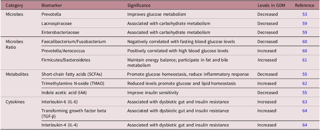
Their integration with other biological layers significantly enhances the translational potential of microbiome-based biomarkers (Table 1). Combining microbiome, metabolome, and epigenome data in multi-omics approaches may make predictions more accurate and help identify people at risk of GDM early on. Reference Karami, Mousavi, Rafiee, Heidari and Shahrokhi52,Reference Bokoliya, McClellan, Zhou and Fan53 Nevertheless, existing biomarker candidates have not been validated in various populations and clinical environments, restricting their immediate utility. Future initiatives should concentrate on the creation of standardized, reproducible biomarker panels and the assessment of their clinical efficacy in extensive prospective cohorts. These kinds of progress could lead to precision medicine, where personalized microbial and epigenetic profiles help with early diagnosis, prevention, and targeted treatments.
Microbiome-based biomarkers for gestational diabetes mellitus

Table 1. Long description
A table with five columns and eight rows, including a header row. The columns are labeled Category, Biomarker, Significance, Levels in GDM, and Reference. The table compares various microbiome-based biomarkers for gestational diabetes mellitus. Row 1: Category, Biomarker, Significance, Levels in GDM, Reference. Row 2: Microbes, Prevotella, Improves glucose metabolism, Decreased, 53. Row 3: Microbes, Lacnospiraceae, Associated with carbohydrate metabolism, Decreased, 59. Row 4: Microbes, Enterobacteriaceae, Associated with carbohydrate metabolism, Decreased, 59. Row 5: Microbes Ratio, Faecalibacterium/Fusobacterium, Negatively correlated with fasting blood glucose levels, Decreased, 60. Row 6: Microbes Ratio, Prevotella/Aerococcus, Positively correlated with high blood glucose levels, Increased, 60. Row 7: Microbes Ratio, Firmicutes/Bacteroidetes, Maintain energy balance; participate in fat and bile metabolism, Increased, 61. Row 8: Metabolites, Short-chain fatty acids (SCFAs), Promote glucose homeostasis, reduce inflammatory response, Decreased, 55. Row 9: Metabolites, Trimethylamine N-oxide (TMAO), Reduced levels promote glucose and lipid homeostasis, Increased, 62. Row 10: Metabolites, Indole acetic acid (IAA), Improve insulin sensitivity, Decreased, 55. Row 11: Cytokines, Interleukin-6 (IL-6), Associated with dysbiotic gut and insulin resistance, Increased, 63. Row 12: Cytokines, Transforming growth factor beta (TGF-beta), Associated with dysbiotic gut and insulin resistance, Increased, 64. Row 13: Cytokines, Interleukin-4 (IL-4), Associated with dysbiotic gut and insulin resistance, Increased, 64.
Epigenetics in GDM
The interaction between GDM, epigenetic mechanisms, and the maternal microbiome is increasingly recognized for its significant influence on maternal and fetal health, long-term metabolic outcomes, and intergenerational disease risk. In pregnancies affected by GDM, distinct DNA methylation patterns are evident in both maternal and placental tissues, impacting genes related to metabolic regulation, fetal growth, inflammation, and insulin signaling. Reference Li, Liu and Liu58,Reference Al Bekai, El and Kalout65 Maternal hyperglycemia can leave a lasting imprint on the fetus through epigenetic changes, particularly increased DNA methylation at certain CpG sites in cord blood. Some of these changes have been linked to a greater risk of preterm birth and may also contribute to future metabolic challenges in the child. Reference Wang, Xu and Zhang66 Offspring of mothers with GDM often inherit these altered epigenetic markers, increasing their risk of obesity, insulin resistance, and metabolic syndrome later in life. Reference Al Bekai, El and Kalout65,Reference Wang, Xu and Zhang66
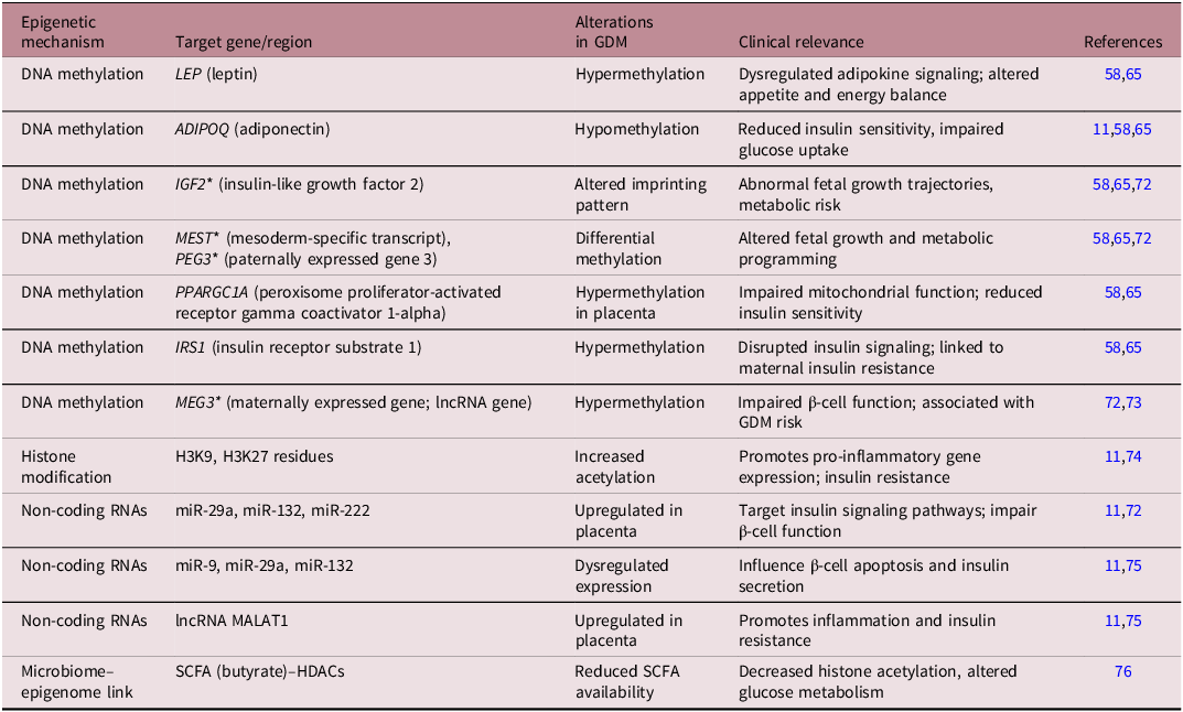
Recent evidence indicates that epigenetic signatures are influenced not only by maternal glycemic status but also by the gut microbiome. Specifically, microbiome-derived metabolites, particularly SCFAs, such as butyrate and propionate, have been demonstrated to modulate the activity of epigenetic enzymes like histone deacetylases (HDACs). Reference Sharma, Li, Stoll and Tollefsbol43 These metabolites facilitate chromatin relaxation, thereby promoting the transcription of genes essential for insulin sensitivity and glucose metabolism. Conversely, microbial byproducts such as trimethylamine N-oxide (TMAO), produced from the metabolism of dietary choline and carnitine, have been linked to pro-inflammatory states and impaired insulin signaling, thereby increasing the risk of GDM. Reference Sharma, Li, Stoll and Tollefsbol43,Reference Jaworska, Kuś and Ufnal67 Beyond SCFAs, the maternal microbiome also provides essential cofactors and substrates, such as folate and B vitamins, which are synthesized by species like Bifidobacteria. These species serve as methyl and acetyl donors for DNA and histone modification processes. Reference Zhang, Liu and Li68 Multi-omic analyses have begun to elucidate how shifts in microbial diversity affect the host metabolite pool and, consequently, influence epigenetic regulation. For example, an increased abundance of Firmicutes in GDM has been linked to hypermethylation of metabolic gene promoters, highlighting the microbiome’s regulatory impact on the host epigenome (Table 2). Notably, the effects of this microbiome–epigenome interaction are not confined to metabolic tissues. Reference Lin, Han and Wang69 Recent studies utilizing single-cell transcriptomics and proteomics have revealed that microbial metabolites may also affect neuroendocrine circuits, suggesting the involvement of the gut–brain axis in GDM pathophysiology. Reference Kim70 Importantly, given that epigenetic changes are potentially reversible, they offer a promising avenue for intervention. Nutritional strategies, prebiotics, probiotics, and other microbiome-modulating therapies may provide a means to reshape the maternal epigenetic landscape and improve outcomes for both mother and child. Reference Fasano, Chassaing and Haller71 However, the heterogeneity in existing studies underscores the need for more robust, long-term, and integrative multi-omics research to clarify these relationships and develop precision-based therapeutic strategies.
Epigenetic alterations associated with GDM and their clinical implications

Table 2. Long description
A table with 10 rows and 5 columns. The columns are labeled Epigenetic mechanism, Target gene/region, Alterations in GDM, Clinical relevance, and References. The table lists various epigenetic mechanisms, their target genes or regions, the alterations observed in gestational diabetes mellitus (GDM), the clinical relevance of these alterations, and the references for each entry. Row 1: DNA methylation, LEP (leptin), Hypermethylation, Dysregulated adipokine signaling; altered appetite and energy balance, 58,65. Row 2: DNA methylation, ADIPOQ (adiponectin), Hypomethylation, Reduced insulin sensitivity, impaired glucose uptake, 11,58,65. Row 3: DNA methylation, IGF2 (insulin-like growth factor 2), Altered imprinting pattern, Abnormal fetal growth trajectories, metabolic risk, 58,65,72. Row 4: DNA methylation, MEST (mesoderm-specific transcript), PEG3 (paternally expressed gene 3), Differential methylation, Altered fetal growth and metabolic programming, 58,65,72. Row 5: DNA methylation, PPARGC1A (peroxisome proliferator-activated receptor gamma coactivator 1-alpha), Hypermethylation in placenta, Impaired mitochondrial function; reduced insulin sensitivity, 58,65. Row 6: DNA methylation, IRS1 (insulin receptor substrate 1), Hypermethylation, Disrupted insulin signaling; linked to maternal insulin resistance, 58,65. Row 7: DNA methylation, MEG3 (maternally expressed gene; lncRNA gene), Hypermethylation, Impaired β-cell function; associated with GDM risk, 72,73. Row 8: Histone modification, H3K9, H3K27 residues, Increased acetylation, Promotes pro-inflammatory gene expression; insulin resistance, 11,74. Row 9: Non-coding RNAs, miR-29a, miR-132, miR-222, Upregulated in placenta, Target insulin signaling pathways; impair β-cell function, 11,72. Row 10: Non-coding RNAs, miR-9, miR-29a, miR-132, Dysregulated expression, Influence β-cell apoptosis and insulin secretion, 11,75. Row 11: Non-coding RNAs, lncRNA MALAT1, Upregulated in placenta, Promotes inflammation and insulin resistance, 11,75. Row 12: Microbiome-epigenome link, SCFA (butyrate)-HDACs, Reduced SCFA availability, Decreased histone acetylation, altered glucose metabolism, 76.
(*imprinted genes).
Maternal diet, probiotics, and therapeutic interventions
Diet can significantly modify host-microbial interactions, potentially improving maternal health. Emerging evidence shows that diet can influence gene activity through epigenetic changes, with lasting effects on metabolism and disease risk. In women with and without GDM, certain gene variants have been linked to an increased risk of GDM, lipid levels, and body mass index. These findings suggest that nutrition-related genetic variations may influence metabolic health during and after pregnancy. Reference Ray, Zeng and Kusi77
Diets rich in fat and refined sugars are consistently linked with reduced microbial diversity and depletion of SCFA-producing bacteria, limiting the production of butyrate, acetate, and propionate that normally support insulin sensitivity, lipid regulation, and beneficial epigenetic modifications. Reference Dreisbach, Morgan, Cochran, Gyamfi, Henderson and Prescott32 In contrast, fiber-rich diets enhance the abundance of Bifidobacterium and Faecalibacterium, boost SCFA production, reduce systemic inflammation, and have been associated with improved glucose tolerance and lower gestational weight gain in pregnancy, making them promising preventive strategies for GDM. Reference Edwards, Cunningham, Dunlop and Corwin23 Beyond diet, targeted microbiome-directed interventions have shown therapeutic potential. Probiotics, particularly Lactobacillus and Bifidobacterium strains, improve microbial balance, reduce inflammatory markers, and modestly lower fasting plasma glucose, HbA1c, and insulin resistance in women at high risk of GDM, as demonstrated in randomized controlled trials. Reference Brown, Al-Memar and Marchesi4,Reference Dolatkhah, Hajifaraji, Abbasalizadeh, Aghamohammadzadeh, Mehrabi and Mesgari Abbasi63 Prebiotics, including inulin-type fructans and galacto-oligosaccharides, further promote the growth of beneficial bacteria, and when combined with probiotics as synbiotics, may deliver synergistic benefits by both introducing and sustaining healthy microbial populations. Preliminary studies suggest that synbiotic supplementation may reduce maternal weight gain and inflammatory cytokines, though evidence in GDM-specific cohorts remains limited Reference Pandey, Naik and Vakil78 (Figure 3). Importantly, these interventions may also modulate epigenetic programming: SCFA-producing bacteria contribute to histone acetylation at insulin-related genes, while prebiotic-derived methyl donors support DNA methylation at metabolic loci such as PPARγ and SLC2A4. Reference Zhang, Liu and Li68 Such microbiome–epigenome interactions highlight the potential for reducing intergenerational metabolic risk. Complementary lifestyle and pharmacological strategies add to this landscape. Exercise enhances insulin sensitivity while enriching SCFA-producing taxa and reducing pro-inflammatory microbes, amplifying the benefits of dietary changes. Reference Gao, Chen and Li26 Metformin, a cornerstone in GDM management, not only improves maternal glucose control but also modifies gut microbiota, enriching Akkermansia muciniphila and reducing pathogenic Clostridia, suggesting that part of its therapeutic effect may be microbiome-mediated. Reference Molina-Vega, Picón-César and Gutiérrez-Repiso79
Influence of diet on microbiome and GDM management (TMAO: Trimethylamine N-oxide; LPS: Lipopolysaccharides; LCFA: Long chain fatty acids; SCFAs: Short-chain fatty acids; Upward Arrow: Increase; Downward Arrow: Decrease).

Conclusion
Gestational diabetes mellitus is not merely a transient metabolic disturbance but a complex condition shaped by interactions between the maternal microbiome, epigenetic regulation, and environmental factors. The microbiome–epigenome axis provides a compelling framework linking maternal metabolic status to fetal programming and long-term disease risk. However, much of the current evidence remains associative, highlighting the need for caution in drawing causal inferences. Future research should prioritize longitudinal, multi-ethnic cohort studies to capture dynamic changes in the microbiome and epigenome throughout pregnancy. Integrative multi-omics approaches combining microbiome, metabolomic, and epigenetic data will be essential to identify robust biomarkers and mechanistic pathways. Standardization of sampling methods, analytical pipelines, and diagnostic criteria is also critical to improve reproducibility across studies. From a translational perspective, advancing microbiome-based diagnostics and personalized interventions holds significant promise. Targeted strategies such as precision nutrition, microbiome-modulating therapies, and epigenetic interventions may enable early risk prediction and improved clinical outcomes. However, their successful implementation will depend on rigorous validation, mechanistic clarity, and long-term clinical evaluation. Ultimately, understanding the maternal microbiome–epigenome interplay offers an opportunity not only to improve GDM management but also to reduce the intergenerational transmission of metabolic disease, shifting the focus from treatment to prevention and precision reproductive health.
Acknowledgements
Authors would like to thank the Department of Personalized and Molecular Medicine, Era University, Lucknow, for providing basic support for this article.
Financial support
This research received no specific grant from any funding agency, commercial or not-for-profit sectors.
Competing interests
None.