Pulmonary venous obstruction is the most crucial complication after correction of total anomalous pulmonary venous drainage. In type II total anomalous pulmonary venous drainage, pulmonary venous obstruction after correction occurs via the new pulmonary venous pathway. Obstruction of the pulmonary venous pathway can occur because of the rigid fibrous tissue around the pericardial roof or the artificial patch, and inflammation of the cut-back site.Reference Lo Rito, Gazzaz and Wilder 1 To resolve this problem, we used a pedicled right atrial flap, which is expected to grow as the body grows. In this case report, we describe this new surgical procedure for type IIa and IIb total anomalous pulmonary venous drainage, including its results and limitations.
Case reports
Case 1
A 10-day-old female neonate, weighing 2.8 kg, underwent physical examination, blood sampling, chest radiography, electrocardiography, and echocardiography. Continuous murmurs were auscultated over the 2nd and 3rd left intercostal spaces and along the sternal border, and tachypnoea was noted during physical examination. Chest radiography revealed that her heart was enlarged and the pulmonary vasculature was engorged. Electrocardiography demonstrated the presence of a normal sinus rhythm with right ventricular hypertrophy and right axis deviation. Echocardiography revealed a dilated right atrium, dilated coronary sinus with total pulmonary venous return, and atrial septal defect. A diagnosis of type IIa total anomalous pulmonary venous drainage was made.
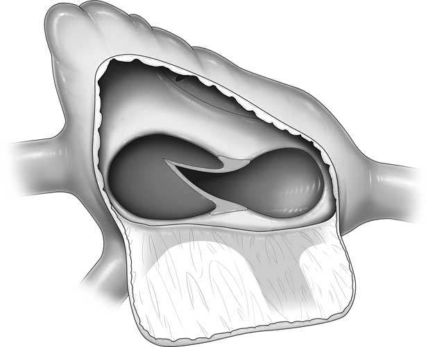
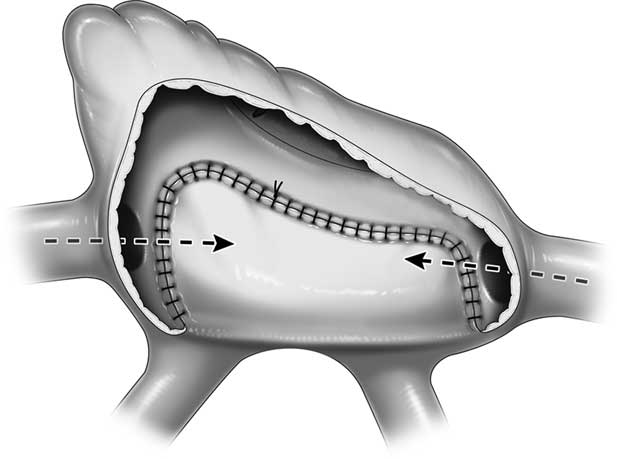
Surgical repair was performed through a median sternotomy. Cardiopulmonary bypass was established after heparinisation, and the incision line was marked at the right atrium. The upper line was towards and 1 cm below the sinus node to avoid injury to the node, the lateral line was parallel to the atrioventricular groove, and the inferior line was made towards the junction of the sulcus terminalis and inferior caval vein. During mild hypothermic cardiopulmonary bypass and aortic cross-clamping, the right atrium was opened along the marked line and the coronary sinus roof was cut back (Fig 1). Subsequently, the pedicled right atrium patch was sutured to cover the atrial septal defect and the cut-back region of the coronary sinus with 6-0 polydioxanone sutures (Fig 2). The left side of the remaining right atrial wall was sutured to the sulcus terminalis, taking care to avoid injuring the sinus node, without additional patch material. After rewarming, the patient was easily weaned from cardiopulmonary bypass without arrhythmia, and her postoperative haemodynamic state was stable. On postoperative day 3, echocardiography revealed good cardiac function and non-obstructed blood flow through the rerouted path. The patient was discharged on postoperative day 10 in good condition.
The roof of the coronary sinus was cut back to enlarge the rerouting pathway.

Type IIa total anomalous pulmonary venous drainage repair is performed. The pedicled right atrial patch was used to cover the atrial septal defect and the cut-back region of the coronary sinus, using 6-0 polydioxanone sutures, taking care to avoid obstruction of the orifices.

Case 2
A 4-month-old boy weighing 5.2 kg was admitted. Tachypnoea and cyanotic lips were noted during physical examination. Chest radiography revealed that his heart was enlarged and the pulmonary vasculature was engorged. Electrocardiography demonstrated the presence of a normal sinus rhythm with right ventricular hypertrophy and right axis deviation. Echocardiography and CT revealed a dilated right atrium. Each of the four pulmonary veins was separately connected to the common pulmonary venous chamber behind the right atrial wall, and small atrial septal defect. The diagnosis of type IIb total anomalous pulmonary venous drainage was made.
Surgical repair was performed in the same manner as in case 1. There were four orifices of pulmonary veins to the right of the atrial chamber and a small patent foramen ovale. The tissue of the atrial septum was resected. The pedicled right atrial flap was then sutured to cover the atrial septal defect and the orifice of the common pulmonary venous chamber with 6-0 polydioxanone sutures, avoiding obstruction of the superior and inferior caval vein. The remaining right atrial wall was sutured to the right atrium to avoid injury of the sinus node, without additional patch material. After rewarming, the patient was easily weaned from the cardiopulmonary bypass without arrhythmia, and his postoperative haemodynamic state was stable. The patient was discharged on postoperative day 12 in good condition.
Discussion
Although type II total anomalous pulmonary venous drainage is not a specific risk factor for postoperative pulmonary venous obstruction, 0–18% of patients with any type of total anomalous pulmonary venous drainage experience postoperative pulmonary venous obstruction.Reference Seale, Uemura and Webber 2 – Reference Ricci, Elliott and Cohen 4 Risk factors for postoperative pulmonary venous obstruction in the anastomosis site between the common pulmonary venous chamber and the left atrium include a direct purse-string effect from the suture line, inflammatory response, and fibrosis of the pulmonary vein tissue.Reference Lo Rito, Gazzaz and Wilder 1 In 2003, Ricci et al demonstrated a higher incidence of postoperative residual obstruction and mortality in patients surgically receiving artificial material for pulmonary venous obstruction repair. Gore-Tex or left atrium tissue enlargement was used to enlarge the anastomosis site or peripheral obstruction. Furthermore, they noted that the use of any native tissue is preferable to artificial material whenever possible.Reference Ricci, Elliott and Cohen 4
In cases of type II total anomalous pulmonary venous drainage, postoperative pulmonary venous obstruction occurs because of an inflammation reaction of the pericardial or artificial patch, or the posterior side of the new pulmonary venous pathway, which consists of the cut-back wall of the coronary sinus or resected atrial septal defect. Our novel pedicled atrial flap, which has growth potential, can cover the anterior roof of the new pulmonary venous pathway. If the atrium is not enlarged, the pedicled atrial patch must be harvested because the most important aspect of this procedure is the use of this patch for rerouting the pathway from the pulmonary venous chamber to the left atrium; moreover, the atrial wall can be closed with additional patch material. There is potential consideration regarding the risk of thrombosis between the atrial flap and the atrial septum; however, in these cases, postoperative echocardiography did not reveal thrombosis or flow constriction in the gap between the patch and atrial wall.
Although there is a need for careful follow-up, we believe that this new method is a surgical option for type II total anomalous pulmonary venous drainage repair.
Financial Support
This research received no specific grant from any funding agency in the public, commercial, or not-for-profit sectors.
Conflicts of Interest
None.
Ethical Standards
All procedures performed in studies involving human participants were in accordance with the ethical standards of the institutional and/or national research committee and with the 1964 Helsinki Declaration and its later amendments or comparable ethical standards.
Acknowledgements
The authors thank Mrs. Kojima, whose language support and suggestions were of inestimable value to this report.