Introduction
Intramedullary spinal cord metastasis (IMSC) is a rare occurrence (1–3%) among the spinal cord tumours with a poor median survival rate, 4–6 months, Reference Lv, Liu, Quan, Li, Dong and Liu1–Reference Hata, Takai and Takahashi5 and seen mostly in patients with lung cancer with brain metastases (associated with carcinomatous meningitis) apart from breast cancer, lymphomas, melanomas and renal cell carcinomas. Advances in imaging techniques like magnetic resonance imaging (MRI) and improvement in treatment methods Reference Potti, Abdel-Raheem, Levitt, Schell and Mehdi2,Reference Wu, Wang, Yang, Jia and Xu3,Reference Hata, Takai and Takahashi5,Reference Ehret, Senger and Kufeld6 including recent immunotherapy and molecular targeted agents have resulted in increase in survival rates for the locally advanced non-small cell lung carcinomas (NSCLC). Leptomeningeal metastases are noted in 25% patients with IMSC metastases as majority of these patients also can have brain metastases. Reference Schiff and O’Neill7 Presentation can vary from weakness, numbness, pain, Brown-Sequard syndrome (hemicord dysfunction) and diagnosed with computed tomography (CT) myelogram, MRI and treatment comprises of palliative radiation therapy apart from corticosteroids and surgery if an operable candidate.
Case Report
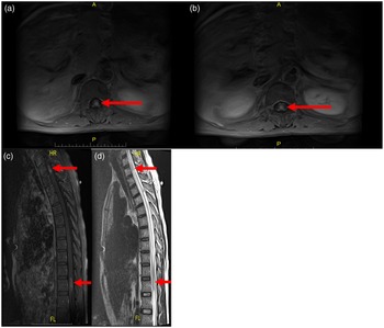
A 57-year-old-male, former smoker, an established case of adenocarcinoma of the right lung and stage IV NSCLC with multiple brain metastases, was treated with palliative external beam radiation therapy (EBRT) to the whole brain (WBRT, to a dose of 3000 centigray (cGy) in 10 fractions over 2 weeks) at an outside facility (outside USA). Liquid biopsy was negative for epidermal growth factor receptor (EGFR) exon 19 deletion, wild type. Four months later, he presented to the emergency room to our facility in USA with bilateral lower extremity weakness, numbness, tingling, difficulty walking, speech problems, back pain with progressive bowel and bladder incontinence. No systemic therapy was administered in the interim. Serum sodium was low, 131 mol/L. An MRI of the thoracic, lumbar spine regions demonstrated a 1·5 × 0·6 cm lesion within spinal cord at T2/T3 and a 2·4 × 0·7 cm lesion at T12 involving the conus, IMSC location (Figure 1A–D). Three enhancing lesions with surrounding vasogenic oedema and carcinomatous meningitis were seen on MRI of the brain. CT scan of the chest, abdomen and pelvis revealed a 7·2 × 6 × 5·5 cm right upper lobe mass with bulky mediastinal, bilateral hilar lymph node metastases and multiple lung nodules bilaterally. A Foley catheter was placed, and a bolus dose of intravenous dexamethasone, 10 mg, was administered followed by round the clock steroids, Keppra for seizure prophylaxis. Multidisciplinary team of neurosurgeon, neurologist, medical oncologist and radiation oncologist assessed the patient, and in view of disseminated stage IV malignancy, surgery was deferred and repeat biopsy of the lung lesion done was positive for poorly differentiated adenocarcinoma. Immunohistochemistry studies done showed tumour cells to be positive for pancytokeratin, CK7, Vimentin and TTF-1 and negative for CK20, p63, synaptophysin, chromogranin and CEA. EGFR, KRAS wild type, HER2 negative and PD-L1 were 80% positive. An urgent palliative radiation therapy to both the IMSC lesions was done to T1–T4 and T11-L1 cord levels to a dose of 3000 cGy in 10 fractions using 300 cGy per fraction over 13 days duration utilising 3-dimensional conformal radiation therapy technique. Daily image guidance with cone beam CT was done prior to treatments for treatment verification accuracy and precision, image-guided radiation therapy. Syndrome of inappropriate antidiuretic hormone secretion was managed conservatively. He tolerated treatments very well and steroid taper was done after completion of RT. Patient refused systemic treatment (chemo-immunotherapy) and was on symptomatic/supportive measures. He died 3 months later while on home hospice.
(A–D) MRI of thoracic spine (with contrast, axial sagittal T1 and T2 weighted images) showing an enhancing mass within the spinal canal at T2/T3 (as in 1C–D), intramedullary location, 1·5 × 0·6 cm, and the second enhancing mass seen distally at the level of T12 involving conus, intramedullary, 2·4 × 0·7 cm (as in 1A–B), central canal is dilated.

Discussion
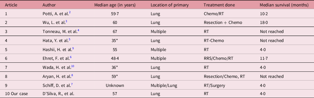
IMSC from NSCLC is a rare diagnosis and different treatment methods comprise surgery (if an operable candidate), radiation therapy to preserve the neurological function (Table 1) or supportive care. Survival is poor in these patients as the recovery from neurological function is poor despite palliative RT and contributes to compromised quality of life and high morbidity. Reference Lv, Liu, Quan, Li, Dong and Liu1,Reference Potti, Abdel-Raheem, Levitt, Schell and Mehdi2,Reference Tonneau, Mouttet-Audouard, Le Tinier, Mirabel and Pasquier4 In this case, patient presented with Brown-Sequard syndrome with disseminated leptomeningeal disease 4 months after palliative WBRT for brain metastases. MRI is diagnostic in many cases and paraneoplastic myelopathies Reference Hata, Takai and Takahashi5,Reference Schiff and O’Neill7 need to be ruled out in lung cancer patients (Lambert-Eaton myasthenic syndrome, cerebellar ataxia, sensory neuropathy, limbic encephalitis, encephalomyelitis, autonomic neuropathy, retinopathy and opsomyoclonus). Thoracic lesions are most common sites for IMSC metastases like in the current case, Reference Lv, Liu, Quan, Li, Dong and Liu1 noted at T2/3 and T12 levels. Few cases also presented with Brown-Sequard syndrome from lung malignancy. Reference Ehret, Senger and Kufeld6–Reference Aryan, Farin, Nakaji, Imbesi and Abshire8 In patients with poor performance status, and with widespread metastatic disease, mainstay of treatment is generally fractionated EBRT Reference Hata, Takai and Takahashi5,Reference Ehret, Senger and Kufeld6,Reference Hashii, Mizumoto and Kanemoto9 which helps to prevent further neurological deterioration and alleviates the back pain. Steroids provide only symptom relief and reduce the radiation-induced oedema.
Case series on IMSC metastases and treatment methods

Abbreviations: IMSC, intramedullary spinal cord metastases; RT, radiation therapy; Chemo, chemotherapy; RRS, robotic radiotherapy surgery.
* Case report.
Conclusions
Prompt diagnosis and treatment of IMSC metastases, with early intervention, by urgent palliative radiation therapy apart from medical management should be considered in inoperable patients with advanced stages of malignancies to preserve the neurological function.
Acknowledgements
None.
Financial Support
This research received no specific grant from any funding agency, commercial or not-for-profit sectors.
Conflicts of Interest
The author(s) declare none.