Introduction
The family Mesometridae (Digenea) includes species that are usually found on the intestine of teleost fishes of the families Sparidae and Acanthuridae, which are generally restricted to the Mediterranean Sea. However, certain species are also present in the Atlantic and Pacific oceans. To date, the family Mesometridae contains only eight recognized species belonging to five genera, named Centroderma, Elstia, Mesometra, Parawardula and Wardula (Jones & Blair, Reference Jones, Blair, Jones, Bray and Gibson2005; Pérez-del Olmo et al., Reference Pérez-del Olmo, Gibson, Fernández, Sanisidro, Raga and Kostadinova2006).
Ultrastructural studies of spermiogenesis and spermatozoa in the Platyhelminthes, and in particular the Digenea, have revealed much diversity in their characteristics. This diversity is of great importance and usefulness for phylogenetic reconstructions (Hendelberg, Reference Hendelberg1969; Euzet et al., Reference Euzet, Świderski and Mokhtar-Maamouri1981; Ehlers, Reference Ehlers1984; Świderski, Reference Świderski1986; Justine, Reference Justine1991, Reference Justine1995, Reference Justine1998, Reference Justine, Littlewood and Bray2001; Bâ & Marchand, Reference Bâ and Marchand1995; Bruňanská, Reference Bruňanská, Lejeune and Delvaux2010; Levron et al., Reference Levron, Miquel, Oros and Scholz2010; Quilichini et al., Reference Quilichini, Foata, Justine, Bray and Marchand2010a, Reference Quilichini, Foata, Justine, Bray and Marchandb). Concerning the family Mesometridae, to date there has been only one ultrastructural study that has focused on Wardula capitellata (see Bakhoum et al., Reference Bakhoum, Ndiaye, Sène, Bâ and Miquel2012). The aim of the present work was to perform an ultrastructural study of spermiogenesis and of the spermatozoon of Elstia stossichianum, a second mesometrid species that belongs to another of the five existing genera of this family.
Materials and methods
Living adult specimens of E. stossichianum (Monticelli, 1892) were collected from the intestine of naturally infected sparid teleosts, Sarpa salpa (Linnaeus, 1758), captured off the coast of Dakar (Senegal).
Adult worms were examined in 0.9% sodium chloride solution and subsequently processed for transmission electron microscopy. Specimens were fixed in cold (4°C) 2.5% glutaraldehyde in a 0.1 m sodium cacodylate buffer at pH 7.4 for 2 h, rinsed in a 0.1 m sodium cacodylate buffer at pH 7.4, post-fixed in cold (4°C) 1% osmium tetroxide in the same buffer for 1 h, rinsed in a 0.1 m sodium cacodylate buffer at pH 7.4, dehydrated in an ethanol series and propylene oxide, and finally embedded in Spurr resin. Ultrathin sections were obtained using a Reichert–Jung Ultracut E ultramicrotome (Reichert Instruments GmbH, Seefeld, Germany), placed on copper grids and double-stained with uranyl acetate and lead citrate according to Reynolds (Reference Reynolds1963).
The presence of glycogen was detected using the method of Thiéry (Reference Thiéry1967). Gold grids were treated in periodic acid, thiocarbohydrazide and silver proteinate (PA–TCH–SP) as follows: 30 min in 10% PA, rinsed in milliQ water, 24 h in TCH, rinsed in acetic solutions and milliQ water, 30 min in 1% SP in the dark, and rinsed in milliQ water.
Finally, both copper and gold grids were examined using a JEOL 1010 transmission electron microscope (Jeol (Europe) SAS, Croissy-sur-Seine, France) at an accelerating voltage of 80 kV.
Results
Spermiogenesis
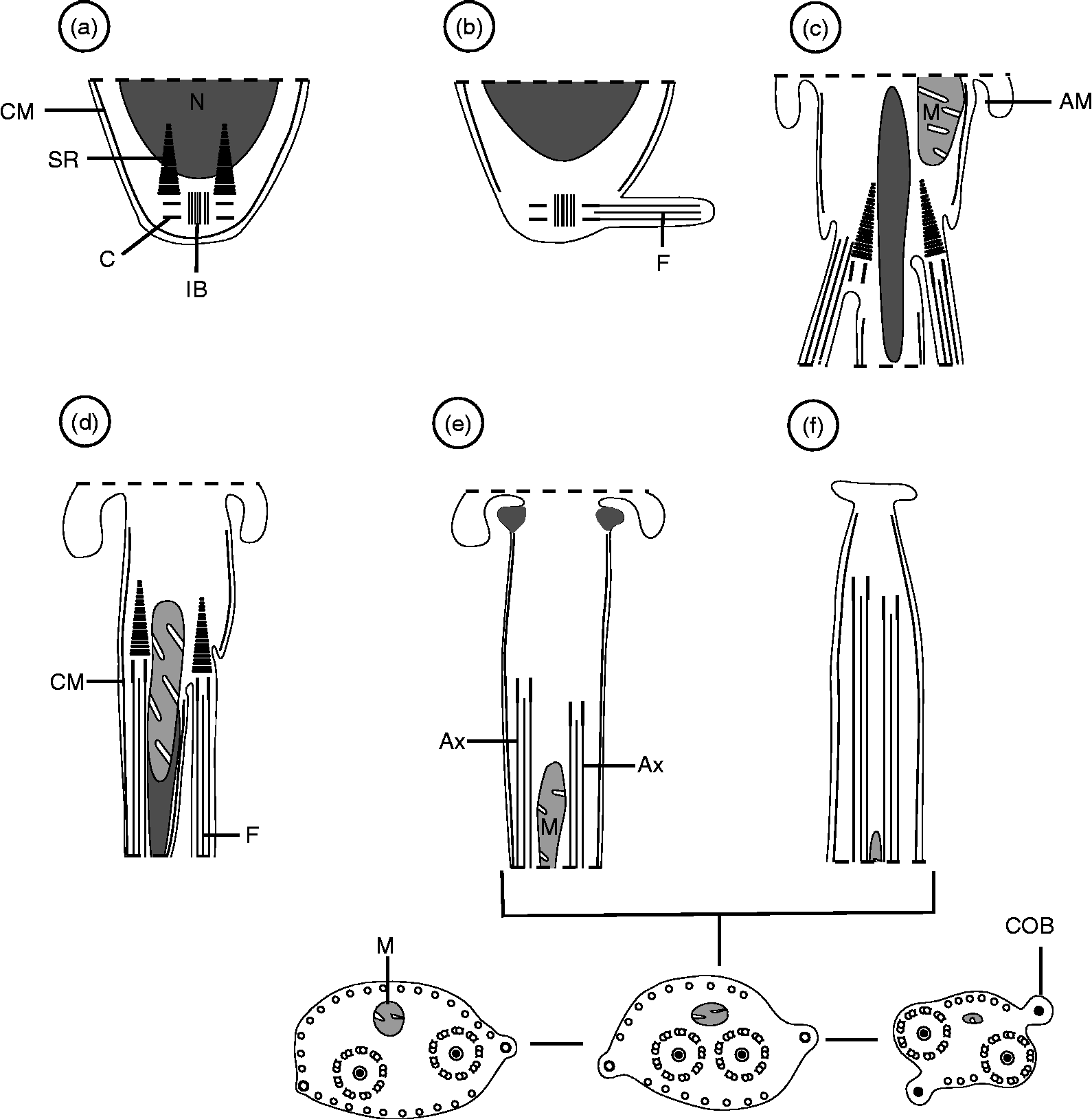
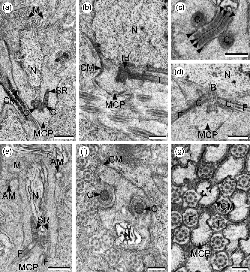
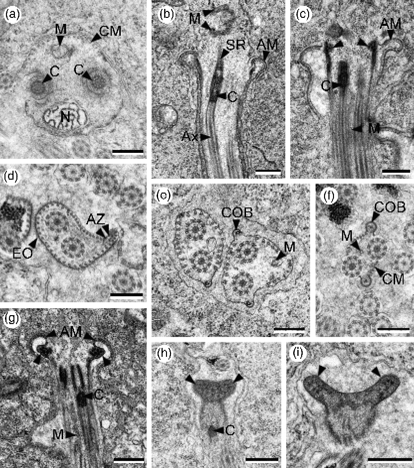
Spermiogenesis in E. stossichianum begins with the formation of a differentiation zone containing a nucleus, several mitochondria, two centrioles associated with striated rootlets and an intercentriolar body (figs 1a and 2a). The intercentriolar body is made up of seven electron-dense layers (fig. 2c). Each centriole develops a free flagellum growing orthogonally to a median cytoplasmic process (figs 1b and 2b). Later, these flagella rotate and become parallel to the median cytoplasmic process (figs 1c and figs 2d–g). During flagellar rotation, the nucleus and mitochondria migrate along the spermatid body (figs 1c, d, 2e, f and 3a). However, the migration of the nucleus occurs before the migration of mitochondria (figs 2f and 3a). Four attachment zones are already visible in the median cytoplasmic process before its fusion with the flagella (fig. 2g). The proximo-distal fusion is asynchronous (figs 1d, 3a). Before the total constriction of the ring of arched membranes the striated rootlets are still present in the spermatid's cytoplasm (fig. 3b). During the final stages of spermiogenesis, both lateral expansion and external ornamentation are observed in the proximal areas of the spermatid (fig. 3d). Additionally, electron-dense material is observed near the ring of arched membranes, and mitochondrial migration is still occurring (figs 1e and 3c). This electron-dense material gives rise to prominent electron-dense structures in the final stage of spermiogenesis (fig. 3g) that form the forked anterior extremity in the future spermatozoon after the constriction of the ring of arched membranes (figs 1f and 3h, i). During the spermiogenesis of E. stossichianum it was also possible to observe for the first time the formation of cytoplasmic ornamented buttons (figs 1e, f and 3e, f).
Reconstruction of the main stages of spermiogenesis of Elstia stossichianum. AM, Arched membrane; Ax, axoneme; C, centriole; CM, cortical microtubules; COB, cytoplasmic ornamented button; F, flagellum; IB, intercentriolar body; M, mitochondrion; N, nucleus; SR, striated rootlet.

Spermiogenesis of Elstia stossichianum. (a) Longitudinal section of a differentiation zone showing centrioles (C) associated with striated rootlets (SR) and intercentriolar body (IB). CM, Cortical microtubules; M, mitochondria; MCP, median cytoplasmic process; N, nucleus. Scale bar: 1 μm. (b) Orthogonal development of one flagellum (F). CM, Cortical microtubules; IB, intercentriolar body; MCP, median cytoplasmic process; N, nucleus. Scale bar: 0.5 μm. (c) Detail of intercentriolar body exhibiting seven electron-dense layers (arrowheads); C, centrioles. Scale bar: 0.3 μm; (d) Longitudinal section in which two flagella (F) rotate toward the median cytoplasmic process (MCP). C, Centrioles; IB, intercentriolar body; N, nucleus. Scale bar: 0.5 μm. (e) Differentiation zone with flagella (F) nearly parallel to the median cytoplasmic process (MCP) and showing the elongation of nucleus (N). AM, Arched membrane; M, mitochondrion; SR, striated rootlet. Scale bar: 0.5 μm. (f) Cross-section of a differentiation zone at the level of centrioles (C) before the proximo-distal fusion of flagella. CM, cortical microtubules; N, nucleus. Scale bar: 0.3 μm. (g) Cross-sections before the proximo-distal fusion showing free flagella (F) and median cytoplasmic processes (MCP). Note the four attachment zones (AZ) (arrowheads); CM, cortical microtubules. Scale bar: 0.3 μm.
Spermiogenesis of Elstia stossichianum. (a) Cross-section of a differentiation zone at the level of centrioles (C) indicating the asynchronous proximo-distal fusion of flagella. CM, cortical microtubules; M, mitochondrion; N, nucleus. Scale bar: 0.3 μm. (b–c) Longitudinal sections after the proximo-distal fusion in which electron-dense material (arrowheads) is observed at the arched membranes (AM) level. Ax, axoneme; C, centriole; M, mitochondrion; SR, striated rootlet. Scale bars: 0.5 μm. (d) Appearance of lateral expansion in spermiogenesis. AZ, attachment zones; EO, external ornamentation. Scale bar: 0.3 μm. (e–f) Cross-sections showing different stages of appearance of cytoplasmic ornamented buttons (COB). M, mitochondrion. Scale bars: 0.3 μm. (g) Longitudinal section showing the constriction of the ring of arched membranes (AM). Note the formation of future anterior forked extremity of the spermatozoon (arrowheads). C, centriole; M, mitochondrion. Scale bar: 0.5 μm; (h–i) Longitudinal sections after liberation of the young spermatozoon showing the progressive development of the forked anterior extremity (arrowheads); C, centriole. Scale bars: 0.5 μm (h), 0.3 μm (i).
Spermatozoon
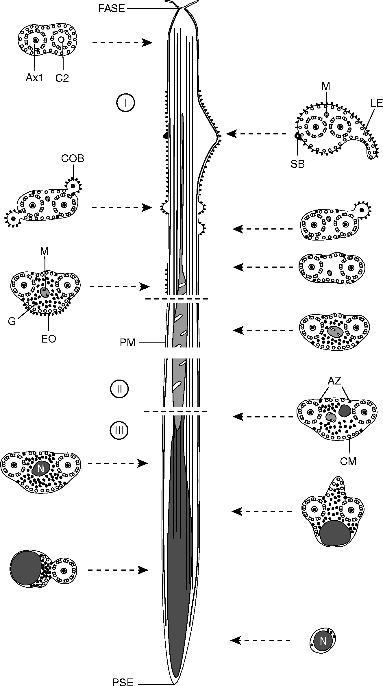
The observation of numerous cross- and longitudinal sections enabled us to establish three regions (I–III) from the anterior to the posterior spermatozoon extremity considering the presence of different ultrastructural characteristics.
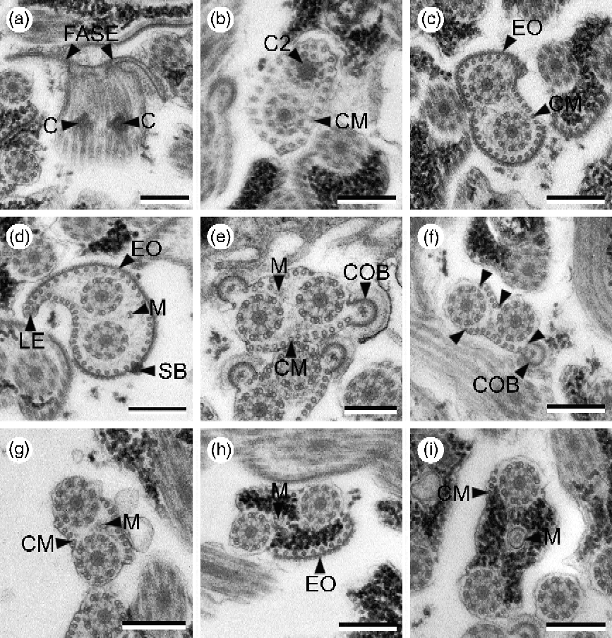
Region I corresponds to the anterior spermatozoon extremity with an external morphology showing a forked anterior tip (figs 4I and 5a). This anterior spermatozoon extremity contains the centrioles of two axonemes which exhibit the typical 9+‘1’ pattern of the trepaxonematan Platyhelminthes and it also exhibits a continuous and submembranous layer of parallel cortical microtubules (fig. 5a and b). In the middle part of region I (the ornamented area), it is possible to observe a mitochondrion, an external ornamentation, a lateral expansion, spine-like bodies and cytoplasmic ornamented buttons (figs 4I and 5c–f). In this area, initially the cortical microtubules describe a continuous layer interrupted by two attachment zones (fig. 5c and d). Later, cross-sections with two or one cytoplasmic ornamented buttons exhibit four attachment zones and, thus, parallel cortical microtubules become arranged into two fields: the first with about 9–13 units and the second with approximately 4–9 units (fig. 5e and f). In the distal part of region I, the mature spermatozoon lacks most of these structures and exhibits only the external ornamentation and two bundles of parallel cortical microtubules (fig. 5g and h). Moreover, this distal part of region I is also characterized by the appearance of a large amount of granules of glycogen (fig. 5h), unequivocally evidenced by the test of Thiéry (Reference Thiéry1967) (fig. 6f). It is interesting to note the presence of a thin part of the mitochondrion in a large area of this region figs 4I and 5d–h).
Schematic reconstruction of mature spermatozoon of Elstia stossichianum; to simplify the drawing, granules of glycogen are not shown in the longitudinal section. Ax1, first axoneme; AZ, attachment zone; C2, second centriole; CM, cortical microtubule; COB, cytoplasmic ornamented button; EO, external ornamentation; FASE, forked anterior spermatozoon extremity; G, glycogen; LE, lateral expansion; M, mitochondrion; N, nucleus; PM, plasma membrane; PSE, posterior spermatozoon extremity; SB, spine-like body.
Spermatozoon of Elstia stossichianum. (a) Forked anterior spermatozoon extremity (FASE) showing centrioles (C) of the two axonemes. Scale bar: 0.3 μm. (b) Cross-section with first complete axoneme and centriole of the second axoneme (C2). Note a continuous layer of cortical microtubules (CM). Scale bar: 0.3 μm. (c) Appearance of external ornamentation (EO) around the two axonemes; CM, cortical microtubules. Scale bar: 0.3 μm. (d) Cross-section showing lateral expansion (LE) accompanied by cortical microtubules (CM), external ornamentation of the plasma membrane (EO) and spine-like body (SB); M, mitochondrion. Scale bar: 0.3 μm. (e–f) Cross-sections showing cytoplasmic ornamented buttons (COB). Note the presence of four attachment zones (arrowheads) and filiform mitochondrion (M). CM, Cortical microtubules. Scale bars: 0.3 μm. (g) Cross-section without cytoplasmic ornamented buttons showing two fields of cortical microtubules (CM); M, mitochondrion. Scale bar: 0.3 μm. (h–i) Cross-sections showing the appearance of granules of glycogen (G) and the progressive increase in the size of the mitochondrion (M). CM, cortical microtubules; EO, external ornamentation. Scale bars: 0.3 μm.
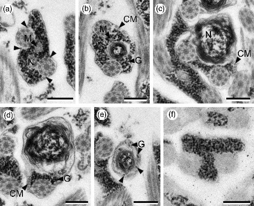
Spermatozoon of Elstia stossichianum. (a–b) Cross-sections at nuclear area exhibiting two axonemes, mitochondrion (M) and nucleus (N). Note the position of the nucleus between axonemes. Arrowheads, attachment zones; CM, cortical microtubules; G, granules of glycogen. Scale bars: 0.3 μm. (c) Cross-section characterized by an eccentric position of the nucleus (N) and disappearance of the mitochondrion. CM, cortical microtubules. Scale bar: 0.3 μm. (d) Cross-section toward posterior spermatozoon extremity. Note the disappearance of the first axoneme. CM, cortical microtubules; G, granules of glycogen. Scale bar: 0.3 μm. (e) Posterior spermatozoon extremity exhibiting only a nucleus and a few granules of glycogen (G). Note the presence of some microtubules (arrowheads). Scale bar: 0.3 μm. (f) Granules of glycogen evidenced by the test of Thiéry (Reference Thiéry1967). Scale bar: 0.3 μm.
Region II is characterized by the increased size of the mitochondrion. This region also exhibits both axonemes, the two bundles of cortical microtubules constituted by a few units and granules of glycogen (figs 4II and 5i).
Region III is the nuclear area of the spermatozoon. The transition between regions II and III is marked by the simultaneous presence of the posterior part of the mitochondrion and the initial part of the nucleus (figs 4III and 6a). Posteriorly, when the mitochondrion disappears the nucleus progressively increases in size (fig. 6b and c). The nuclear region is also characterized by the presence of the axonemes, cortical microtubules and granules of glycogen (figs 4III and 6a–d). Near the posterior spermatozoon extremity both of the axonemes disappear (fig. 6d) one after the other (fig. 6e). Glycogen and cortical microtubules also decrease progressively and, thus, the posterior tip of the spermatozoon contains only the nucleus with a very few granules of glycogen and some microtubules (singlets from the second axoneme or cortical microtubules) (fig. 6e).
Discussion
Spermiogenesis
Spermiogenesis in E. stossichianum follows the general pattern previously described in the Digenea apart from schistosomatids and didymozoids (Justine, Reference Justine, Combes and Jourdane2003). Spermiogenesis begins by the growth of two flagella and the elongation of a median cytoplasmic process from the differentiation zone containing two centrioles associated with striated rootlets, an intercentriolar body and submembranous cortical microtubules. This pattern of spermiogenesis is observed in most digeneans (see Ndiaye et al., Reference Ndiaye, Miquel, Fons and Marchand2003, Reference Ndiaye, Miquel, Bâ and Marchand2004; Levron et al., Reference Levron, Ternengo and Marchand2004; Miquel et al., Reference Miquel, Fournier-Chambrillon, Fournier and Torres2006) and has also been described in W. capitellata, the other mesometrid studied previously (table 1).
Ultrastructural spermatological characters of Mesometridae species.

ASE, anterior spermatozoon extremity; Ax, axoneme; AZ, attachment zones; COB, cytoplasmic ornamented button; EO, external ornamentation; FASE, forked anterior spermatozoon extremity; FR, flagellar rotation; IB, intercentriolar body; LE, lateral expansion; M, number of mitochondria; N, nucleus; PSE, posterior spermatozoon extremity; SB, spine-like body; +, presence of considered character.
During spermiogenesis a flagellar rotation of 90°, described generally in other digenean species (see Foata et al., Reference Foata, Quilichini and Marchand2007; Seck et al., Reference Seck, Marchand and Bâ2007, Reference Seck, Marchand and Bâ2008a, Reference Seck, Marchand and Bâb), is also reported in this study. However, in some digeneans a flagellar rotation greater than 90° has been reported. This was the case in Fasciola hepatica, Helicometra fasciata, Monorchis parvus, Dicrocoelium hospes and Nicolla wisniewskii (Levron et al., Reference Levron, Ternengo and Marchand2003, Reference Levron, Ternengo and Marchand2004; Ndiaye et al., Reference Ndiaye, Miquel, Fons and Marchand2003; Agostini et al., Reference Agostini, Miquel, Ndiaye and Marchand2005; Quilichini et al., Reference Quilichini, Foata, Orsini and Marchand2007c). The importance of the variability in the angle of rotation has been discussed by several authors, who considered that it would be useful in the interpretation of relationships within the Platyhelminthes in general and within the digeneans in particular (Miquel et al., Reference Miquel, Fournier-Chambrillon, Fournier and Torres2006; Bakhoum et al., Reference Bakhoum, Bâ, Shimalov, Torres and Miquel2011a, Reference Bakhoum, Torres, Shimalov, Bâ and Miquelb).
With respect to the intercentriolar body, the phylogenetic interest of this characteristic is related to its progressive reduction in evolved taxa of Platyhelminthes. It is constituted by 11 electron-dense layers in aspidogastreans (sister group of digeneans) and by nine, seven, six or five layers in digeneans (see Bakhoum et al., Reference Bakhoum, Bâ, Shimalov, Torres and Miquel2011a, Reference Bakhoum, Torres, Shimalov, Bâ and Miquelb). Nevertheless, the most frequent situation in digeneans is the presence of seven electron-dense layers, as occurs in E. stossichianum.
Apart from the previously described structures, spermiogenesis in E. stossichianum and also in W. capitellata (Bakhoum et al., Reference Bakhoum, Ndiaye, Sène, Bâ and Miquel2012) is characterized by the presence of cytoplasmic ornamented buttons described for the first time in these species. Indeed, the formation of these buttons is observed when both flagella have already fused with the median cytoplasmic process in the advanced stages of spermiogenesis. In E. stossichianum, during migration of mitochondria, these buttons appear to be slightly protruding. Later, when the mitochondrion has completed its migration, the ornamented buttons increase in size and show their final morphology, and at this stage only remnants of mitochondrial migration are observed in these sections. The description of the formation of these cytoplasmic ornamented buttons is very important for understanding and interpreting their presence in the mature spermatozoon (whether related or not to lateral expansions).
Spermatozoon
In digenean spermatozoa four ultrastructural characteristics appear common to all species studied, except those belonging to the schistosomatids and didymozoids. These characteristics are: (1) two axonemes of the 9+‘1’ trepaxonematan pattern; (2) nucleus; (3) one or more mitochondria; and (4) parallel cortical microtubules. In addition to these four classical structures, several characteristics have been described and proposed recently as interesting tools for phylogenetic purposes: lateral expansions, external ornamentation, spine-like bodies, cytoplasmic ornamented buttons and the morphologies of anterior and posterior spermatozoon extremities (see Miquel et al., Reference Miquel, Fournier-Chambrillon, Fournier and Torres2006; Quilichini et al., Reference Quilichini, Foata, Justine, Bray and Marchand2010b, Reference Quilichini, Foata, Justine, Bray and Marchand2011; Bakhoum et al., Reference Bakhoum, Ndiaye, Sène, Bâ and Miquel2012).
The forked anterior spermatozoon tip, which was observed and described for the first time in the present study in E. stossichianum, has not been observed in W. capitellata (Bakhoum et al., Reference Bakhoum, Ndiaye, Sène, Bâ and Miquel2012) (table 1). However, considering this anterior spermatozoon extremity, both of the studied mesometrids (W. capitellata and E. stossichianum; Bakhoum et al., Reference Bakhoum, Ndiaye, Sène, Bâ and Miquel2012; present study) exhibit two axonemes slightly displaced longitudinally. Such axoneme disposition has been reported in other digenean species, namely Haematoloechus sp., Echinostoma caproni, N. testiobliquum, N. wisniewskii and Rubenstrema exasperatum (Justine & Mattei, Reference Justine and Mattei1982; Iomini & Justine, Reference Iomini and Justine1997; Quilichini et al., Reference Quilichini, Foata and Marchand2007a, c; Bakhoum et al., Reference Bakhoum, Bâ, Shimalov, Torres and Miquel2011a). However, another difference is observed in the anterior spermatozoon areas of the mesometrids studied until now. In fact, two lateral expansions have been observed in the anterior spermatozoon region in W. capitellata (Bakhoum et al., Reference Bakhoum, Ndiaye, Sène, Bâ and Miquel2012), while in E. stossichianum only one lateral expansion has been noted. One lateral expansion has been reported in other digenean species belonging to different families, e.g. Echinostomatidae (Iomini & Justine, Reference Iomini and Justine1997), Fasciolidae (Ndiaye et al., Reference Ndiaye, Miquel, Fons and Marchand2003, Reference Ndiaye, Miquel, Bâ and Marchand2004), Paramphistomidae (Seck et al., Reference Seck, Marchand and Bâ2008a, Reference Seck, Marchand and Bâb), Pronocephalidae (Ndiaye et al., Reference Ndiaye, Quilichini, Sène, Bâ and Marchand2011) and Troglotrematidae (Miquel et al., Reference Miquel, Fournier-Chambrillon, Fournier and Torres2006).
The external ornamentation of the plasma membrane, localized in the anterior spermatozoon region, is another interesting characteristic for phylogenetic purposes. These structures evidenced in E. stossichianum are also present in W. capitellata (Bakhoum et al., Reference Bakhoum, Ndiaye, Sène, Bâ and Miquel2012) and they have been reported in most digenean spermatozoa (see Iomini & Justine, Reference Iomini and Justine1997; Ndiaye et al., Reference Ndiaye, Miquel, Fons and Marchand2003, Reference Ndiaye, Miquel, Bâ and Marchand2004, Reference Ndiaye, Quilichini, Sène, Bâ and Marchand2011; Quilichini et al., Reference Quilichini, Foata, Justine, Bray and Marchand2010a; Bakhoum et al., Reference Bakhoum, Bâ, Shimalov, Torres and Miquel2011a, Reference Bakhoum, Torres, Shimalov, Bâ and Miquelb). Nevertheless, different external ornamented areas have been distinguished in the two mesometrids studied until now: (1) external ornamentation at the level of the lateral expansions; (2) ornamentation associated with the cytoplasmic buttons; and (3) more posterior ornamentation covering only the ventral side of the spermatozoon. In this respect, Quilichini et al. (Reference Quilichini, Foata and Marchand2007b) have proposed two groups of digeneans according to the ornamentation's location: (1) digeneans presenting an anterior external ornamentation; and (2) those exhibiting posterior external ornamentation, generally observed in the mitochondrial side of the spermatozoon. In the present study we describe a discontinuous external ornamentation, as occurs in the case of the other mesometrid that has been studied, W. capitellata (see Bakhoum et al., Reference Bakhoum, Ndiaye, Sène, Bâ and Miquel2012). This discontinuity in the external ornamentation of the plasma membrane is quite exceptional, but it is interesting to remark that Justine & Mattei (Reference Justine and Mattei1982) described two types of external ornamentations showing a discontinuity in the spermatozoon of Haematoloechus sp.
In addition to lateral expansions and external ornamentation, spine-like bodies represent another characteristic of phylogenetic interest described in the anterior region of the spermatozoa of many species (see Bakhoum et al., Reference Bakhoum, Bâ, Shimalov, Torres and Miquel2011a, Reference Bakhoum, Torres, Shimalov, Bâ and Miquelb). Considering the representatives of Mesometridae, spine-like bodies are observed in the area containing lateral expansion both in W. capitellata (Bakhoum et al., Reference Bakhoum, Ndiaye, Sène, Bâ and Miquel2012) and in E. stossichianum (present study). This is also the case with many digenean species, particularly those belonging to the families Diplodiscidae, Gastrothylacidae, Paramphistomidae and Pronocephalidae (see Seck et al., Reference Seck, Marchand and Bâ2007, Reference Seck, Marchand and Bâ2008a, Reference Cribb, Bray, Littlewood, Pichelin, Herniou, Littlewood and Brayb; Bakhoum et al., Reference Bakhoum, Torres, Shimalov, Bâ and Miquel2011b; Ndiaye et al., Reference Ndiaye, Quilichini, Sène, Bâ and Marchand2011). The association of lateral expansion, external ornamentation and spine-like bodies observed in the above-mentioned families has been discussed by Ndiaye et al. (Reference Ndiaye, Quilichini, Sène, Bâ and Marchand2011). In future, this association may be useful when establishing spermatological patterns within digeneans.
In anterior areas of the spermatozoa of the two mesometrids studied until now there is an interesting structure, cytoplasmic ornamented buttons, which were described for the first time in W. capitellata (Bakhoum et al., Reference Bakhoum, Ndiaye, Sène, Bâ and Miquel2012). In the latter study, this characteristic was interpreted as a possible reduction of the two lateral expansions, thus giving rise to cytoplasmic ornamented buttons. However, in E. stossichianum it was possible to observe (1) the formation of cytoplasmic ornamented buttons during spermiogenesis; and (2) the presence of only one lateral expansion in the mature spermatozoon. These observations seem to indicate that cytoplasmic ornamented buttons and lateral expansions are unrelated structures. Thus, in our opinion, the cytoplasmic ornamented buttons, to date described only in the representatives of the family Mesometridae, should be considered new ultrastructural characteristics in digenean spermatozoa. In future, it would be particularly interesting to perform ultrastructural spermiological studies on the other genera of Mesometridae (Mesometra, Centroderma and Parawardula), and also on representatives of their closely related family, Microscasphidiidae.
A posterior spermatozoon extremity exhibiting only a nucleus is described in W. capitellata and in E. stossichianum, and has also been reported in many other digenean species, particularly in those belonging to Paramphistomoidea (Bakhoum et al., Reference Bakhoum, Torres, Shimalov, Bâ and Miquel2011b), Faciolidae (Ndiaye et al., Reference Ndiaye, Miquel, Fons and Marchand2003, Reference Ndiaye, Miquel, Bâ and Marchand2004) and Pronocephalidae (Ndiaye et al., Reference Ndiaye, Quilichini, Sène, Bâ and Marchand2011). In addition, a posterior spermatozoon extremity with a nucleus has also been reported in the aspidogastreans, which is considered a sister group of digeneans. Such similarities allow us to consider the presence of a nucleus in the posterior spermatozoon extremity as a basal characteristic in digeneans. However, more attention and studies are needed, because determination of a posterior spermatozoon extremity in digeneans has been a matter of discussion. Some authors, such as Quilichini et al. (Reference Quilichini, Foata, Justine, Bray and Marchand2010b), have proposed ending sequences for posterior spermatozoon extremities in digeneans, while in most descriptions only the posterior element is considered.
Present spermatological knowledge reveals many similarities between ultrastructural characteristics of the two mesometrids W. capitellata and E. stossichianum (see Bakhoum et al., Reference Bakhoum, Ndiaye, Sène, Bâ and Miquel2012; present study) and those of species described earlier belonging to the superfamily Paramphistomoidea (see Bakhoum et al., Reference Bakhoum, Torres, Shimalov, Bâ and Miquel2011b). In fact, several authors had nested Mesometridae in the superfamily Paramphistomoidea (see Cribb et al., Reference Cribb, Bray, Littlewood, Pichelin, Herniou, Littlewood and Bray2001). However, the systematic position of mesometrids remains controversial (see Cribb et al., Reference Cribb, Bray, Littlewood, Pichelin, Herniou, Littlewood and Bray2001; Jones & Blair, Reference Jones, Blair, Jones, Bray and Gibson2005). While the presence of lateral expansion could link the Mesometridae to the Paramphistomoidea, the presence of cytoplasmic ornamented buttons would be a valuable and additional argument for excluding mesometrids from the superfamily Paramphistomoidea.
Acknowledgements
The authors would like to thank Núria Cortalellas and Almudena García from ‘Unitat de Microscòpia, Facultat de Medicina, Centres Científics i Tecnològics de la Universitat de Barcelona (CCiTUB)’ for their support in the preparation of samples. The present study was partially supported by the PCI projects (no. A/023428/09 and no. A/030039/10) of the ‘Agencia Española de Cooperación Internacional para el Desarrollo (AECID)’. A.J.S.B. benefits from a MAEC-AECID doctoral grant (2010-11, no. 0000538055).