Introduction
Administration of melatonin to advance the breeding season in sheep has been widely used, since this hormone conveys the photoperiodic signal – which in turn mediates seasonality in sheep (Yeates, Reference Yeates1949) – to the reproductive neuroendocrine axis (Bittman et al., Reference Bittman, Dempsey and Karsch1983). An increased lambing percentage has been reported following such treatment during anoestrus (Haresign et al., Reference Haresign, Peters and Staples1990; Chemineau et al., Reference Chemineau, Vandaele, Brice and Jardon1991), which could be mediated through a higher rate of embryonic survival, either by an improvement in luteal function or by a reduction in the antiluteolytic mechanisms.
In ruminants, the onset and maintenance of pregnancy is established from signalling by the embryo to the maternal system, and progesterone secreted from the corpus luteum is essential in the initial stages of pregnancy. The maternal recognition of pregnancy has been defined as the process through which the regression of the corpus luteum (luteolysis) is blocked in early pregnancy (Short, Reference Short1969). The luteolytic mechanism in ruminants includes a positive feedback system involving the endometrial prostaglandin (PG) F2α, and oxytocin. On the other hand, oestradiol and progesterone are the main modulators of uterine function, and both of them are implicated in the timing of luteolysis via their nuclear receptors (Lamming et al., Reference Lamming, Wathes, Flint, Payne, Stevenson and Vallet1995; Meikle et al., Reference Meikle, Kulcsar, Chilliard, Febel, Delavaud, Cavestany and Chilibroste2004). The embryo generates a signal (interferon-tau, IFNτ) into the uterine horns during elongation, which alters uterine gene expression, modifying the episodic PGF2α release, which is responsible for the luteal regression (review, Spencer et al. (Reference Spencer, Burghardt, Johnson and Bazer2004)).
In this context, works describing the effect of exogenous melatonin on the establishment of pregnancy and on embryo development are scarce. For instance, Gimeno et al. (Reference Gimeno, Fuentes, Landa, Sterinspeziale, Cardinali and Gimeno1980) reported that melatonin blocks in vitro generation of PGF2α by the uterus, and it has been found that melatonin can modify PG biosynthesis in the hypothalamus (Bojanowska and Forsling, Reference Bojanowska and Forsling1997). On the other hand, an effect of melatonin on progesterone secretion by the corpus luteum has been reported in sheep (Durotoye et al., Reference Durotoye, Webley and Rodway1997), cattle (Webley and Luck, Reference Webley and Luck1986) and humans (Webley and Luck, Reference Webley and Luck1986; Webley et al., Reference Webley, Luck and Hearn1988), and the presence of melatonin receptors has been demonstrated in human granulosa cells (Yie et al., Reference Yie, Niles and Younglai1995) and in the uterine endometrium of oestrous rats (Zhao et al., Reference Zhao, Poon and Pang2000).
Little is known about the direct effect of melatonin on the embryo. McEvoy et al. (Reference McEvoy, Robinson, Aitken and Robertson1998) did not observe differences at medium term in embryo production between melatonin-treated (April) or non-treated donor ewes, and in the ability of transferable embryos to survive the transfer. There is evidence of melatonin receptors in the embryonic retina of chicks (Iuvone and Gan, Reference Iuvone and Gan1994) and of melatonin content in the retina of chick embryos from the 7th day of embryonic age (Espinar et al., Reference Espinar, Osuna, Feliu and Guerrero1994), but there is no record about the presence of melatonin or its receptors in sheep blastocysts.
The aim of this article is to review the body of knowledge on the effect of melatonin on the ovine ovary, the embryo and the establishment of pregnancy.
Effect of melatonin on the ovine ovary
Ovulation rate
It is widely known that the highest ovulation rate is achieved a few weeks after the onset of the breeding season, and lower ovulation rates have been reported in both seasonal anoestrus (for reduced seasonality breeds) and at the beginning and at the end of the breeding season (Forcada et al., Reference Forcada, Abecia and Sierra1992; Rondon et al., Reference Rondon, Forcada, Zarazaga, Abecia and Lozano1996). Therefore, most of the references in the literature deal with the effects at medium term of melatonin treatments during seasonal anoestrus. Several authors have reported an increased ovulation rate after treatment with melatonin implants in both seasonal (Robinson et al., Reference Robinson, Wigzell, Aitken, Wallace, Ireland and Robertson1991; Haresign, Reference Haresign1992) and Mediterranean ewes (Forcada et al., Reference Forcada, Zarazaga and Abecia1995; Zuniga et al., Reference Zúñiga, Forcada and Abecia2002). Some studies using synchronized ewes have found that a medium-term treatment with melatonin does not improve ovulation rate by modifying the timing of follicle emergence, but increasing the number of ovulatory follicles by decreasing the atresia of medium and large follicles (Bister et al., Reference Bister, Noel, Perrad, Mandiki, Mbayahaga and Paquay1999; Noel et al., Reference Noël, Mandiki, Perrad, Bister and Paquay1999). However, melatonin treatments do not induce significant increases in ovulation rate following a superovulatory treatment, either in anoestrus (Forcada et al., Reference Forcada, Abecia, Cebrián-Perez, Muiño-Blanco, Valares, Palacín and Casao2006) or during the breeding season (McEvoy et al., Reference McEvoy, Robinson, Aitken and Robertson1998), suggesting that a protocol of several FSH injections probably recruits all the follicles independently of atresia mechanisms (McEvoy et al., Reference McEvoy, Robinson, Aitken and Robertson1998). Likewise, no differences have been reported between ovulation rates consecutive to superovulatory treatments performed in anoestrus or the reproductive season (Mitchell et al., Reference Mitchell, Dingwall, Mylne, Hunton, Matthews, Gebbie, McCallum and McEvoy2002; Gonzalez-Bulnes et al., Reference Gonzalez-Bulnes, Garcia-Garcia, Santiago-Moreno, Dominguez, Lopez-Sebastian and Cocero2003).
Oocyte production and competence
No differences in the number and in vitro maturation (IVM) rates of oocytes from ovaries recovered from slaughtered ewes were found between the anoestrus and the breeding season (Rao et al., Reference Rao, Naidu, Amarnath, Vagdevi, Rao, Brahmaiah and Rao2002). Moreover, Luther et al. (Reference Luther, Redmer, Reynolds, Choi, Pant, Navanukraw, Arnold, Scheaffer, Borowicz, Kirsch, Weigl, Kraft and Grazul-Bilska2005) reported that melatonin did not improve both parameters in implanted ewes when compared with controls during the seasonal anestrous period. Previous results from our laboratory show that the addition of melatonin to the IVM medium does not improve maturation rate in oocytes from sheep ovaries recovered either in anoestrus or in the breeding season (Casao et al., unpublished results). In contrast, Dimitriadis et al. (Reference Dimitriadis, Papanikolau, Vainas, Amiridis, Valasi, Samartzi and Rekkas2005) reported a positive effect of supplementation with melatonin in the IVM medium on the number of bovine oocytes reaching the metaphase-II stage.
Luteal activity
The premature demise of the corpora lutea after exogenous gonadotropin stimulation is a poorly understood cause of embryo recovery failure that is reported following gonadotropin-releasing hormone (GnRH)-induced ovulation in anestrous ewes (McLeod et al., Reference McLeod, Haresign and Lamming1982), although progestagen priming before ovulation induction eliminates premature luteolysis in those females (McLeod et al., Reference McLeod, Capel, Lamming, Haresign and Peters1984; Southee et al., Reference Southee, Hunter and Haresign1988). Inadequate luteal function has also been reported in superovulated ewes following oestrous synchronization using a dual prostaglandin injection treatment (Trounson et al., Reference Trounson, Willadsen and Moor1976; Schiewe et al., Reference Schiewe, Fitz, Brown, Stuart and Wildt1991). In embryo donor ewes, the incidence of premature luteal regression after a progestagen protocol and superovulation seems to be higher in the seasonal anoestrus than in the breeding season, even at temperate latitudes (Jabbour et al., Reference Jabbour, Ryan, Evans and Maxwell1991; Ryan et al., Reference Ryan, Hunton and Maxwell1991). Corpora lutea from ewes undergoing premature luteolysis have a reduced population of small luteal cells compared to normal corpora lutea (Schiewe et al., Reference Schiewe, Fitz, Brown, Stuart and Wildt1991). Subnormal luteal function is a result of the premature release of PGF2α from the uterus because of the presence of endometrial oxytocin receptors, which in turn is up-regulated by high levels of oestradiol (Ishwar and Memon, Reference Ishwar and Memon1996). The incidence of ewes with regressed corpora lutea seems to be related to excessive levels of oestrogen, bringing about luteolysis and to a lower endometrial concentration of progesterone receptors (which down-regulate oestrogen endometrial receptors expression in anestrous ewes (Robinson et al., Reference Robinson, Wigzell, Aitken, Wallace, Ireland and Robertson1991) than in normal cycles (Bister et al., Reference Bister, Noel, Perrad, Mandiki, Mbayahaga and Paquay1999).
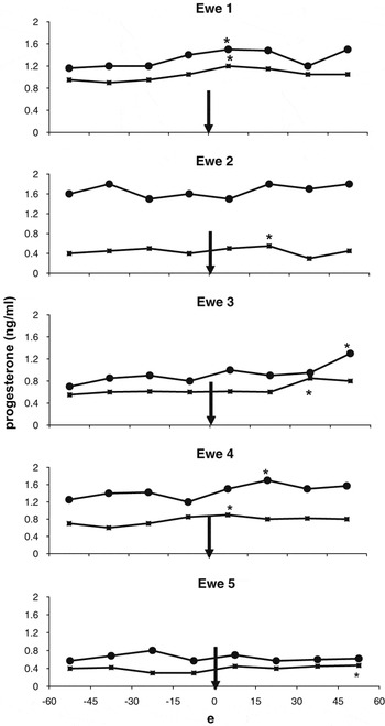
An acute luteotrophic effect of melatonin infusion during either anoestrus or the breeding season has been reported (Durotoye et al., Reference Durotoye, Webley and Rodway1997), leading to an increase in plasma progesterone concentrations. These results were confirmed in vivo in another experiment (Abecia et al., Reference Abecia, Forcada and Zuniga2002); during the luteal phase of a synchronized sexual cycle (7 and 15 days after pessary removal), five ewes were challenged with an intravenous melatonin injection (3 μg/kg BW0.75), and the response of the corpus luteum, in terms of plasma progesterone concentration after the challenge, was measured. All the ewes on day 7 and three of five on day 10 showed a progesterone response at some time during the hour following the melatonin challenge (Figure 1). The paired t-test revealed that melatonin increased the overall plasma progesterone concentrations, both on day 7 (pre, 0.61 ± 0.11; post, 0.73 ± 0.13 ng/ml; P < 0.01) and on day 10 (pre, 1.16 ± 0.19; post, 1.30 ± 0.20 ng/ml; P < 0.01). These results provided evidence for an effect of melatonin on progesterone secretion in vivo. Since the experimental procedures were performed at a very short term, this effect cannot be attributed to the endogenous rhythm of melatonin secretion.
Profiles of mean plasma progesterone response on day 7 (▪) and day 10 (●) after pessary withdrawal. The time of melatonin injection (3 μg/kg BW0.75) is indicated by arrow. *Indicates a progesterone response, defined as an increase at two points during the post-treatment period above the mean ± 2 s.d. of the values in the pre-treatment period (adapted from Abecia et al., Reference Abecia, Forcada and Zuniga2002).

Those acute luteotrophic effects of melatonin seem to be coincident with the results reported after medium-term treatments during anoestrus to advance the breeding season and to improve the ovulation rate. Wallace et al. (Reference Wallace, Robinson, Wigzell and Aitken1988) observed that melatonin administered daily improved progesterone concentrations in the latter half of the luteal phase. Although melatonin implants during the anoestrus associated with a male-effect management did not modify the secretion of luteinising hormone (LH) before or following the introduction of rams (Gómez Brunet et al., Reference Gómez Brunet, López Sebastian, Picazo, Cabellos and Goddard1995), results from our laboratory have reported that the pineal hormone induces a significant decrease of the proportion of ewes exhibiting a short cycle in response to the ram effect (Abecia et al., Reference Abecia, Palacin, Forcada and Valares2006). This short cycle is the consequence of a subnormal corpus luteum that has a short life span and regresses around day 7 after ovulation. However, no effect of melatonin implants on luteal function has been elucidated after superovulation during the seasonal anoestrous period (Forcada et al., Reference Forcada, Abecia, Cebrián-Perez, Muiño-Blanco, Valares, Palacín and Casao2006).
Effect of melatonin on the embryo
Fertilization rate
Season has an effect on fertilization rate, and some authors have reported a higher number of fertilized oocytes recovered from superovulated ewes in the breeding season than during the anoestrus (Mitchell et al., Reference Mitchell, Dingwall, Mylne, Hunton, Matthews, Gebbie, McCallum and McEvoy2002; Gonzalez-Bulnes et al., Reference Gonzalez-Bulnes, Garcia-Garcia, Santiago-Moreno, Dominguez, Lopez-Sebastian and Cocero2003). However, the treatment with melatonin in anoestrus did not improve fertilization rate in superovulated ewes when compared with controls (McEvoy et al., Reference McEvoy, Robinson, Aitken and Robertson1998; Forcada et al., Reference Forcada, Abecia, Cebrián-Perez, Muiño-Blanco, Valares, Palacín and Casao2006). Likewise, Stenbak et al. (Reference Stenbak, Redmer, Berginski, Erickson, Navanukraw, Toutges, Bilski, Kirsch, Kraft, Reynolds and Grazul-Bilska2001) reported a higher in vitro fertilization (IVF) of oocytes recovered from superovulated ewes in the breeding season than during the anoestrus, whereas no differences in fertilization rate were elucidated after IVF of oocytes recovered from superovulated ewes treated or not with melatonin in anoestrus (Luther et al., Reference Luther, Redmer, Reynolds, Choi, Pant, Navanukraw, Arnold, Scheaffer, Borowicz, Kirsch, Weigl, Kraft and Grazul-Bilska2005).
It would be advisable to highlight that the ram is the other agent involved in the fertilization process. Although the effects of melatonin on semen production and quality are not fully known, it seems that the treatment with pineal hormone can improve sperm motility (Kaya et al., Reference Kaya, Baspinar, Yildiz, Kurtoglu, Ataman and Haliloglu2000) and even modify semen plasma characteristics (Garde López-Brea et al., Reference Garde López-Brea, Pérez-Guzmán, Pérez Garnelo, Garzón Sigler and Montoro Angulo1996).
Embryo survival and development
Some studies have suggested that there is no effect of season on the number of recovered embryos from superovulated ewes, particularly in Mediterranean genotypes (Lopez Sebastian et al., Reference Lopez Sebastian, Cognie, Cocero, Delafuente and Poulin1990; Gonzalez-Bulnes et al., Reference Gonzalez-Bulnes, Garcia-Garcia, Santiago-Moreno, Dominguez, Lopez-Sebastian and Cocero2003). However, embryos from superovulated ewes during the seasonal anoestrus seem to have a lower viability, as indicated by a higher percentage of retarded and degenerated embryos when compared with those recovered during the reproductive season (Mitchell et al., Reference Mitchell, Dingwall, Mylne, Hunton, Matthews, Gebbie, McCallum and McEvoy2002). Daily treatment with oral melatonin for 45 days to superovulated ewes in anoestrus does not seem to modify embryo production or survival (McEvoy et al., Reference McEvoy, Robinson, Aitken and Robertson1998); however, Forcada et al. (Reference Forcada, Abecia, Cebrián-Perez, Muiño-Blanco, Valares, Palacín and Casao2006) recently reported that melatonin implants tended to improve the survival of embryos collected from Rasa Aragonesa ewes after superovulation in anoestrus. More specifically, melatonin induced a significant reduction in the number and rate of non-viable (degenerate and retarded) embryos.
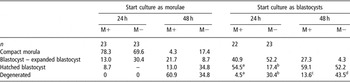
In order to determine whether melatonin could improve embryo development in vitro, 91 thawed sheep embryos (46 morulae and 45 blastocysts) were cultured with or without melatonin (Abecia et al., Reference Abecia, Forcada and Zuniga2002). If the embryos were blastocysts when the culture started, melatonin increased the percentage that had hatched after 24 h of culture (P < 0.01), and there was a lower percentage of degenerated embryos at the end of the incubation period (P < 0.05). However, melatonin seemed to exert no effect on the in vitro development of morulae, a similar percentage of embryos reaching the stage of blastocyst or hatched blastocyst when compared with controls after 24 or 48 h of culture (Table 1). As we were aware, this was the first report of a direct effect of melatonin on embryonic development, using an in vitro model. Further research in our laboratory has recently elucidated a lower percentage of degenerated embryos after an in vitro culture of fertilized ova associated with the presence of melatonin in the culture medium when compared with controls, cultured in the absence of melatonin after fertilization (Casao et al., unpublished results).
Percentage of embryos in the different stages of development when incubated with (M+) or without (M−) melatonin (1 μg/ml) in the culture medium (adapted from Abecia et al., Reference Abecia, Forcada and Zuniga2002)

a,bP < 0.01.
c,dP < 0.05.
It remains to determine whether melatonin is able to exert effects at the uterine level, thus modifying embryo environment in vivo. Some preliminary data from our laboratory (Sosa, unpublished results) suggest that the uterine sensitivity to progesterone – in terms of progesterone receptor expresssion – of superovulated ewes could be reduced by melatonin treatment. We have recently demonstrated that melatonin-treated ewes during anoestrus had higher plasma concentrations of progesterone during the male effect-induced luteal phase than untreated ewes (Abecia et al., Reference Abecia, Palacin, Forcada and Valares2006), and this could have been reflected in a stronger down-regulation of progesterone receptor (PR) in the melatonin group. Taking into account the results of that preliminary trial, and the fact that steroid-modulated melatonin receptors have been found in rat uterus (Zhao et al., Reference Zhao, Poon and Pang2000 and Reference Zhao, Pang and Poon2002), a role of melatonin in the endometrial physiology could be envisioned, although there is still a wide field to explore.
Effect of melatonin on maternal pregnancy recognition mechanisms
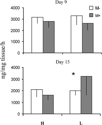
Abecia et al. (Reference Abecia, Forcada and Lozano1999), in a study involving the effect of both dietary energy and melatonin on the in vitro production of PGF2α by endometrial tissue on days 9 and 15 of pregnancy, observed that melatonin affected PGF2α secretion in vitro only in undernourished ewes on day 15 of pregnancy, when the signals of maternal recognition of pregnancy are being established. Neither the plane of nutrition nor the inclusion of melatonin in the culture medium modified progesterone production on day 9 of pregnancy. On day 15 of pregnancy, the corpora lutea of undernourished ewes tended to secrete more progesterone in the presence of melatonin than in its absence (Figure 2). These ewes had a lower pregnancy rate and higher secretion of PGF2α, and this apparent reduction in endometrial luteolytic hormone suggested that melatonin treatment might improve embryonic survival in undernourished ewes through a reduction of the luteolytic signals.
Mean (±s.e.) progesterone secretion in vitro (ng/mg tissue per h) by endometrial tissue, with (M+) or without (M−) melatonin in the culture medium (10 ng/ml), on days 9 and 15 of pregnancy, in ewes fed 1.5 × (H) and 0.5 × (L) maintenance requirements (adapted from Abecia et al., Reference Abecia, Forcada and Lozano1999).

Regarding an indirect effect of melatonin on embryo development Cagnacci et al. (Reference Cagnacci, Kräuchi, WirzJustice and Volpe1997), in a study on the effect of melatonin on the core body temperature in humans, postulated that this relationship is modified in the luteal menstrual phase; this modification may represent a mechanism for keeping the core body temperature higher at night to promote better embryonic implantation and survival. Moreover, McElhinny et al. (Reference McElhinny, Davis and Warner1996) concluded that although melatonin is administered to treat a range of conditions in a mouse strain, it would not adversely affect the embryo.
Overall, results of these reviewed experiments seem to indicate a direct effect of melatonin on the ovine ovary, the embryo and some of the mechanisms involved in the establishment of pregnancy. Although the improvement of ovulation rate seems to be related to its seasonal pattern, an acute, non-seasonal direct effect of melatonin on luteal functionality and embryo development has been demonstrated. Thus, it can be concluded that the success of exogenous melatonin as a means to improve lamb production of sheep is due, at least in part, to an improvement of luteal support and embryonic survival.
Acknowledgements
Some of the results presented have been derived from grants AGL2004-00432 from CICYT and A26 and PIP164/2005 from DGA (Spain).