Introduction
Microtubule-severing proteins (MTSPs) generate internal breaks in microtubules (MTs). MTSPs are conserved in eukaryotes from ciliates to mammals, and their function is important in diverse cellular processes ranging from cilia biogenesis to cell division, phototropism, and neurogenesis. MTSPs are categorized into the ‘meiotic’ subfamily of ATPases associated with the diverse cellular activity (AAA) superfamily members (Vale, Reference Vale2000; Monroe and Hill, Reference Monroe and Hill2016). All MTSPs have an AAA domain that can bind ATP, which is hydrolyzed, to sever a MT along its length (Vale, Reference Vale2000; Roll-Mecak and Vale, Reference Roll-Mecak and Vale2008; Johjima et al., Reference Johjima, Noi, Nishikori, Ogi, Esaki and Ogura2015; Monroe and Hill, Reference Monroe and Hill2016). Therefore, MT severing is an alternative and distinct form of MTs reorganization from end-limited MT depolymerization by kinesins (Wordeman, Reference Wordeman2005; Howard and Hyman, Reference Howard and Hyman2007; Roll-Mecak and McNally, Reference Roll-Mecak and McNally2010; Friel and Howard, Reference Friel and Howard2011; Tanenbaum et al., Reference Tanenbaum, Macurek, van der Vaart, Galli, Akhmanova and Medema2011; Sharp and Ross, Reference Sharp and Ross2012).
Abnormal chromosome segregation during mitosis can lead to aneuploidy, which is one of the main causes of tumorigenesis (Meraldi et al., Reference Meraldi, Honda and Nigg2004; Lee, Reference Lee2014). Many studies have shown that MTSPs play important roles in mitosis through their MT-severing activity. Katanin, the first discovered MTSP, targets the centrosome and promotes the aggregation of γ-tubulin on the spindle, therefore promoting the formation of the microtubule organizing centre (MTOC) (Hartman et al., Reference Hartman, Mahr, McNally, Okawa, Iwamatsu, Thomas, Cheesman, Heuser, Vale and McNally1998; Buster et al., Reference Buster, McNally and McNally2002). The second identified MTSP member, spastin, was found to be the most common variant to cause autosomal dominant hereditary spastic paraplegia (Hazan et al., Reference Hazan, Fonknechten, Mavel, Paternotte, Samson, Artiguenave, Davoine, Cruaud, Dürr, Wincker, Brottier, Cattolico, Barbe, Burgunder, Prud’homme, Brice, Fontaine, Heilig and Weissenbach1999). Spastin was later shown to be localized to the centrosome during mitosis and to play an important role in centrosome-based mitosis (Errico et al., Reference Errico, Claudiani, D’Addio and Rugarli2004; Zhang et al., Reference Zhang, Rogers, Buster and Sharp2007). Fidgetin (Zhang et al., Reference Zhang, Rogers, Buster and Sharp2007; Mukherjee et al., Reference Mukherjee, Diaz Valencia, Stewman, Metz, Monnier, Rath, Asenjo, Charafeddine, Sosa, Ross, Ma and Sharp2012), fidgetin-like 1 (Luke-Glaser et al., Reference Luke-Glaser, Pintard, Tyers and Peter2007), katanin-like 1 (Sonbuchner et al., Reference Sonbuchner, Rath and Sharp2010) and katanin-like 2 (Ververis et al., Reference Ververis, Christodoulou, Christoforou, Kamilari, Lederer and Santama2016) are also considered essential for mitosis. They show different localization patterns in cells, and the absence of these proteins affects the assembly of spindle microtubules to varying degrees.
Compared with that of mitosis, the process of meiosis is unique. Meiosis consists of one DNA replication and two rounds of cell division, forming haploid gametes. There is no typical centrosome in oocytes or spermatocytes, but some centrosome components have been found at the two poles of the spindle. There may be different regulatory mechanisms in meiosis and mitosis. Normal meiosis is essential for genomic stability, and the main cause of human genetic diseases and early abortion is aneuploidy caused by dysfunction of oocyte division (Kogo et al., Reference Kogo, Kowa-Sugiyama, Yamada, Bolor, Tsutsumi, Ohye, Inagaki, Taniguchi, Toda and Kurahashi2010; Filges et al., Reference Filges, Manokhina, Peñaherrera, McFadden, Louie, Nosova, Friedman and Robinson2015; Feng et al., Reference Feng, Sang, Kuang, Sun, Yan, Zhang, Shi, Tian, Luchniak, Fukuda, Li, Yu, Chen, Xu, Guo, Qu, Wang, Sun, Liu, Shi, Wang, Feng, Shao, Chai, Li, Xing, Zhang, Nogales, Jin, He, Gupta, Cowan and Wang2016). In Caenorhabditis elegans oocytes, katanin, which is composed of MEI-1 and MEI-2 subunits, has been shown to promote spindle assembly through microtubule-severing activities (Clark-Maguire and Mains, Reference Clark-Maguire and Mains1994; Srayko et al., Reference Srayko, Buster, Bazirgan and Mains2000, Reference Srayko, O’Toole, Hyman and Müller-Reichert2006; McNally et al., Reference McNally, Berg, Cortes and McNally2014; Beard et al., Reference Beard, Smit, Chan and Mains2016; Joly et al., Reference Joly, Martino, Gigant and Pintard2016). Loss of katanin in C. elegans also results in a complete loss of spindle bipolarity and a complete failure to organize chromosomes into a metaphase plate and a complete failure to segregate chromosomes, with severely abnormal polar body extrusion (Clark-Maguire and Mains, Reference Clark-Maguire and Mains1994; Srayko et al., Reference Srayko, Buster, Bazirgan and Mains2000, Reference Srayko, O’Toole, Hyman and Müller-Reichert2006; McNally et al., Reference McNally, Berg, Cortes and McNally2014; Beard et al., Reference Beard, Smit, Chan and Mains2016; Joly et al., Reference Joly, Martino, Gigant and Pintard2016). The role of MTSPs in meiosis in female mammals has been rarely reported, and mammalian meiosis is usually used as a model of human meiosis.
As a member of the microtubule-severing protein family, spastin also plays important roles in cell division and neurite growth, which are closely related to MTs (Ahmad et al., Reference Ahmad, Yu, McNally and Baas1999; Karabay et al., Reference Karabay, Yu, Solowska, Baird and Baas2004). Spastin gene mutations were found to be the most common cause of autosomal dominant hereditary spastic paraplegia (HSP) (Hazan et al.,Reference Hazan, Fonknechten, Mavel, Paternotte, Samson, Artiguenave, Davoine, Cruaud, Dürr, Wincker, Brottier, Cattolico, Barbe, Burgunder, Prud’homme, Brice, Fontaine, Heilig and Weissenbach1999). In the axons of nerve cells, spastin tends to sever long microtubules into short segments, releasing additional aggregated terminals. This severing activity makes the microtubules more vigorous, promotes the formation of neurite branches, and promotes the growth and regeneration of neurites. Spastin overexpression significantly increased the formation of neurite branches, while spastin depletion resulted in a significant decrease in the formation of axonal collateral branches but did not significantly reduce the length of the axons (Qiang et al., Reference Qiang, Yu, Andreadis, Luo and Baas2006; Yu, et al., Reference Yu, Qiang, Solowska, Karabay, Korulu and Baas2008). The results showed that the expression of spastin in neurons was regulated by the transcription factor ELK1. ELK1 binds to the key promoter regions of katanin-60 and spastin, and methylation in this region can lead to a decrease in ELK1 binding and an increase in the transcription of the corresponding genes (Kelle et al., Reference Kelle, Kırımtay, Selçuk and Karabay2019). In addition, spastin has been shown to interact with collapsin response mediator protein 5 (CRMP5), which provides structural support and promotes microtubule aggregation, which forms transportation routes. Spastin interacts with CRMP5 to promote the outward growth of neurites by controlling microtubule dynamics (Ji et al., Reference Ji, Zhang, Chen and Guo2018). The microtubule-binding activity-related site has been located in the 59th amino acid residue of spastin (N-terminal region in the AAA domain), which is known as the microtubule-binding domain (MTBD) (Eckert et al., Reference Eckert, Le, Link, Friedmann and Woehlke2012). The presence of CRMP5 at this spastin region can enhance the microtubule-severing activity of spastin. During somatic mitosis, spastin is concentrated at the centrosome of cells. This localization is regulated by the centrosome protein NA14 (Zhang et al., Reference Zhang, Rogers, Buster and Sharp2007). Spastin can add free ends at the centrosome through its microtubule-severing activities, which promotes the depolymerization of microtubules at spindle poles, enabling chromosomes to separate from each other (Errico et al., Reference Errico, Claudiani, D’Addio and Rugarli2004; Zhang et al., Reference Zhang, Rogers, Buster and Sharp2007).
However, spastin has never been studied during the meiosis of mammalian germ cells. In this study, we focus on the distribution and function of spastin in mouse oocytes to determine if they are consistent with those of somatic cells. Does the mechanism of spastin protein expression and spastin function regulation coincide with that suggested by previous studies?
Materials and methods
General chemicals, reagents cells and animals
Unless otherwise specified, chemicals and reagents were purchased from Sigma-Aldrich; Merck KGaA. The NIH3T3 cell line was purchased from the American Type Culture Collection (CRL-1658, cat. no. C2006). In total, 265 3-week-old female specific pathogen-free ICR mice (weighing 18–20 g) used in this study were obtained from Vital River Experimental Animal Technical Co., Ltd. Animals were housed at a temperature of 20–26°C and a humidity of 40–70% with a 12 h light/dark cycle. The mice were fed in feeding boxes, and the frequency of food replacement was two times a week, and the frequency of water bottle replacement was three times a week. All animal experiments were approved by the Animal Protection and Utilization Committee of Nanjing Medical University (Nanjing, China) and conducted in accordance with the institutional guidelines.
Antibodies
Mouse monoclonal anti-β-actin (cat. no. A5316-100) antibody was obtained from Sigma-Aldrich; Merck KGaA. Mouse monoclonal anti-SPASTIN (A-4) (cat. no. sc-398264) and mouse monoclonal anti-β-tubulin (cat. no. sc-5274) antibodies were purchased from Santa Cruz Biotechnology, Inc. Human anti-centromere CREST antibody (cat. no. 15-234) was purchased from Antibodies Incorporated. Rabbit polyclonal anti-β-actin (cat. no. 30102ES40) was purchased from Yeasen Biotech Co., Ltd. Rabbit polyclonal anti-CRMP5 (cat. no. ab36203) was purchased from Abcam. Cy2-conjugated donkey anti-mouse IgG (code no. 715-225-150), rhodamine (TRITC)-conjugated donkey anti-goat IgG (code no. 705-025-147), and Alexa Fluor 647-conjugated donkey anti-human IgG (code no. 709-605-149) were purchased from Jackson ImmunoResearch Laboratories, Inc. Horseradish peroxidase (HRP)-conjugated rabbit anti-goat IgG (cat. no. 31402) and HRP-conjugated goat anti-mouse IgG (cat. no. 31430) were purchased from Invitrogen; Thermo Fisher Scientific, Inc.
Oocyte collection and culture
Immature oocytes at the germinal vesicle (GV) stage were obtained from the ovaries of 3-week-old female ICR mice. The mice were euthanized with carbon dioxide. The ovaries were removed and placed in HEPES containing 2.5 nm milrinone and 10% FBS (GIBCO, Thermo Fisher Scientific, Inc.). The follicles were punctured with injection acupuncture and oocytes were washed out from the cumulus–oocyte complexes. Next, 50 separated shelled oocytes were placed in 100-µl culture medium drops, under the mineral oil in plastic containers (BD Biosciences). The medium was MEM+ [containing 0.01 mm EDTA, 0.23 mm Na-pyruvate, 0.2 mm penicillin/streptomycin, 3 mg/ml bovine serum albumin (BSA) and 20% FBS]. The oocytes were cultured at 37.0°C, 5% O2 and 5% CO2 in air. Before in vitro maturation (IVM), 2.5 nM milrinone was added to all media to prevent meiosis recovery.
siRNA production and microinjection
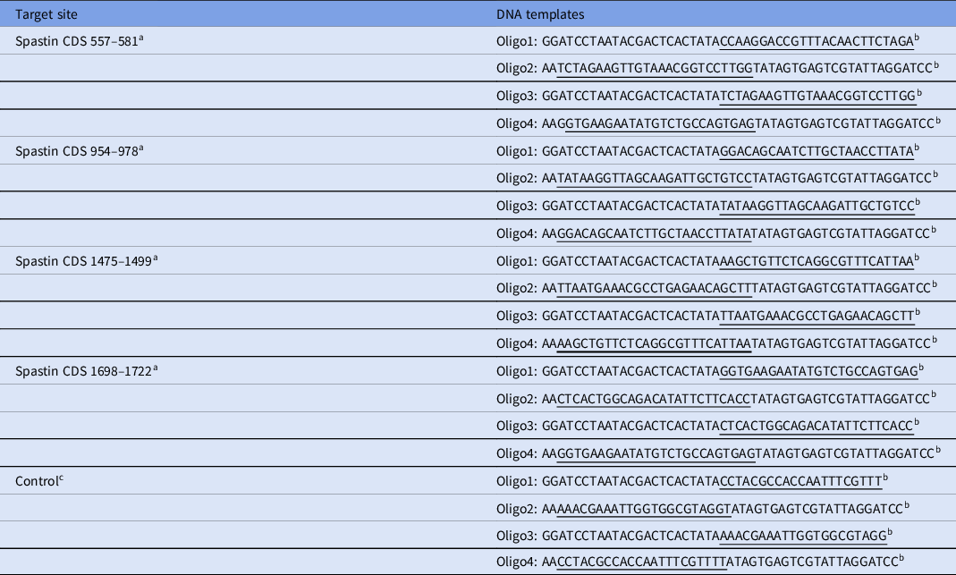
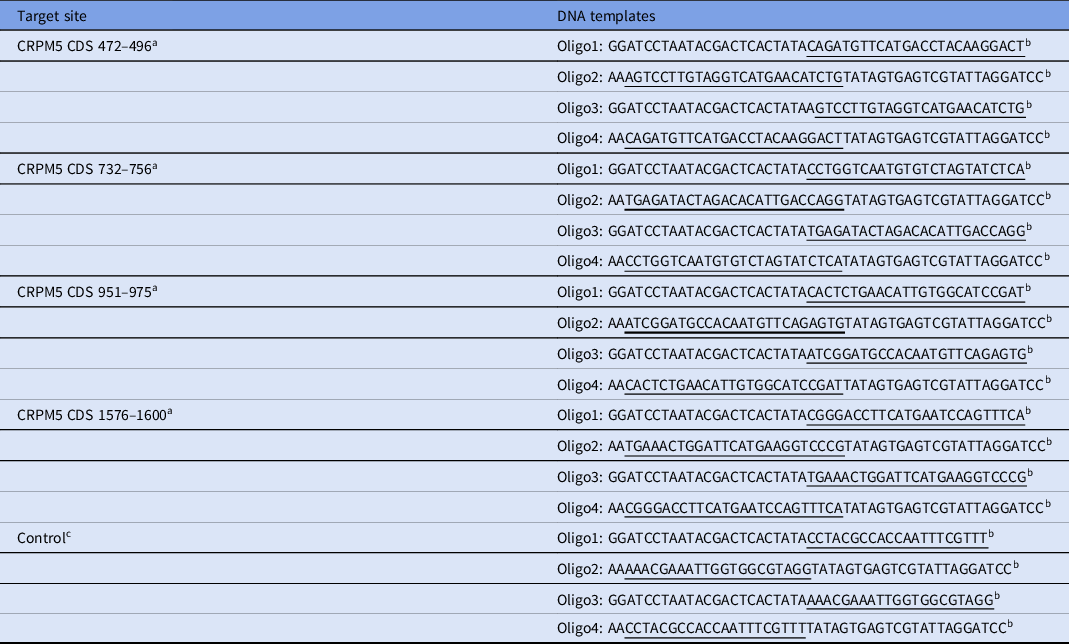
Table 1 and Table 2 list the sequences of all DNA templates used to produce siRNA for spastin and CRMP5, respectively. The sequence of the control template is a simulated sequence and does not specifically bind to any mRNA in the mouse genome. By blocking the DNA template of four different DNA coding regions (amino acid sequence coding in protein, CDS) of spastin and CRMP5 mRNA, the template was designed online through BLOCK-iT™ RNAi Designer (http://rnaidesigner.invitrogen.com/rnaiexpress/) with some modifications. Sequence specificity was verified by BLAST (http://blast.ncbi.nlm.nih.gov/Blast.cgi) homology search.
DNA oligos for siRNA production

a The numbers are the starting and ending position of the target sites in spastin CDS (NM_001162870.1 in NCBI).
b Two pairs of DNA oligos are needed for each double-stand siRNA. Oligo2 is complementary to Oligo1 except for an ‘AA’ overhang at the 5′ end; Oligo3 is complementary with Oligo4 except for an ‘AA’ overhang at the 5′ end. In each oligo, gene-specific sequences are underlined, other sequences are for recognition and binding by T7 RNA polymerase.
c Control siRNA does not target any mRNA sequence in mouse.
DNA oligos for siRNA production

a The numbers are the starting and ending position of the target sites in CRMP5 CDS (NM_001356948.1 in NCBI).
b Two pairs of DNA oligos are needed for each double-stand siRNA. Oligo2 is complementary to Oligo1 except for an ‘AA’ overhang at the 5′ end; Oligo3 is complementary to Oligo4 except for an ‘AA’ overhang at the 5′ end. In each oligo, gene-specific sequences are underlined, other sequences are for recognition and binding by T7 RNA polymerase.
c Control siRNA does not target any mRNA sequence in mouse.
siRNAs were made using the T7 RiboMAX™ Express RNAi System (Promega Corporation), according to the manufacturer’s instructions. In short, for a double-stranded siRNA in four spastin and CRMP5 CDS regions, two pairs of synthesized complementary single-stranded DNA oligonucleotides were first annealed to form two double-stranded DNA templates. Then, two complementary single-stranded siRNA were synthesized according to the two templates and annealed to form the final double-stranded siRNA. Next, the siRNA was purified by conventional phenol/chloroform/isopropanol precipitation, and then checked on the agarose gel. The siRNA was then calibrated and stored at −80°C. A ready-to-use siRNA mixture was prepared by mixing siRNA with four targets at an equal molar ratio of 5 μm to the final concentration.
Microinjection of siRNA into the cytoplasm of fully grown immature oocytes was used to knock down spastin and CRMP5. After injections, oocytes were arrested at the GV stage with 2.5 μM milrinone for 20 h, and then were cultured in milrinone-free M2 medium for maturation.
Immunofluorescence
The oocytes were washed in PBS with 0.05% polyvinylpyrrolidone (PVP) for a short time, then infiltrated in 0.5% Triton X-100/PHEM (60 mM PIPES, 25 mM HEPES pH 6.9, 10 mM EGTA, 8 mM MgSO4) for 5 min, and washed in PBS/PVP three times. Then, the oocytes were fixed in 3.7% PFA/PHEM for 20 min at room temperature, washed in PBS/PVP three times (10 min each time), and blocking buffer (1% BSA/PHEM with 100 mM glycine) was used at room temperature. The oocytes were incubated overnight at 4°C with a primary antibody diluted in blocking buffer, washed three times (10 min each time) in PBS with 0.05% Tween-20 (PBST), incubated in closed buffer for 45 min with a secondary antibody diluted in blocking buffer (1:750 in all cases) and washed three times (10 min each time) in PBST. Finally, the oocytes were stained with 10 μg/ml Hoechst 33258 (Sigma-Aldrich; Merck KGaA) at room temperature for 10 min. Then the oocytes were installed on a slide with mounting medium (0.5% propyl gallate, 0.1 M Tris–HCl, pH 7.4, 88% glycerol) and covered with a layer of glass (thickness 0.13–0.17 μm). To maintain the dimension of oocytes, two double-sticky bands (90-μm thick) were placed between the slide and the cover slide. The main antibodies were diluted as follows: anti-spastin (cat. no. sc-398264, Santa Cruz Biotechnology, Inc.), 1:200; anti-tubulin (cat. no. sc-5274, Santa Cruz Biotechnology, Inc.), 1:500; anti-human centromere CREST, (cat. no. 15-234, Antibodies Incorporated), 1:500. Oocytes were examined using an Andor Revolution spinning disc confocal workstation (Oxford Instruments).
Western blotting
In total, 100 oocytes were dissolved in Laemmli sample buffer (Bio-Rad Laboratories, Inc.) containing protease inhibitors and boiled for 5 min before being subjected to 10% SDS-PAGE. The separated protein was transferred to a PVDF membrane, then blocked with TBST (TBS containing 0.05% Tween-20) containing 5% nonfat dried milk for 1 h at room temperature. Then the PVDF membrane was separated and incubated with the following main antibodies at 4°C overnight: mouse monoclonal anti-β-actin (cat. no. A5316-100) was diluted with a blocking buffer (TBS containing 0.05% Tween-20) at a ratio of 1:1000; mouse monoclonal anti-SPASTIN (A-4) (cat. no. sc-398264) was diluted with a blocking buffer at a ratio of 1:500; rabbit polyclonal anti-CRMP5 (cat. no. ab36203) was diluted with a blocking buffer at a ratio of 1:2000. After washing in TBST, the membranes were incubated with HRP-conjugated rabbit anti-goat IgG or HRP-conjugated goat anti-mouse IgG (diluted with a blocking buffer at 1:1000) for 1 h at room temperature and then treated with ECL Plus Western Blotting Detection System (Vazyme). In the experiment, ImageJ software v.1.8.0 was used as data analysis software.
Image area measurement and fluorescence intensity
A series of images taken under the confocal laser microscope along the Z axis was imported into ImageJ software and the images were superimposed by the Z stacks function of the software. The icon marked with ‘line chart’ in ImageJ software was selected to enclose the spindle outline with a closed line chart. The average fluorescence intensity in the line graph could be obtained by using the shortcut key ‘control + m’. The same method could be used to measure the average fluorescence intensity of the background around the spindle. The former subtracts the latter, which is the average fluorescence intensity of spindle microtubules. The measurements were repeated three times and the average value taken.
Data analysis and statistics
All experiments were repeated at least three times. Measurements on confocal images were performed using ImageJ software (National Institutes of Health). Results were expressed as mean ± standard error of the mean (SEM). Multigroup comparisons of the means were carried out by one-way analysis of variance (ANOVA) test with post hoc contrasts using Student–Newman–Keuls test. P < 0.05 was considered to indicate a significant difference.
Results
Spastin is distributed in the spindle microtubules of metaphase I (MI) and MII oocytes
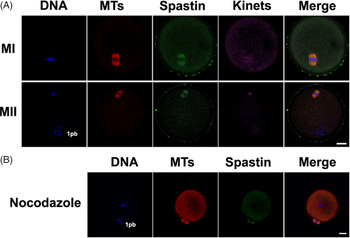
First, we determined the distribution of spastin in the oocytes by immunofluorescence. In the MI and MII stage oocytes, spastin aggregated on the spindle and colocalized with microtubules (Fig. 1A). When the depolymerized microtubules were treated with nocodazole, the spastin disappeared (Fig. 1B), which indicated that the location of spastin depended on the microtubules, and it was not difficult to infer that its role was closely related to spindle microtubules.
Spastin is located at the spindle microtubules of MI and MII oocytes. (A) Immunofluorescence revealed that spastin was located at the spindle microtubule of the MI and MII mouse oocytes. (B) With nocodazole-treated oocytes depolymerized microtubules, the immunofluorescence indicating the location of spastin disappeared with the disappearance of the microtubules. 100 oocytes were used in each group. Three independent experiments were performed for each result. Kinets refers to the kinetochore. 1pb refers to the first polar body of MII. Tubulin is displayed in red, Spastin is shown in green, kinetochores are in magenta, and DNA is in blue. Scale bars, 20 μm.

Absence of spastin results in a maturation disorder and abnormal fertilization of the oocytes
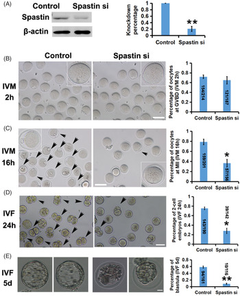
To explore the function of spastin in MI and MII oocytes, we designed specific siRNAs and injected them into developmentally inhibited GV oocytes to knock down spastin. Western blot analysis showed that approximately 80% of the spastin was deleted (Fig. 2A). IVM experiments showed that oocytes lacking spastin showed obvious developmental disorders not shown by the control group. When IVM was assessed for 2 h, the proportion of ruptured germinal vesicle (germinal vesicle breakdown, GVBD) in the knockdown group was basically the same as that in the control group (Fig. 2B), but when IVM was assessed for 16 h, the elimination rate of the first polar body in the spastin-depleted group was significantly lower than that in the control group (Fig. 2C). The oocytes with a first polar body were fertilized in vitro with the sperm of normal male mice, and the early embryos were cultured in vitro. The two-cell (Fig. 2D) and blastocyst development rates (Fig. 2E) in the spastin-depleted group showed a significant decline. This finding indicated that the absence of spastin leads to the obstruction of oocyte maturation, and the fertilization ability and early embryo development among the so-called matured oocytes, with a first polar body, were also significantly reduced without spastin.
Knocking down spastin leads to severe maturation and fertilization abnormalities in oocytes. (A) Western blot and related statistical analysis showed that the expression of spastin decreased by c. 80% under the specific siRNA. β-actin was used as a control. 100 oocytes were used in each group. (B) In vitro development of oocytes and related statistical analysis showed that there was no significant change in the ratio of GVBD of oocytes after 2 h of in vitro development. (C) In vitro development of oocytes and related statistical analysis showed that the ratio of 1 pb (first polar body) oocytes (indicated by black arrow) decreased significantly after 16 h of in vitro development. (D, E) In vitro fertilization (IVF) of oocytes and related statistical analysis showed that the proportion of 2-cell embryos (D, indicated by black arrow) formed after the specific siRNA knockdown of spastin and 24 h of fertilization and development of normal male sperm was significantly reduced, the proportion of blastocysts formed after 5 days of in vitro development was also significantly reduced. Scale bar, 100 μm. *P < 0.05 and **P < 0.01 indicate significant differences. The number of oocytes or early embryos analyzed has been added to the statistical chart.

Absence of spastin resulted in a significant decrease in the number of spindle microtubules in the MI and MII oocytes
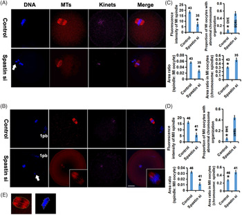
To determine how spastin affects oocyte development, we developed spastin-deletion oocytes into MI and MII stages in vitro. We found that the fluorescence intensity of spindle microtubule staining in the spastin-deficient MI (Fig. 3A) and MII (Fig. 3B) oocytes was significantly lower than that in the control group, indicating that microtubule density decreased significantly at this time. The results of immunofluorescence imaging showed that the ratio of spindle area to cytoplasmic area of the oocytes lacking spastin was significantly smaller than that of the control group, while the ratio of chromosome area to spindle area was significantly increased (Fig. 3C, D), indicating that deletion of spastin also led to a reduction in spindle size. In addition, it was found that deleting spastin in both the MI (Fig. 3A) and MII (Fig. 3B) oocytes led to abnormal chromosome organization (Fig. 3, arrows). This finding suggests that the abnormal structure of the spindle caused by the lack of spastin affects the function of the spindle. Chromosome abnormalities during meiosis may eventually affect the number of chromosomes in daughter cells, which is fatal to cells. The decrease in the number of spindle microtubules inevitably affects the structure and function of the spindle. It is easy to explain the meiosis and maturation obstacles in these oocytes.
Knocking down spastin causes a significant decrease in the number and volume of spindle microtubules in MI and MII oocytes. (A, B) In MI stage (A) and MII stage (B) oocytes, when spastin was knocked down by specific siRNA, the fluorescence intensity of the spindle microtubule staining was significantly lower than that in the control group. In addition, in both the MI (A, arrow) and MII (B, arrow) oocytes, the numbers of oocytes with abnormal chromosome organization were found to increase in the spastin-knockdown oocytes. Kinets refers to the kinetochore. 1pb refers to the first polar body of MII. Tubulin is displayed in red, kinetochores are shown in magenta, and DNA is in blue. Scale bar, 20 μm. (C, D) Measurement and statistics of the fluorescence intensity of the spindles, proportion of oocytes with abnormal chromosome organization, the spindle:oocyte area ratio and the chromosome:spindle area ratio are shown in (C) (MI) and D (MII). The number of oocytes has been marked in the statistical chart. (E) The method used to measure the spindle and chromosome area is shown in (E). *P < 0.05 indicates significant differences.

Exogenous spastin can rescue the oocyte dysplasia caused by CRMP5 deletion
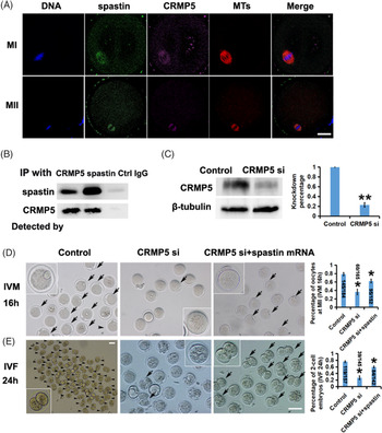
In neurons, CRMP5 can bind to spastin at specific sites to promote microtubule-severing activities. We know through experiments that this effect also exists in meiotic oocytes. First, we demonstrated by immunofluorescence that CRMP5 was distributed in spindle microtubules in oocytes and that it colocalized with spastin (Fig. 4A). Co-IP experiments show that both spastin and CRMP5 co-precipitate and interact with each other (Fig. 4B). We knocked down CRMP5 in the oocytes with specific siRNA (the knockdown percentage is shown in Fig. 4C) and then performed an IVM test. The results showed that the elimination rate of the first polar body in the knockdown group was significantly lower than that in the control group after IVM monitoring for 16 h (Fig. 4D). In vitro fertilization and early embryo culture experiments showed that the rate of two-cell embryo formation from CRMP5-deficient oocytes fertilized with normal sperm was much lower than that of the control group (Fig. 4E). The deletion of CRMP5 in the oocytes has an effect similar to that of spastin knockdown. When we knocked down CRMP5 with specific siRNA and then injected transcribed spastin mRNA in vitro into the oocytes, the rate of oocyte maturation (Fig. 4D) and two-cell embryo formation (Fig. 4E) induced by in vitro fertilization increased. Exogenous spastin can rescue oocyte dysplasia caused by CRMP5 deletion. To detect the validity of exogenous enhanced green fluorescent protein (eGFP)–spastin mRNA, exogenous eGFP–spastin mRNA was injected into CRMP5-knockdown oocytes and then immunofluorescence experiments were carried out. As shown in Fig. S1, CRMP5 was almost colourless while the green fluorescence of EGFP showed the position of the spindle, which was consistent with the location of spastin.
Exogenous spastin can rescue oocyte maturity disorders and abnormal fertilization ability caused by CRMP5 deletion. (A) Immunofluorescence showing that CRMP5 was distributed in spindle microtubules in the oocytes and colocalized with spastin. (B) Co-IP results showed that CRMP5 antibody could precipitate CRMP5 and spastin. Spastin antibody could precipitate spastin and CRMP5. Control IgG could not precipitate CRMP5 or spastin. 300 oocytes were used in each group. (C) Western blot and related statistical analysis showed that the expression of CRMP5 decreased by c. 80% under the specific siRNA. β-Actin was used as a control. 100 oocytes were used in each group. (D) After 16 h of IVM, there was a significant difference in the percentage of 1 pb (first polar body) oocytes (indicated by black arrow) in the CRMP5-knockdown and control groups. The ‘CRMP5 siRNA+spastin mRNA’ group showed partial recovery of the percentage of 1 pb oocytes (indicated by black arrows). (E) IVF revealed that spastin depletion induced a significant decrease in the two-cell embryo (indicated by black arrows) rate. The ‘CRMP5 siRNA + spastin mRNA’ group showed partial recovery of the first two-cell embryo rate. Scale bars, 100 μm. The number of oocytes or early embryos analyzed has been added to the statistical chart. *P < 0.05 and **P < 0.01 indicates significant differences.

Exogenous spastin can reverse the decrease in the spindle density of oocytes caused by CRMP5 deletion
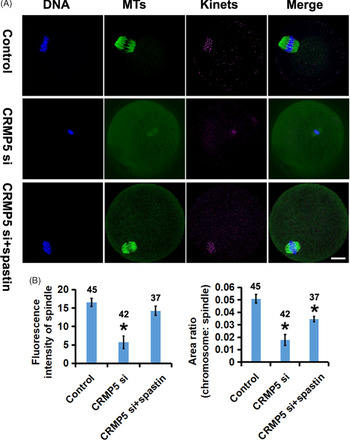
In these experiments, we found that the deletion of CRMP5 can lead to abnormal oocyte development and that it is related to spastin. We wondered whether CRMP5 also affected spindle assembly. Our next experiments proved that it affected spindle assembly. CRMP5-knockdown oocytes were allowed to develop to the MI and MII stages in vitro, and the fluorescence intensity of the spindle microtubule staining was significantly reduced (Fig. 5A and Fig. 5B left). When the spastin mRNA was microinjected into the oocyte to scavenge CRMP5, the fluorescence intensity of the spindle microtubules was significantly restored (Fig. 5A and Fig. 5B left). Similar to the phenotype of spastin knockdown, the lack of CRMP5 in oocytes also showed a significant decrease in the ratio of spindle area to cytoplasmic area (Fig. 5A and Fig. 5B right). When exogenous spastin mRNA was added to the culture, the ratio increased to an extent, and the difference was significant compared with the change in the control group (Fig. 5A and Fig. 5B right). It was observed that additional spastin mRNA can reverse the decrease in spindle MT density and spindle volume of the oocytes that had been caused by CRMP5 deletion in whole or in part. The role of spastin in promoting spindle assembly and cell maturation during oocyte development may be influenced and regulated by CRMP5.
Exogenous spastin can reverse the decrease in the spindle density and volume of the oocytes caused by CRMP5 deletion. (A) The fluorescence intensity of the spindle microtubule staining and spindle area in the CRMP5-deficient oocytes was significantly lower than that in the control group. The ‘CRMP5 siRNA + spastin mRNA’ group showed partial recovery of the above two indicators. (B) Measurement and statistics of the fluorescence intensity of the spindle and area ratio (chromosome:spindle). The number of oocytes analyzed has been added to the statistical chart. Scale bar, 20 μm. *P < 0.05 indicates significant differences.

Discussion
The process of oocyte development and maturation is essentially the process of oocyte meiosis. Oocytes only undergo accurate meiosis to achieve homologous chromosome recombination and separation and transmit complete genetic information to offspring accurately and steadily to successfully complete fertilization and support embryo development, eventually producing healthy offspring. If an abnormal meiosis process results in an obstacle to oocyte maturation or aneuploidy due to the uneven distribution of genetic material, hereditary diseases, such as Down’s syndrome, may manifest in the offspring (Nogales and Wang, Reference Nogales and Wang2006; Gardner et al., Reference Gardner, Zanic and Howard2013; Brouhard, Reference Brouhard2015). Early embryo development disorders caused by various reasons can cause embryo development to stagnate and eventually abort. Studies have shown that abnormal oocyte development is the main cause of early embryonic sterility (Han et al., Reference Han, Wen, Ju, Chen, Ke, Zhou, Li, Xia, Tang and Liang2017). The spindle composed of microtubules is a very important structure for meiosis. The key to the whole meiosis process is the correct separation of homologous chromosomes under the traction of the spindle microtubules. This process requires the precise regulation of many proteins.
As an important group of microtubule-associated proteins (MAPs), microtubule-severing protein (MTSP) plays an important role in regulating MT activity (Vale, Reference Vale2000; Frickey and Lupas, Reference Frickey and Lupas2004). MTSP mainly utilizes the energy released by ATP hydrolysis to remove the protective caps of both the positive and negative poles of the MTs, therefore exposing the free MT ends to MT polymerase or depolymerase and enabling the dynamic changes of MT elongation or shortening (Buster et al., Reference Buster, McNally and McNally2002; Sharp and Ross, Reference Sharp and Ross2012). Another major function of MTSP is ‘nucleation’, in which a long MT is severed into several short segments, and the subsequent exposure of the free end of each MT segment can generate a new long MT through the polymerase, thereby promoting the MT density of some cell structures (Srayko et al., Reference Srayko, O’Toole, Hyman and Müller-Reichert2006). Notably, the mutation or deletion of MTSP can lead to severe hereditary diseases, neurological diseases, developmental disorders and low fertility in mice (Errico et al., Reference Errico, Ballabio and Rugarli2002; Mao et al., Reference Mao, Xiong, Xiong, Wang, Zhang and Jin2014; Mishra-Gorur et al., Reference Mishra-Gorur, Caglayan, Schaffer, Chabu, Henegariu and Vonhoff2014; Banks et al., Reference Banks, Lassi, Hoerder-Suabedissen, Tinarelli, Simon and Wilcox2018). Although the MTSP family shares a conservative AAA domain, the other regions of each member are completely different. Therefore, each MTSP has both synergistic and separate functional roles (Sharp and Ross, Reference Sharp and Ross2012).
In this study, it was found that, in mouse oocytes, the microtubule-severing protein spastin was located in the spindle microtubule, which differs from its localization in the centrosome in somatic cells. Spastin is located in the centrosome of somatic cells and plays a key role in the process of chromosome separation in the anaphase of mitosis by severing microtubules to change the dynamics of the microtubules. Does spindle microtubule-based spastin play a role in oocyte meiosis? The immunofluorescence results showed that the spindle microtubule density and spindle area of the oocytes without spastin was significantly decreased. This is similar to the results in C. elegans oocytes (Srayko et al., Reference Srayko, Buster, Bazirgan and Mains2000, Reference Srayko, O’Toole, Hyman and Müller-Reichert2006; Beard et al., Reference Beard, Smit, Chan and Mains2016; Joly et al., Reference Joly, Martino, Gigant and Pintard2016). Microtubule severing has been known for decades to be required for oocyte meiotic spindle assembly in C. elegans, with numerous publications documenting the requirements for the AAA ATPase katanin, another member of MTSP family, which in C. elegans is compose of the two subunits MEI-1 and MEI-2 (Clark-Maguire and Mains, Reference Clark-Maguire and Mains1994; Srayko et al., Reference Srayko, Buster, Bazirgan and Mains2000, Reference Srayko, O’Toole, Hyman and Müller-Reichert2006; McNally et al., Reference McNally, Berg, Cortes and McNally2014; Beard et al., Reference Beard, Smit, Chan and Mains2016; Joly et al., Reference Joly, Martino, Gigant and Pintard2016). The results showed that the mutation of MEI-1/MEI-2 in C. elegans resulted in a significant decrease in the microtubule level during meiosis (Srayko et al., Reference Srayko, Buster, Bazirgan and Mains2000, Reference Srayko, O’Toole, Hyman and Müller-Reichert2006; McNally et al., Reference McNally, Berg, Cortes and McNally2014; Joly et al., Reference Joly, Martino, Gigant and Pintard2016). Obviously, this is closely related to the ‘nucleation’ effect of MTSPs. After spastin or katanin is inhibited, the ‘nucleation’ effect is weakened, resulting in a significant decrease in the density of spindle microtubules and structural abnormalities. The abnormal structure of the spindle caused by the lack of MTSPs affected the function of the spindle.
Due to the lack of spastin, the structure of the spindle is abnormal, which affects the function of the spindle. Our study showed that a high proportion of abnormal chromosome organization appeared in spastin-deficient oocytes. Disordered chromosome arrangement during meiosis may eventually affect the number of chromosomes in daughter cells, which will greatly affect the ultimate fate of the oocyte. This is consistent with the study of C. elegans (Srayko et al., Reference Srayko, Buster, Bazirgan and Mains2000, Reference Srayko, O’Toole, Hyman and Müller-Reichert2006; McNally et al., Reference McNally, Berg, Cortes and McNally2014; Joly et al., Reference Joly, Martino, Gigant and Pintard2016). Loss of katanin in C. elegans results in a complete failure to organize chromosomes into a metaphase plate and a complete failure to segregate chromosomes (Srayko et al., Reference Srayko, Buster, Bazirgan and Mains2000, Reference Srayko, O’Toole, Hyman and Müller-Reichert2006; McNally et al., Reference McNally, Berg, Cortes and McNally2014; Joly et al., Reference Joly, Martino, Gigant and Pintard2016). Whether the abnormal organization of chromosomes in this study reflects a defect in anaphase or an earlier and more severe defect in chromosome organization prior to metaphase, including congression to a metaphase plate, remains to be further proved by living cell experiment. However, it has been well proved that spastin knockdown can lead to the failure of oocyte maturation, abnormal fertilization and early embryo development.
Our results showed that the oocytes with deleted spastin showed obvious developmental disorder after IVM for 16 h, which showed that the excretion rate of the first polar body decreased. This is similar to the phenotype of katanin mutation in C. elegans oocytes (Srayko et al., Reference Srayko, Buster, Bazirgan and Mains2000, Reference Srayko, O’Toole, Hyman and Müller-Reichert2006; Beard et al., Reference Beard, Smit, Chan and Mains2016; Joly et al., Reference Joly, Martino, Gigant and Pintard2016). It has been suggested that the persistence of MT around extruded chromatin could cause the large polar body phenotype in MEI-1 and MEI-2 mutants. In cases in which no polar body is extruded, the rudimentary meiotic spindles may be pushed too far away from the cortex (Srayko et al., Reference Srayko, Buster, Bazirgan and Mains2000, Reference Srayko, O’Toole, Hyman and Müller-Reichert2006; Beard et al., Reference Beard, Smit, Chan and Mains2016; Joly et al., Reference Joly, Martino, Gigant and Pintard2016). Another possibility is that MEI-1/MEI-2 prevent the formation of astral MTs, therefore allowing the meiotic spindle to remain in close proximity to the anterior cortex for polar body extrusion (Srayko et al., Reference Srayko, Buster, Bazirgan and Mains2000, Reference Srayko, O’Toole, Hyman and Müller-Reichert2006; Beard et al., Reference Beard, Smit, Chan and Mains2016; Joly et al., Reference Joly, Martino, Gigant and Pintard2016). However, the oocytes with spastin knockdown did not show obvious bipolar loss, and there was no significant increase in astral MTs, which made us more inclined to the direct result of the decrease of spindle microtubule density and chromosomal abnormalities. Of course, this also needs further research in the future to prove. In addition, when the oocytes with the first polar body were fertilized with the sperm of normal male mice and cultured in vitro, the proportion of two-cell embryos and blastocysts of the oocytes with deleted spastin decreased significantly. This suggests that spastin promotes spindle assembly by regulating spindle microtubule density, therefore ensuring normal meiosis and oocyte development.
In somatic cells, spastin gathers in the centrosome at the two poles of the spindle, that is, the region of the MTOC. Through its severing action, long microtubules are cut into short segments to generate more active ends. Microtubule polymerases can bind at the active ends where they play an enzymatic role to promote microtubules elongation, therefore increasing the density of the spindle microtubules, which leads to the nucleation activity of the MTSPs. In the meiosis of oocytes, the spindle lacks the centrosome structure, and the MTOC of the spindle is relatively scattered. While mouse oocyte spindles are acentriolar, the mouse oocyte spindles are bipolar and pole components have been shown to coalesce from a more dispersed distribution to a more clearly bipolar organization (Jin et al., Reference Jin, Suk and Kim2019). In C. elegans oocytes that are completely defective, as in katanin mutants, a similar coalescence happens. At this time, the bipolar spindle was absent, and MT staining was only found around the mitotic chromatin (Srayko et al., Reference Srayko, Buster, Bazirgan and Mains2000, Reference Srayko, O’Toole, Hyman and Müller-Reichert2006; Beard et al., Reference Beard, Smit, Chan and Mains2016; Joly et al., Reference Joly, Martino, Gigant and Pintard2016). However, in our experiment, the spindle morphology of mouse oocytes with spastin knockdown seemed to be normal, and there was no obvious bipolar deficiency. We speculate that MEI-1 and MEI-2 in C. elegans oocytes gather near the chromosome and the two poles of the spindle, and their mutation has a greater effect on the spindle assembly in these special parts, so the probability of bipolar deletion of the spindle is higher. The mammalian MTSP family includes many members. Spastin is widely distributed in the spindle microtubules. It severs MT at the given position of microtubules and provides the additional active end of microtubules, therefore increasing the density of microtubules and promoting the assembly of spindles. In addition, the knockdown effect can only significantly reduce spastin but not completely inactivate it. Perhaps, therefore, it only shows a significant decrease in spindle density without obvious bipolar loss. In future research, the establishment of spastin knockout mice or mutants, the dynamic observation of spindle polarity markers and the comparison of the effects of various proteins in the MTSP family may better explain this problem.
As described above, in nerve cells, CRMP5 binds to spastin in the MTBD, regulating the microtubule-severing effect of spastin and promoting the outward growth of neurites by controlling the microtubule dynamics. We wondered whether the microtubule-severing activity of spastin was regulated by CRMP5 in oocytes. Our results showed that CRMP5 was colocalized with spastin in the spindle microtubules of oocytes. The Co-IP experiment proved the interaction between them. When CRMP5 was knocked down, the density of the spindle microtubules decreased, the development of the oocytes was impaired and fertilization was abnormal, such that findings were consistent with the expression of oocytes in the absence of spastin. Moreover, the introduction of exogenous spastin can partially preserve these phenotypes, which also shows that CRMP5 can regulate the microtubule-cutting activity of spastin in oocytes in a manner similar to its action in nerve cells.
In summary, this study reveals, for the first time, that spastin, a member of the MTSP family, is concentrated on spindle MTs during meiosis in mouse oocytes. Knocking down spastin results in a decrease in spindle microtubule density, which leads to oocyte maturation failure and abnormal fertilization. The microtubule-severing effect of spastin was regulated by CRMP5. The specific process by which spastin affects spindle assembly by severing microtubules and the detailed mechanism by which CRMP5 regulates spastin action need further study.
Supplementary material
To view supplementary material for this article, please visit https://doi.org/10.1017/S0967199421000344
Acknowledgements
We thank Dr Zhang (Hangzhou Medical College) for providing us with plasmids.
Ethics approval and consent to participate
All animal experiments were approved by the Animal Care and Use Committee of Nanjing Medical University (Nanjing, China) and performed in accordance with institutional guidelines.
Consent for publication
Not applicable
Availability of data and material
The datasets used and/or analyzed during the current study available from the corresponding author on reasonable request.
Competing interests
The authors declare that they have no competing interests.
Funding
This present study was supported by the Grant for Zhejiang Provincial Science and Technology Department (LQ19H040006). Zhejiang Provincial Foundation Committee provided financial support for this research and the preparation of the article. The funding bodies played no role in the design of the study and collection, analysis, and interpretation of data and in writing the manuscript.
Authors’ contributions
HFS and LLG conceived and designed the experiments. LLG, ZJ, JWL, SSJ, WYC and YS performed the experiments. LLG analyzed the data. JWL, SSJ, HFS and LLG contributed the reagents/materials/analysis tools. HFS and LLG wrote the paper. All authors read and approved the manuscript and agree to be accountable for all aspects of the research in ensuring that the accuracy or integrity of any part of the work are appropriately investigated and resolved.