Introduction
Congenital cholesteatoma comprises an epidermoid cyst derived from a congenital remnant of the keratinising squamous epithelium of the temporal bone.Reference Mevio, Gorini, Sbrocca, Artesi, Lenzi and Lecce1 It is known to develop from the ectoderm of the primitive notochord.Reference Rashad, Hawthorne, Kumar and Welsh2
Congenital cholesteatoma is generally benign and slowly progressive, but characteristically forms an expanding mass which destroys surrounding tissues. Typically, patients do not complain of otorrhoea or give a history of tympanic membrane perforation or of middle-ear disease or middle-ear surgery.Reference Mevio, Gorini, Sbrocca, Artesi, Lenzi and Lecce1, Reference Kazahaya and Potsic3 Congenital cholesteatoma may develop from various temporal bone sites, including the petrous apex, cerebellopontine angle, middle-ear cavity, mastoid process and external auditory canal. The most common site of origin is the middle-ear cavity, and the most rare is the mastoid process.Reference Mevio, Gorini, Sbrocca, Artesi, Lenzi and Lecce1, Reference Rashad, Hawthorne, Kumar and Welsh2, Reference Cureoglu, Osma, Oktay, Nazaroglu, Meric and Topcu4, Reference Luntz, Telischi, Bowen, Ress and Balkany5
We here report a case of congenital cholesteatoma of mastoid origin; only a few such cases have been reported worldwide. The patient was diagnosed by clinical examination, radiological evaluation and surgical findings.
Case report
A 57-year-old man was admitted to Chuncheon Sacred Hospital with a one-month history of dizziness, as well as tinnitus in the right ear. He also complained of headache, right-sided hearing disturbance, right neck pain and retroauricular pain, and intermittent otorrhoea. The patient was receiving medical treatment for hypertension, diabetes and asthma.
On physical examination, bulging of the posterior wall of the external auditory canal was observed, together with a normal tympanic membrane with no ear discharge. Vestibular function was normal.
On pure tone audiometry, a mixed type hearing loss was noted on the right side. Computed tomography (CT) of the temporal bone (axial view) showed destruction of the posterior wall of the external auditory canal by a lesion of soft tissue density in the right mastoid cavity, and also destruction of the bony plates of the posterior fossa and the sigmoid sinus. Destruction of the mastoid tegmen was seen on the coronal view (Figure 1).
Pre-operative computed tomography of the right mastoid area. (a) Axial view showing an expansile, lytic lesion eroding the external auditory canal posterior wall, the bony plate of the posterior fossa and the bony plate covering the sigmoid sinus. (b) Coronal view showing the lesion destroying the bony plate of the mastoid tegmen.

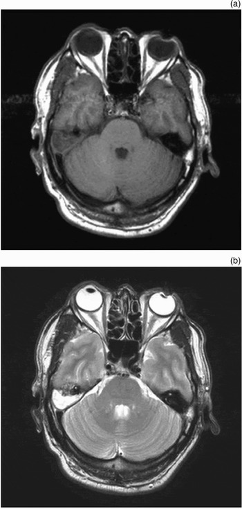
On T1-weighted magnetic resonance imaging (MRI), a hypointense, mass-like lesion appeared, which was hyperintense on the T2-weighted image (Figure 2).
Pre-operative, axial magnetic resonance imaging of the right mastoid area. (a) T1-weighted image showing a fairly uniform, non-enhancing, hypointense mass of signal intensity intermediate between that of cerebrospinal fluid and grey matter. (b) T2-weighted image showing a hyperintense mass of signal intensity similar to that of cerebrospinal fluid.

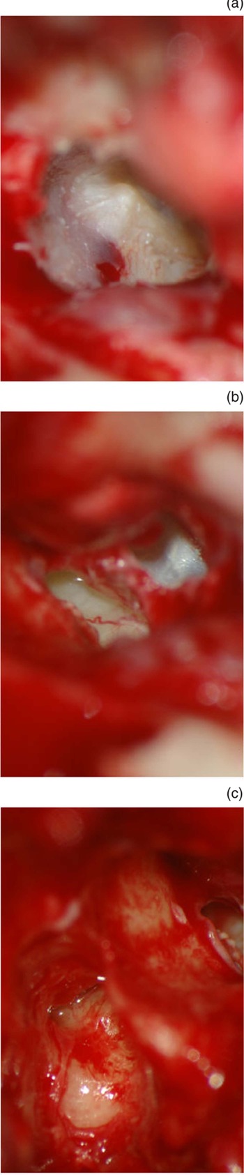
At surgery, following exposure of the mastoid portion via a retroauricular approach, the posterior wall of the external auditory canal was found to be destroyed in two separate places. After removal of mastoid cortical bone, a huge cholesteatoma sac was observed in the mastoid cavity, containing a large amount of keratinous material; this seemed to be the cause of the external auditory canal posterior wall destruction (Figure 3a and 3b). The tegmen mastoideum, the bony plate of the posterior fossa, and the bony plate covering the sigmoid sinus were also observed to be destroyed (Figure 3c). The external auditory canal skin and tympanic membrane were intact, and the middle ear and the aditus ad antrum mucosa were normal (Figure 4).
Operative findings. (a) Destruction of the external auditory canal posterior wall (arrow). (b) The huge cholesteatoma sac, visible in the mastoid cavity. (c) Destruction of the tegmen mastoideum (arrow) and the bony plate covering the sigmoid sinus (arrow head), also showing the results of surgical removal of the superior and posterior semicircular canals (asterisk).

Further operative findings. (a) The intact tympanic membrane. (b) The normal middle-ear mucosa. (c) The normal aditus ad antrum mucosa.

Using a partial translabyrinthine approach, eradicating the superior and posterior semicircular canals, the huge cholesteatoma sac (extending to the petrous apex over the arcuate eminence) was completely excised. In addition, the long process of the incus was found to be fused with the lateral attic wall, although this did not appear to be associated with the cholesteatoma.
Conchal cartilage was used to reconstruct the external auditory canal posterior wall. Finally, abdominal fat, Surgicel (Oxidised Regenerated Cellulose, 2005 Johnson & Johnson Medical Ltd, Gargrave, North Yorkshire, UK) and glue (Human Plasma Fibrinogen, Greenplast, Green Cross Care, South Korea) were used for mastoid obliteration.
Post-operatively, the patient received daily dressing changes, along with intravenous antibiotic therapy prior to hospital discharge to reduce the pus-like otorrhoea which persisted for several weeks.
Six months later, MRI showed that the cholesteatoma had not recurred (Figure 5). Pure tone audiometry indicated that the patient's hearing had been saved. At the time of writing, it was planned that the patient undergo ossiculoplasty in the near future.
Six-month post-operative, axial magnetic resonance imaging of the right mastoid area. (a) T1-weighted and (b) T2-weighted images showing no recurrence; fat (with a hyperintense signal) is visible.

Discussion
Congenital cholesteatoma was first reported in the literature by Luchae in 1885. In 1965, Derlacki and Clemis described the condition as comprising a pearly white mass in the medial portion of the intact tympanic membrane, without otorrhoea or tympanic membrane perforation or any history of previous otological procedures.Reference Kazahaya and Potsic3
Congenital cholesteatomas commonly appear in the petrous apex, cerebellopontine angle, mastoid, middle ear and external auditory canal. They may be asymptomatic in their early stages, but show various symptoms, according to their site of origin, as they progress.Reference Mevio, Gorini, Sbrocca, Artesi, Lenzi and Lecce1, Reference Rashad, Hawthorne, Kumar and Welsh2, Reference Cureoglu, Osma, Oktay, Nazaroglu, Meric and Topcu4 Congenital cholesteatoma of the middle ear, the most common site, generally appears during childhood and may cause hearing disturbance due to erosion of the ossicular chain.
However, congenital cholesteatoma of the petrous apex of the temporal bone generally occurs during adulthood, and can compress the facial nerve, causing facial nerve weakness, and also the brainstem.Reference Mevio, Gorini, Sbrocca, Artesi, Lenzi and Lecce1, Reference Rashad, Hawthorne, Kumar and Welsh2
Rarely, the cholesteatoma arises from the mastoid, presenting with symptoms including neck pain, retroauricular swelling and pain, and dizziness. The neck pain is usually caused by inflammation of the muscle insertion into the mastoid process. The retroauricular swelling and pain are caused by outgrowth or reactive inflammation of the mass, at more advanced stages.Reference Mevio, Gorini, Sbrocca, Artesi, Lenzi and Lecce1 Luntz et al. Reference Luntz, Telischi, Bowen, Ress and Balkany5 and Mevio et al. Reference Mevio, Gorini, Sbrocca, Artesi, Lenzi and Lecce1 have stated that compression of the ipsilateral cerebellar hemisphere can lead to dizziness, and Derlacki and ClemisReference Derlacki and Clemis6 have reported cases of bony labyrinth erosion causing dizziness. However, the dizziness reported in the present case, which subsided after surgery, was considered atypical, since there was no particular evidence explaining its causation.
Although the intermittent otorrhoea seen in the present case did not fit the definition of congenital cholesteatoma described above, Rashad et al. Reference Rashad, Hawthorne, Kumar and Welsh2 have also reported a congenital cholesteatoma of mastoid origin which presented with ear discharge, due to a small fistula of the external auditory canal posterior wall.
In the diagnosis of congenital cholesteatoma, the most important tools are CT and MRI; they are accurate, detailed, comparatively non-invasive, and supply useful pre-operative information.Reference Cureoglu, Osma, Oktay, Nazaroglu, Meric and Topcu4
Luntz et al. Reference Luntz, Telischi, Bowen, Ress and Balkany5 have summarised the symptoms and radiological findings of congenital cholesteatoma of mastoid origin as follows (note the similarity to the present case). Firstly, the initial presentation is neck pain. Secondly, CT scanning shows a cystic, expansile lesion occupying the mastoid process without involving the middle ear; due to its space-occupying effect, it may compress the cerebellum and the dural sinuses adjacent to it and/or erode the bony labyrinth. Thirdly, MRI scanning shows hyperintensity on T2-weighted images, with little or no peripheral enhancement on postcontrast T1-weighted images, typical of congenital cholesteatoma involving the petrous apex.
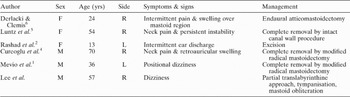
From the worldwide literature, we were able to identify only a few reported cases of congenital cholesteatoma of mastoid origin (Table I). On careful review, these patients were mostly adults, and both sexes were equally affected. Various symptoms were reported, as mentioned above. In most cases, total excision of the cholesteatoma sac via mastoidectomy was performed. Operative findings showed destruction of the tegmen mastoideum, the bony plate of the posterior fossa and the bony plate covering the sigmoid sinus; the middle ear and aditus ad antrum were clear.
Reported cases of congenital cholesteatoma of mastoid origin*

* Including the present case. Yrs = years; F = female; M = male; R = right; L = left
In the present case, the long process of the incus was fused with the lateral attic wall. This finding may be an aspect of a congenital anomaly of the ossicular chain. However, the patient had no history of middle-ear problems, and there was no connection between the ossicular chain and the cholesteatoma.
Conclusion
We have presented a clinical case of congenital cholesteatoma of mastoid origin, of which only a few cases have been reported worldwide. The diagnosis was based on clinical examination, radiological evaluation and surgical findings.