Implications
Current findings indicate that it might be possible to feed phytoestrogens to gilts during critical stages of their development in order to enhance mammary development after puberty. Further work is needed to determine the optimal conditions of such a treatment (i.e. ideal dose, timing and duration), the potential effects on future lactation performance as well as the mechanisms of action. Feeding genistein to growing gilts, however, has no impact on bone resorption. Mammary development in gilts also does not appear to be stimulated solely by ingesting a standard diet containing soya compared with a soya-free diet.
Introduction
Sow milk yield is a major determinant of piglet growth and is largely affected by the number of milk secretory cells present at the onset of lactation (Head et al., Reference Head, Bruce and Williams1991). Feeding management of gilts during the early phase of mammary cell accretion (from 90 days of age until puberty; Sorensen et al., Reference Stokes, Davis and Koch2002) has an impact on mammary development (Farmer et al., Reference Farmer, Petitclerc, Sorensen, Vignola and Dourmad2004; Sorensen et al., 2006), and it is likely that specific feed ingredients may be useful in stimulating this process of mammogenesis. Phytoestrogens have gained increasing interest in recent years because of their potential health benefits in prevention of mammary cancer (review by Warri et al., Reference Warri, Saarinen, Makela and Hilakivi-Clarke2008) and other benefits such as alleviation of bone loss in postmenopausal women (review by Lister et al., Reference Lister, Skinner and Hunter2007). These naturally occurring plant compounds with estrogenic or anti-estrogenic activities (depending on the concentration of endogenous estrogens present; Sun Hwang et al., Reference Sun Hwang, Seok Kwak, Jae Lim, Hee Lee, Soon Kang, Boo Choe, Gil Hur and Ok Han2006) comprise three major classes. They are isoflavones (comprising genistein), lignans and coumestans, and isoflavones are present in large amounts in soya, which is a major component of swine feed.
In rodents, it was shown that neonatal exposure to genistein stimulates mammary development and differentiation (review by Jefferson et al., Reference Jefferson, Padilla-Banks and Newbold2007) and there is one report in swine suggesting that postpubertal exposure to genistein also enhances mammogenesis (Ford, Reference Ford2003). Because of its structural similarity to estradiol-17β, many of the actions of genistein are mediated through estradiol receptors alpha (ERα) and ER beta (ERβ; Nilsson et al., Reference Nilsson, Mäkelä, Treuter, Tujague, Thomsen, Andersson, Enmark, Pettersson, Warner and Gustafsson2001). Indeed, genistein was shown to enhance mammary gland differentiation in prepubertal rats by increasing epidermal growth factor receptors (EGFRs) and transforming growth factor alpha (TGFα) in mammary terminal ductal structures (Brown et al., Reference Brown, Wang, Cotroneo, Zhao and Lamartiniere1998). More recently, it was reported that neonatal exposure to genistein results in increased levels of ERβ mRNA in the mice mammary gland, whereas ERα protein and mRNA are decreased (Padilla-Banks et al., Reference Padilla-Banks, Jefferson and Newbold2006). The insulin-like growth factor 1 (IGF1) is essential for mammogenesis (Ruan and Kleinberg, Reference Ruan and Kleinberg1999) and was found to be a paracrine mediator of estrogen action in the bovine mammary gland (Li and Capuco, Reference Li and Capuco2008). Interestingly, the IGF1 receptor is essential for mediating genistein effects in human breast cancer cells and the IGF1 receptor promoter activity, induced by genistein, requires the action ER (Chen et al., Reference Chen, Gao and Wong2007), thus suggesting a cross-talk between the ER and IGF1 receptor signaling pathways in mammary cells. Mundy (Reference Mundy2006) also suggested that genistein may prevent bone resorption by promoting bone formation, and this can be measured using specific markers, such as cross-linked N-telopeptide of type I collagen (NTx; Weiler et al., Reference Weiler, Finsler, Wehr and Claus2001), present in the circulation. This study was, therefore, undertaken to determine whether dietary supplementation with the phytoestrogen genistein during the prepubertal period affects mammogenesis and/or bone resorption in gilts and if feeding soya alone impacts these variables.
Material and methods
Animals and treatments
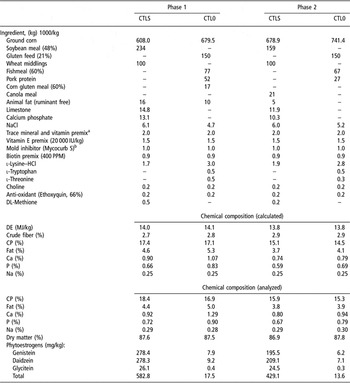
Forty-five crossbred gilts ((Yorkshire × Landrace) × Landrace) were fed a commercial diet until 89 days of age. The next day, they were equally divided into the following 3 nutritional regimens: (i) diet containing no soya (CTL0, n = 15), (ii) soya-based commercial diet (CTLS, n = 15) and (iii) CTLS with 2.3 g daily supplementation of genistein (GEN, n = 15). The genistein was offered once daily to gilts (at 1400) after 6 h of feed removal and was premixed in 15 g of corn flour. Gilts from all treatment groups received 15 g of corn flour in the same manner, and it was included in the compositional analyses of the diets. There was a 10-day adaptation period from 80 days of age, during which all gilts received 15 g of finely ground corn without genistein. All dietary regimens were isonitrogenous and isocaloric based on calculated values (Table 1), which were used in formulating diets that were fed ad libitum over two phases of growth, namely from 75 to 119 days of age (phase 1) and from 120 days to slaughter at 182 ± 1 days (phase 2). Compositions of all diets are shown in Table 1. Representative feed samples were taken twice weekly throughout the experiment for compositional analyses (shown in Table 1). Throughout the experiment, all gilts were housed in individual stalls (0.6 × 2.1 m) from 1500 to 0800 and were given fresh feed once daily (1500). Individual feed consumption was recorded daily from 90 days of age until slaughter. At 0730, gilts were roused and encouraged to stand up and eat any leftover feed, they were then housed in groups of 15 (5 from each dietary treatment group) from 0800 until 1400 (surface area of 29.7 m2). As of 140 days of age, gilts were moved to a similar room with a boar present and estrous behavior was evaluated twice daily by allowing physical contact between gilts and the boar. Date and duration of first estrus were recorded, and all gilts had started cycling before slaughter. Gilts were weighed and their backfat thickness measured ultrasonically at the last rib (Scanmatic SM-1, Medimatic, Hellerup, Denmark) on days 89, 150, and 183, following an overnight fast. The experiment took place between September 2005 and February 2006. Animals were cared for according to a recommended code of practice (Agriculture and Agri-Food Canada, 1993).
Actual ingredients, calculated and analyzed composition of the two basal diets on an as-fed basis, which were fed from days 75 to 119 (phase 1) or days 120 to slaughter at 183 ± 1 days (phase 2)

Standard soya-based diet: CTLS, diet without soya: CTL0
aProvided the following per kilogram of diet: Cu, 25 mg; Zn, 145 mg; Se, 0.3 mg; Mn, 30 mg; Fe, 100 mg; I, 1.0 mg; Vitamin A, 3000 IU; Vitamin D, 900 IU; Vitamin E, 30 IU; Vitamin K, 1.5 mg; Vitamin B12, 20 μg; thiamin, 2.0 mg; riboflavin, 3.5 mg; pantothenic acid, 15 mg; niacin, 20 mg; folate, 0.45 mg; biotin, 50 μg; pyridoxine, 2.0 mg.
bKemin Industries, Des Moines, IA.
Blood sampling and assays
Jugular blood samples were obtained at 0730, following an overnight fast, on 89 days of age and 10 ± 1 days following the last heat before slaughter (average age of 176.4 days at second blood sampling, ranging from 164 to 184 days, slaughter being at 183 ± 1days) to determine concentrations of estradiol, progesterone, prolactin, IGF1, isoflavone metabolites (at last sampling before slaughter only) and NTx (at last sampling before slaughter only). Samples collected for isoflavones, progesterone and prolactin measurements were left at room temperature for 4 h, stored overnight at 4°C, centrifuged the following day and serum was harvested. Samples for estradiol and IGF1 assays were collected in EDTA-tubes (Becton Dickinson and Cie, Rutherford, NJ, USA) and those for NTx assays in heparinized tubes (Becton Dickinson and Cie); they were all put on ice and centrifuged within 20 min, and plasma was immediately recovered. Serum and plasma samples were frozen at −20°C until they were assayed.
The quantitative analysis of isoflavones and their metabolites in serum samples was completed with a method using liquid chromatography/mass spectrometry combined with photodiode array detection, as developed in Dr Gilani’s laboratory (Sepehr et al., Reference Setchell and Lydeking-Olsen2006).
Concentrations of prolactin were determined using a previously described radioimmunoassay (RIA; Robert et al., Reference Robert, de Passillé, St-Pierre, Dubreuil, Pelletier, Petitclerc and Brazeau1989). The radioinert prolactin was purchased from A.F. Parlow (US National Hormone and Pituitary Program, National Institute of Diabetes and Digestive and Kidney Diseases, Torrence, CA, USA) and the first antibody to prolactin was purchased from Research Products International (Mount Prospect, IL, USA). Parallelism of a pool of serum from sows was demonstrated. Average recovery, calculated by addition of various doses of radioinert hormone to 50 μl of a pooled sample, was 103.4%. Sensitivity of the assay was 1.5 ng/ml. Six samples of a representative pool of serum were carried in duplicates in all assays in order to calculate coefficients of variation (CV). The intra-assay CV, calculated from the mean values of the pools within each assay, was 6.1%. The interassay CV, calculated from the mean values of the pools obtained for different assays, was 8.9%. Concentrations of IGF1 were determined with a previously described RIA (Abribat et al., Reference Abribat, Brazeau, Davignon and Garrel1993). The IGF1 was extracted using the formic acid-acetone method. The first antibody in the IGF1 assay was purchased from A.F. Parlow (US National Hormone and Pituitary Program). The radioinert IGF1 was purchased from GROPEP (Adelaide, SA, Australia). Parallelism of a plasma pool from sows was demonstrated. Average recovery was 101.6% and assay sensitivity was 62.5 pg/ml. Intra-assay CV was 5.4% and all tubes were analyzed in one assay. Estradiol (Diagnostic Systems Laboratories Inc., Webster, TX, USA) and progesterone (ICN Pharmaceuticals Inc., Costa Mesa, CA, USA) were quantified using commercial RIA kits. All samples were assayed in a single assay with an intra-assay CV of 1.2% for estradiol and 4.5% for progesterone. Concentrations of NTx were determined using a commercial ELISA kit (Osteomark NTx serum ELISA assay, Wampole Laboratories, Princeton, NJ, USA) validated for swine serum. The assay was performed as recommended by the suppliers except that samples were diluted 1 : 75 in assay buffer. The accuracy of the kit controls was 97% of expected values and agreement between duplicate samples ranged from 91% to 97%.
Mammary gland measurements and organ sampling
At slaughter, mammary glands were excised from the abdominal wall of all gilts. Parenchymal tissue from the fourth anterior mammary glands was used for mRNA measurements of ERα and ERβ, IGF1, epidermal growth factor (EGF), EGFR, and TGFα. These samples were stored at −80°C until analysis. The remaining mammary glands were then stored at −20°C until dissection and analyses for tissue composition. Frozen mammary glands were trimmed of skin and teats and subsequently stored at −20°C. They were then sawed into 2-cm slices and mammary parenchymal tissue from each slice was dissected from surrounding adipose (i.e. extraparenchymal tissue) at 4°C and both parenchymal and extraparenchymal tissue weights were recorded. Mammary parenchyma was defined as the region containing epithelial tissue, but also contained a large amount of adipose and connective tissues because mammary ducts and alveolar tissues are embedded within loose connective tissue of the mammary fat pad in gilts of this age. Parenchymal tissue from all dissected and sliced glands was homogenized and a representative sample was used for determination of composition by chemical analysis. The DNA content of parenchymal tissue was evaluated using a method based on fluorescence of a DNA stain (Labarca and Paigen, Reference Labarca and Paigen1980). Dry matter (DM), protein, and lipid contents were also measured (AOAC, 2004). Six gilts per treatment group received an i.v. injection of 5 mg/kg (diluted in saline at a concentration of 20 mg/ml and pH of 8 to 8.5) of 5-bromo-2′-deoxyuridine (BrdU; Sigma Chemical, Saint Louis, MO, USA) in the marginal ear vein, 2 to 3 h before slaughter. Three cubes of parenchyma (∼ 3 × 3 mm) from these gilts were obtained from two regions within the fourth anterior mammary gland and used to quantify the portion of epithelial cells that were labeled with BrdU, as an index of proliferation rate. Tissues were collected from the second anterior mammary gland and were fixed in 10% formalin in phosphate-buffered saline at 4°C for 24 h before being transferred to 70% ethanol until further processing. They were then dehydrated through a graded series of ethanol to 100% ethanol and embedded in paraffin according to standard techniques. Embedded tissue was later sectioned at 5 μm for immunohistochemical staining and evaluation. Ovaries were collected from all gilts and the number of corpora lutea counted.
Immunohistochemistry
Slides were prepared as described by Farmer et al. (Reference Farmer, Petit, Weiler and Capuco2007) and bright field microscopic detection of BrdU-labeled cells was performed as described by Capuco et al. (Reference Capuco, Ellis, Wood, Akers and Garrett2002). Immunohistochemical localization of ERα was performed on 3 randomly selected gilts per dietary treatment using a rabbit polyclonal antibody (sc-787 at 1 : 100 dilution, Santa Cruz Biotechnology Inc., Santa Cruz, CA, USA). Tissue sections were prepared for immunohistochemistry as described above. Following overnight incubation with primary antibody, sections were washed in phosphate-buffered saline (PBS; 3 times, 2 min at room temperature) and then incubated with second antibody-horseradish peroxidase conjugate (30 min at room temperature). Sections were washed in PBS and then were incubated with diaminobenzidine, counter stained with hematoxylin, and mounted with Permasilip.
To quantify the number of BrdU-labeled epithelial cells and ER-positive epithelial cells, photographs of stained tissue sections were captured as digital images. For quantifying the BrdU-labeled cells, a slide was prepared and processed for histochemistry from each of two parenchymal regions per gilt. For each slide, 10 tissue areas were photographed with a Spot® digital camera (Diagnostic Instruments Inc., Sterling Heights, MI, USA) on a Zeiss Axioskop microscope (Carl Zeiss Inc., Thornwood, NY, USA) with the ×40 objective. The number of BrdU-labeled epithelial cells and total epithelial cells were counted manually. At least 1100 epithelial cells were scored per gilt (average ± s.e. = 2753 ± 143). For quantifying ER-positive cells, slides were processed, images obtained and the number of ER-positive epithelial cells and total epithelial cells analogously determined. At least 1300 epithelial cells were scored per gilt (average ± s.e. = 2801 ± 141).
Real-time PCR analyses of selected genes
Total RNA was extracted from parenchymal tissue using Trizol™ reagent (Invitrogen, Grand Island, NY, USA) and according to the manufacturer’s instructions. Extracted RNA was then treated with DNAse I (Amplification grade; Invitrogen) to remove contaminating DNA. First-strand complementary DNA was synthesized using oligo dT (13 to 18) primers (Invitrogen) and the Superscript II preamplification system (Invitrogen) following the manufacturer’s instructions.
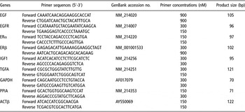
The relative mRNA abundance of selected genes was determined using real-time PCR analyses. Primers were designed based on available porcine sequence (Table 2) and were selected using the Primer Express software 3.0 (PE Applied BioSystems, Foster City, CA, USA). Amplification of peptidylprolyl isomerase A, glyceraldehyde-3-phosphate dehydrogenase and beta actin (ACTβ) housekeeping genes (HKG) was also performed using porcine specific primers described in Table 2. The PCR amplifications were performed in a 10-μl reaction volume consisting of corresponding concentrations of each primer (Table 2), 5 μl of 2 × Power SYBRGreen Master Mix (PE Applied BioSystems), 3 μl of 15 × diluted cDNA, and 0.05 μl of UNG AmpErase (PE Applied BioSystems). Cycling conditions were 2 min at 50°C to activate the amperase, 10 min at 95°C, followed by 40 cycles of 3 s at 95°C and annealing and polymerization at 60°C for 30 s. Amplification, detection, and data analysis were performed with an ABI 7500 Fast Real-Time PCR System (PE Applied BioSystems). Specificity of amplified fragments was verified with the melting curve analysis. Amplifications were performed in triplicate and standard curves were established in duplicate for each gene. Selected genes and HKG amplifications were run in separate assays. A pool of parenchymal tissue cDNA was used to create a standard curve for quantification of transcripts using the relative standard curve method as described by Applied BioSystems (1997). For each experimental sample, the amount of target gene mRNA relative to HKG mRNA was determined from the corresponding standard curves. Relative quantity ratios were then obtained by dividing the relative quantity unit of selected genes by those of HKG. Mean values from triplicates were used to perform statistical analyses.
Oligonucleotide primers used for real-time PCR amplifications of selected genes

EGF = epidermal growth factor; EGFR = epidermal growth factor receptor; ERα = estradiol receptor alpha; ERβ = estradiol receptor beta; IGF1 = insulin-like growth factor 1; TGFA = transforming growth factor alpha; GAPDH = glyceraldehyde-3-phosphate dehydrogenase; PPIA = peptidylprolyl isomerase A; ACTβ = beta actin.
Statistical analyses
The MIXED procedure of Statistical Analysis Systems Institute (SAS, Reference Sepehr, Robertson, Gilani, Cooke and Lau1998) was used for statistical analyses. The ANOVA used for isoflavones, NTx, mammary gland and ovarian variables included the effect of nutritional treatments with the residual error being the error term used to test main effects of treatment. Analyses on weights of mammary extraparenchymal and parenchymal tissues were done using BW of gilts at slaughter as a covariate. Analyses were performed on weight gains and total feed intakes over each of the three growth periods studied (days 90 to 119, days 120 to 149, days 150 to 180). Repeated measures ANOVA with the factors treatment (the error term being gilt within treatment), day of age and the treatment × day of age interaction (the residual error being the error term for these last two factors) were carried out on backfat thickness and hormonal data. When the treatment × day interaction was significant, separate analyses were also performed for each day. Data were corrected with a square root or a logarithmic transformation (using natural logarithms) when appropriate and, in those cases, means presented in tables are back-transformed values. Real-time PCR data were analyzed using the same one-way ANOVA model. For all analyses, differences between means were evaluated through pair-wise multiple comparisons with a Tukey adjustment. The extent of histological development in mammary parenchymal tissue slices was scored using categorical data (visual scoring index from 1 to 5) and was, therefore, analyzed with the Cochran-Mantel–Haenszel test (Stokes et al., Reference Sorensen, Sejrsen and Purup1995). Data in tables are presented as least squares means ± SEM unless indicated otherwise.
Results
Performance, hormonal, NTx and isoflavone data
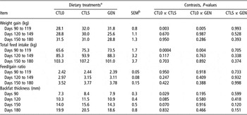
Total feed intake of gilts was affected by treatments (P < 0.001) during the first growth period only, specifically from 90 to 119 days of age; values were lowest for CTL0 compared with the CTLS and GEN gilts (P < 0.01, Table 3). The same relationship held true for weight gains (Table 3), with values being altered by treatments from days 90 to 119 (P < 0.01), and those of CTL0 gilts being lower than those of CTLS and GEN gilts (P < 0.01). Feed conversion, however, was not affected by diets (P > 0.1, Table 3). There was a treatment × day interaction on backfat thickness of gilts (P < 0.05) and analyses for each day showed lower backfat in CTL0 than CTLS and GEN gilts on day 90 (P < 0.05) but only a tendency for values to be lower in these same animals on days 120 and 150 (P < 0.1), and no significant treatment effect on day 180 (P > 0.1, Table 3).
Growth and feed intake performances of gilts fed one of three diets from 90 to 183 days of age

Diets were: diet without soya, CTL0; standard corn-soya diet, CTLS; and standard corn-soya with 2.3 g of supplementary genistein daily, GEN.
aSignificant overall treatment effect on weight gain and total feed intake for days 90 to 119 (P < 0.01) and on backfat on day 90 (P < 0.05).
Tendency for an overall treatment effect on backfat on days 120 and 150 (P < 0.1).
bMaximum value of the standard error of the means.
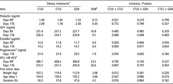
Circulating concentrations of isoflavone metabolites at the end of the experimental period are shown in Table 4. There was a significant effect of diets on all isoflavones measured (P < 0.001) with values being lower in gilts fed the CTL0 diet compared with either the CTLS (P < 0.001) or the GEN (P < 0.001) diets. Concentrations of genistein were also significantly increased (P < 0.001) in the GEN diet compared with the CTLS diet (P < 0.001). Diets had no impact (P > 0.1) on circulating concentrations of prolactin, IGF1, estradiol, progesterone or NTx in gilts at the end of the experimental period (Table 5), yet weight (but not age) of gilts at first estrus was lower (P < 0.05) for CTL0 than CTLS gilts, and duration of the first estrus also tended to be longer in CTL0 than in CTLS gilts (P < 0.1, Table 5).
Concentrations of isoflavone metabolites (μmol/l) at 176 days of age in serum of gilts that were fed one of three diets from 90 to 183 days of age

Diets were: diet without soya, CTL0; standard corn-soya diet, CTLS; and standard corn-soya with 2.3 g of supplementary genistein daily, GEN.
aSignificant overall treatment effect on all variables (P < 0.0001).
Endocrine and first estrus data for gilts fed one of three diets from 90 to 183 days of age

IGF1 = insulin-like growth factor 1; NTx = N-telopeptide of type I collagen.
Diets were: diet without soya, CTL0; standard corn-soya diet, CTLS; and standard corn-soya with 2.3 g of supplementary genistein daily, GEN.
aSignificant overall treatment effect on weight at first estrus (P < 0.05) and tendency for duration of first heat (P < 0.1).
bMaximum value of the standard error of the means.
cSignificant effect of day on prolactin (P < 0.05), estradiol (P < 0.001) and NTx (P < 0.001).
dAll values on day 89 were below assay sensibility (i.e. <0.15 ng/ml) so are not presented.
eBone collagen equivalent.
Mammary gland data
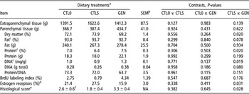
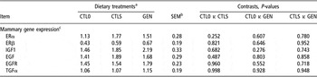
Composition of mammary tissue at slaughter and BrdU incorporation are shown in Table 6. While amount of mammary parenchymal and extraparenchymal tissues were not altered by dietary treatments (P > 0.1), composition of parenchymal tissue was affected. More specifically, parenchymal tissue from GEN gilts contained less DM (P < 0.05), tended to have a lower percentage of fat (P < 0.1), had a greater percentage of protein (P < 0.05), greater concentrations of DNA (P < 0.05) and tended to have more total DNA (P < 0.1) than that of CTLS gilts. There was a significant reduction in number of ER-positive cells (P < 0.05) in these same animals. Representative micrographs of tissue sections depicting mammary gland microanatomy and BrdU labeling at slaughter are shown in Figure 1. Micrographs depict increased mammary development evident by greater histological score of GEN v. CTLS gilts (P < 0.05, Table 6), and lack of difference in histological score between CTL0 and GEN (P > 0.1, Table 6). Immunodetection of BrdU-labeled cells suggests reduced BrdU labeling of mammary epithelium in tissue from CTLS gilts, consistent with the numerical, but non-significant (P > 0.1), differences in means for BrdU labeling index (Table 6). Micrographs of BrdU-stained sections also clearly show heavy nuclear labeling with the BrdU. The relative abundance of transcripts for ERα, ERβ, IGF1, EGF, EGFR and TGFα genes in mammary parenchymal tissue at slaughter was not affected by diets (Table 7).
Composition and BrdU incorporation of mammary glands at slaughter (day 183) in gilts fed one of three diets from 90 to 183 days of age

BrdU = 5-bromo-2′-deoxyuridine.
Diets were: diet without soya, CTL0; standard corn-soya diet, CTLS; and standard corn-soya with 2.3 g of supplementary genistein daily, GEN.
aSignificant overall treatment effect on percentage of dry matter, percentage of protein, and DNA in mg/g (P < 0.05). Tendency for: gram of extraparenchymal tissue, % fat, total DNA and protein/DNA (P < 0.1).
bMaximum value of the standard error of the means.
cExpressed on a dry matter basis.
dRepresents the percent of epithelial cells that are positive for estrogen receptor alpha.
eHistological score of mammary development utilizing a scale from 1 (simple ducts) to 5 (extensive lobular development). Data were analyzed using the non-parametric Mann–Whitney test.
fMean ± s.e.
Mammary gland microanatomy and 5-bromo-2′-deoxyuridine (BrdU) labeling at slaughter (day 183) in gilts fed one of three diets from 90 to 183 days of age. Diets were: diet without soya, CTL0; standard corn-soya diet, CTLS; and standard corn-soya with 2.3 g of supplementary genistein daily, GEN. Representative micrographs of tissue sections from CTL0 (a), CTLS (b) and GEN (c) stained with hematoxylin and eosin depict increased mammary development in GEN v. CTLS gilts (P < 0.03), and lack of difference between CTL0 and GEN (P > 0.1). Immunolocalization of BrdU-labeled cells are depicted in micrographs from CTL0 (d), CTLS (e) and GEN (f) groups and suggest reduced BrdU labeling of mammary epithelium in tissue from CTLS gilts, consistent with numerical, but nonsignificant (P > 0.1), differences in means. BrdU immunohistochemistry also indicates heavy nuclear labeling with BrdU.

Relative mRNA levels of selected genes in mammary parenchymal tissue at slaughter (day 183) in gilts fed one of three diets from 90 to 183 days of age

ERα = estradiol receptor alpha; ERβ = estradiol receptor beta; IGF1 = insulin-like growth factor 1; EGF = epidermal growth factor; EGFR = epidermal growth factor receptor; TGFα = transforming growth factor alpha.
Diets were: diet without soya, CTL0; standard corn-soya diet, CTLS; and standard corn-soya with 2.3 g of supplementary genistein daily, GEN.
aOverall treatment effect not significant for any of the measured genes (P > 0.1).
bMaximum value of the standard error of the means.
cmRNA levels. Values correspond to the relative abundance of studied genes mRNA.
Discussion
The formulation of a diet without soya led to an unconventional diet that was not as appetizing as the other diets, as evidenced by the lower feed intake and decreased weight gain during the first growth period in CTL0 gilts compared with gilts from the other two groups. Yet, there seemed to be an adaptation to this diet because total feed intake and weight gain were not affected by dietary treatments in the second and third growth periods. The absence of treatment effects on growth and feed/gain ratio corroborates findings of Kuhn et al. (Reference Kuhn, Hennig, Kalbe, Rehfeldt, Ren, Moors and Degen2004), who saw no effects of feeding diets differing in isoflavone content (soyabean meal v. soya protein concentrate) on the performance of weanling pigs. Furthermore, these authors reported no treatment differences in carcass composition, meat quality as well as expression of IGF1 receptor mRNA in the muscle of pigs. Current results also showed no effect of treatment on circulating IGF1 concentrations, yet circulating concentrations of isoflavones were increased in gilts fed a standard soya diet compared with a diet without soya. It therefore appears that these changes were not sufficient to elicit significant alterations in any of the growth or physiologic variables measured.
It is well documented that neonatal exposure of mice to genistein disrupts ovarian function during adulthood and reduces fertility (review by Jefferson et al., Reference Jefferson, Padilla-Banks and Newbold2007). To the best of our knowledge, only two studies looked at the effects of phytoestrogens on reproduction in swine. Drane et al. (Reference Drane, Wrathall, Patterson and Hebert1981) fed either a 20% soyameal diet or a soya-free diet to gilts from 10 to 24 weeks of age, at which time they slaughtered the animals and examined their reproductive tissues. Vulval enlargement was noticed in treated gilts, but there were no effects on the reproductive tracts. However, it is important to point out that these animals were prepubertal and it is unknown whether effects would have been apparent had they been evaluated after the onset of puberty. Indeed, in a more recent study, Ford et al. (Reference Ford, Clark, Walters, Wheeler and Hurley2006) demonstrated that exogenous genistein does affect reproductive tissues of ovariectomized postpubertal gilts. More specifically, i.m. injections of increasing doses of genistein (varying from 50 to 400 mg/day) for 10 days stimulated uterine and cervical tissue growth in a dose-dependent manner. The highest dose of genistein (400 mg/day) also increased the number of cells that stained positive for progesterone receptor and for proliferation in the uterus and cervix. Current findings showed a decrease in BW at puberty onset and a tendency for an increase in duration of first estrus in gilts fed the diet without soya compared with gilts fed the commercial soya diet, but there was no effect of added genistein. This is contrary to our expectation because mice treated neonatally with genistein generally show an increased length of the estrous cycle (review by Jefferson et al., Reference Jefferson, Padilla-Banks and Newbold2007) and accelerated onset of puberty (Lewis et al., Reference Lewis, Brooks, Milburn, Soames, Stone, Hall and Ashby2003). This discrepancy between our results and those reported in mice could be due to the difference in age at treatment (i.e. prepubertal v. neonatal) or to the fact that age at first estrus is very robust to dietary manipulation in gilts (Friend et al., Reference Friend, Lodge and Elliot1981). It is also very likely that discrepancies between results in rat and swine studies are related to interspecies differences in isoflavone metabolism. Indeed, Gu et al. (Reference Gu, House, Prior, Fang, Ronis, Clarkson, Wilson and Badger2006) reported that the overall metabolism of isoflavones in pigs is closer to that of human than to that of rats or monkeys. Dosage could also be involved in differences between current results and those of Lewis et al. (Reference Lewis, Brooks, Milburn, Soames, Stone, Hall and Ashby2003), as these last authors saw an effect of treating rats with 40 mg/kg but not 4 mg/kg BW of genistein per day. It is obviously difficult to extrapolate the amount provided per day in the current trial to that provided in the rat trials as average weights of the gilts at the onset and end of treatments differed (i.e. 47.6 and 137.8 kg, respectively) but quantity of ingested genistein was not altered. Thus, 2 g of genistein per day in this study was equivalent to approximately 42.0 and 14.5 mg/kg BW, respectively, at the onset and conclusion of the experiment. The purpose in selecting this dose was to ensure that pigs obtained at least 40 mg/kg BW of genistein (as used by Lewis et al., Reference Lewis, Brooks, Milburn, Soames, Stone, Hall and Ashby2003) at the onset of treatment. Nevertheless, it is obvious that more information on metabolism and tissue uptake of isoflavones in swine are needed to establish optimal dosage in that species. Circulating concentrations of isoflavone metabolites in this study showed that genistein, but not daidzen, glycitein or equol, increased in the blood of swine receiving an oral dose of genistein, but was unaffected solely by providing phytoestrogens via a standard soya diet. The undetectable levels of equol and greater concentrations of genistein and daidzen found in CTL0 gilts are in accordance with previous findings in piglets (Gu et al., Reference Gu, House, Prior, Fang, Ronis, Clarkson, Wilson and Badger2006); however, the increase in concentrations of equol in CTLS gilts is in contradiction with findings from these last authors. Differences in diet or age of animals could be involved in this discrepancy because Gu et al. (Reference Gu, House, Prior, Fang, Ronis, Clarkson, Wilson and Badger2006) measured isoflavones in 28-day old piglets that had been fed a liquid diet based on soya protein isolate as sole protein source. Indeed, Adlercreutz et al. (Reference Adlercreutz, Höckerstedt, Bannwart, Bloigu, Hämäläinen, Fotsis and Ollus1987) established that diet composition (i.e. fat and fiber intakes) can alter the metabolism of estrogens and phytoestrogens. Furthermore, the greater gastrointestinal tract development of pubertal gilts (in this trial) compared with that of suckling piglets could have come into play as intestinal bacteria are known to be involved in metabolism and absorption of phytoestrogens (Adlercreutz et al., Reference Adlercreutz, Höckerstedt, Bannwart, Bloigu, Hämäläinen, Fotsis and Ollus1987).
The lack of treatment effects on circulating concentrations of estradiol, progesterone and prolactin contradicts data in rats where concentrations of LH and prolactin were greater in adult male offspring from dams that were fed soya, compared with dams fed milk and eggs, as a protein source during two reproductive cycles (Pastuszewska et al., Reference Pastuszewska, Taciak, Ochtabinska, Tusnio, Misztal, Romanowicz and Morawski2008). However, it is in agreement with another study where 3 s.c. injections of genistein to prepubertal rats had a biological effect on mammary tissue, but did not alter circulating concentrations of estradiol-17β or progesterone after puberty (Murrill et al., Reference Murrill, Brown, Zhang, Manzolillo, Barnes and Lamartinière1996). On the other hand, Rachon et al. (Reference Rachon, Menche, Vortherms, Seidlova-Wuttke and Wuttke2008) noted increased concentrations of estradiol and prolactin in the circulation of ovariectomized rats receiving a high dose (i.e. 400 mg/kg feed) of the phytoestrogen, equol, for a 3-month period; but they did not look at the effect of equol on intact animals.
Neonatal exposure of mice to genistein affects the mammary glands. Indeed, advanced mammary gland development and differentiation were observed with exposure to genistein, moreover, the dose and timing of exposure has an impact on these effects (review by Jefferson et al., Reference Jefferson, Padilla-Banks and Newbold2007). Mice injected s.c. with lower doses of genistein (0.5 and 5 mg/kg) showed advanced mammary gland morphogenesis at puberty, whereas stunted mammary development was observed at the highest dose (50 mg/kg; Padilla-Banks et al., Reference Padilla-Banks, Jefferson and Newbold2006). The authors noted that morphological changes observed at the lower doses were due to expansion of the mammary gland through the fat pad at a faster rate. Rowell et al. (Reference Rowell, Carpenter and Lamartinière2005) looked at the mechanism of action of genistein on mammary tissue in rats and found that early prepubertal exposure to genistein enhanced cell proliferation by up-regulating the protein guanosine triphosphate-cyclohydrolase 1 and the EGF-signaling pathway. It is not known if that is the case in swine.
There is only one published report on the effect of genistein on mammogenesis in swine. Ford (Reference Ford2003) noted that i.m. injections of 400 mg/day of genistein to postpubertal ovariectomized gilts for 10 days enhanced mammary gland development. There was increased lobular development and alveolar cells, and less dense connective tissue and less fat in the stroma. This is consistent with our current findings of increased DNA in mammary parenchymal tissue and a tendency for lower fat content. Although there have been reports in the literature of genotoxic effects of genistein, these have been restricted to in vitro studies utilizing pharmacological concentrations of genistein that exceeded 5 μM (Klein and King, Reference Klein and King2007). Evaluation of data from this study does not support a genotoxic effect of genistein. Tissue morphology and BrdU-labeling indices of mammary tissue from GEN gilts were not impaired compared with that of CTL0 or CTLS gilts and there was more extensive lobular development in GEN gilts. Finally, it should be noted that BrdU labeling was heavy and thus the labeling indices were not likely to have been compromised by labeling due to unscheduled DNA synthesis. Such repair processes result in incorporation of nucleotides and nucleotide analogs (BrdU) that are a minor fraction of the incorporation that occurs during the semiconservative DNA synthesis necessary for cell proliferation (Valentin-Severin et al., Reference Valentin-Severin, Thybaud, Le Bon, Lhuguenot and Chagnon2004).
The reduced number of ERα-positive mammary epithelial cells in GEN gilts from this study is in agreement with findings of Padilla-Banks et al. (Reference Padilla-Banks, Jefferson and Newbold2006) who observed lower ERα expression in mammary tissue of pubertal mice following neonatal exposure to three doses of genistein (s.c. injections of 0.5, 5 or 50 mg/kg BW per day). An increased expression of ERβ was also noted by Padilla-Banks et al. (Reference Padilla-Banks, Jefferson and Newbold2006) at the lowest dose of genistein, but this was not accompanied by increased protein expression of ERβ in the mammary ductal epithelium, as determined by histochemistry. The negative effect of genistein on ERα may be indicative of negative feedback, but the direct consequence of this lower expression remains unclear in light of the mammary hyperplasia observed. Even though not significant, numerically, there was a 60% increase in BrdU labeling index in GEN v. CTL0 gilts in this study. Conversely, the numerically lower value for CTLS gilts compared with CTL0 gilts was surprising. Consistent with our current observations of increased DNA in mammary parenchyma and reduced number of ERα-positive mammary epithelial cells in GEN gilts, Capuco and colleagues (Capuco et al., Reference Capuco, Ellis, Wood, Akers and Garrett2002; Meyer et al., Reference Meyer, Capuco, Boisclair and Van Amburgh2006) noted that exogenous estrogen stimulates the in vivo proliferation of mammary cells in dairy heifers while decreasing expression of ERα.
Human epidemiological data and in vitro animal studies suggest that genistein may possess breast cancer preventive properties (review by Warri et al., Reference Warri, Saarinen, Makela and Hilakivi-Clarke2008) by altering expression of BRACA1 and PTEN and perhaps by epigenetic mechanisms. Similarly, in vitro data support an effect on mammary tumorogenesis, as exposure of human mammary cells to genistein altered the expression of certain cancer-related genes (Moon et al., Reference Moon, Brazeau and Morris2007). The lack of effect of genistein on the mRNA expression of selected genes in this study could be due to an inadequate dose, yet positive effects on mammary development were observed. This suggests that the enhanced mammogenesis was not related to expression of the genes evaluated. It is of interest to note, however, that it was only at high concentrations (100 μM and not 1 or 10 μM) that genistein and daidzen affected (i.e. decreased) IGF- and EGF-associated gene expression in porcine skeletal muscle tissue in vitro (Kalbe et al., Reference Kalbe, Mau and Rehfeldt2008) and reduced protein synthesis and total DNA in semimembranosus muscle cell cultures from neonatal pigs (Mau et al., Reference Mau, Kalbe, Viergutz, Nurnberg and Rehfeldt2008). Rehfeldt et al. (Reference Rehfeldt, Kalbe, Nürnberg and Mau2009) also reported dose-dependent effects of genistein on protein metabolism in porcine skeletal muscle cells in vitro, with protein degradation being reduced with 0.1 nmol/l of genistein and protein synthesis being reduced with 100 μmol/l of genistein. In vivo and in vitro studies indicate that effects of phytoestrogens are highly dose dependent. Insufficient dose could therefore be related to the lack of effect on mammary gene expression in our study. Additionally, the timing of in vivo exposure could be important as Luijten et al. (Reference Luijten, Verhoef, Dormans, Beems, Cremers, Nagelkerke, Adlercreutz, Penalvo and Piersma2007) reported that the impact of phytoestrogen exposure on mammary tumorogenesis in mice appears to depend on the timing of exposure within the life cycle. The optimal time for phytoestrogen exposure to alter mammogenesis in swine is not known.
A recent review discussing the effects of phytochemicals on bone and joint health (Lister et al., Reference Lister, Skinner and Hunter2007) stated that there has been considerable investigation on the use of phytoestrogens to improve bone health and prevent osteoporosis. Studies in humans have shown promising, yet variable results. Consumption of isoflavones for up to a year increased expression of beneficial bone biomarkers, such as markers for bone mineral density (Williamson and Manach, Reference Williamson and Manach2005). On the other hand, feeding 1 mg of genistein to humans for a year did not alter bone density or bone resorption but increased markers of bone formation (Atkinson et al., Reference Atkinson, Compston, Day, Dowsett and Bingham2004). As hypothesized by Mundy (Reference Mundy2006) genistein may prevent bone resorption by promoting bone formation. An impact on bone resorption was not apparent in this study as concentrations of NTx, which is a highly-specific parameter of bone collagen degradation and serves as an indicator of bone resorption (Weiler et al., Reference Weiler, Finsler, Wehr and Claus2001), were not altered. This may be due to the shorter duration of treatment (i.e. 93 days) in our study compared with studies performed in humans. Indeed, Setchell and Lydeking-Olsen (Reference Sorensen, Farmer, Vestergaard, Purup and Sejrsen2003) in their review stated that diets rich in phytoestrogens have bone sparing effects in the long-term but that the magnitude of the effects are still speculative. Kaludjerovic and Ward (Reference Kaludjerovic and Ward2009) recently demonstrated that neonatal exposure of rats to genistein modulates bone development in adulthood. It is not known whether that would also be the case in swine.
Conclusions
In conclusion, it is apparent that exposure of growing gilts to the phytoestrogen genistein has a stimulatory effect on mammary development measured after puberty. However, this is not accompanied by changes in abundance of transcripts for selected genes in mammary tissue, or in circulating hormone levels. These results also show that mammary development in gilts was not stimulated solely by feeding a standard diet containing soya compared with a soya-free diet.
Acknowledgements
The authors thank L. Thibault, S. Horth, D. Beaudry and P. Robertson for their invaluable technical assistance, A. Hummel for assistance with immunohistochemistry, the staff of the Swine Complex, especially D. Morissette, J. Phaneuf, J. Marois and T. Marsh, for care and slaughter of the animals, S. Méthot for statistical analyses and the ‘Centre d’Insémination Porcine du Québec’ (CIPQ) for supplying the Landrace semen. Sherbrooke AAC contribution no. 1021.