I. INTRODUCTION
The fabrication of artificial low-dimensional structures is one of the most challenging research fields in the solid-state technology. These nanoobjects are prepared by physical and chemical or by combined methods and they have attracted great interest due to their scientific peculiarity and technical significance. Nanostructures are applied both in bulk and low-dimensional materials for composite materials, energy production, catalysis, optoelectronics, nanomagnetic materials, and for biomedicine.Reference Bergman and Jung de Andrade1 One of the most challenging methods of nanostructure production is the phenomenon of self-assembly, which has been applied in the case of compound and group IV semiconductors and a wide range of material and substrate combinations.Reference Barabási2
During self-assembly phenomena, the natural laws are used as instruments to produce the nanostructures. The strain induced self-assembled growth is a basic physical method for the preparation of nanostructures. In the case of thin film growth, three basic modes may occur, depending on the surface and interface free energies: layer-by-layer growth (Frank-van der Merwe type), island growth (Volmer-Weber type), several complete monolayer formation, and subsequently three-dimensional clusters growth on the top of the first monolayers (Stranski–Krastanov type).Reference Reichelt3 The Stranski–Krastanov growth occurs mainly during epitaxial growth of films on monocrystalline substrates, as a consequence of lattice distortions due to lattice mismatch. The general concept of domain matching epitaxy is applicable across the whole misfit scale.Reference Narayan and Larson4 During the growth of strained layers, the film is able to remain planar up to a critical thickness that can be calculated based on the misfit between the strained layer and film according to the domain matching epitaxy model. Above a critical thickness, three-dimensional, dislocation free islands may form.Reference Tersoff and LeGoues5
A special type of epitaxial growth appears during solid-phase thin film reactions, where the reaction product grows epitaxially on the substrate. Metal silicide layers and nanostructures are known to develop such kinds of epitaxial structures. The growth mode during the thin film reaction modifies the epitaxial growth. Sometimes, the Stranski–Krastanov type growth occurs during the growth of epitaxial metal silicide films and silicide nanostructures.Reference Nogami, Liu, Katkov, Ohbuchi and Birge6
Silicide layers are fabricated mainly from a metallic thin film (which is generally polycrystalline) and from silicon substrate in conventional solid-phase reaction. During the growth, different formation modes are observable. The most important type is the diffusion-controlled growth kinetics. All diffusion-controlled reactions have an initial stage under a certain thickness, where they exhibit reaction-controlled growth kinetics.Reference Gösele and Tu7 The next category of growth kinetics is the nucleation-controlled reaction, where the nucleation of the new silicide phase is so difficult that it dominates the process of the phase formation.Reference Anderson, Baglin, Dempsey, Hammer, d’Heurle and Petersson8,Reference d'Heurle9 The nucleation-controlled reactions generally take place suddenly, producing rough surface structures, sometimes in peculiar, circular configurations.
Semiconducting β-FeSi2 is one of the possible materials for future Si-based optoelectronic devices and new generation thin film solar cells, which have to use abundantly available, nontoxic and environmentally friendly chemical elements.Reference Alharbi, Bass, Salhi, Alyamani, Kim and Miller10 Efforts have been made to fabricate iron silicide-based photovoltaic devices, because β-FeSi2 has 23% theoretical efficiency in solar cells, and both its layer and nanoparticle forms have potential applications in photovoltaic technology.Reference Makita, Nakayama, Fukuzawa, Wang, Otogawa, Suzuki, Liu, Osamura, Ootsuka, Mise and Tanoue11–Reference Buonassisi, Istratov, Marcus, Lai, Cai, Heald and Weber17 Terasawa and coworkers proposed a composite β-FeSi2–Si film for photovoltaic use, where iron silicide nanoparticles are embedded in silicon. In this case, photocarriers are generated in the iron silicide particles, which have high photoabsorption coefficient, while carrier transport happens in silicon. This kind of material may result in a superior solar cell as a consequence of its high photoabsorption coefficient and high carrier mobility.Reference Terasawa, Inoue and Ihara18
Basically, β-FeSi2 is an indirect semiconductor but, due to lattice distortions in epitaxial configurations on a silicon substrate, it shows a direct band gap.Reference Migas and Miglio19,Reference Yamaguchi and Mizushima20 During solid-phase thin film reactions, the following phases of the Fe–Si equilibrium phase diagram have been found on Si substratesReference Mäder, von Känel and Baldereschi21–Reference Villars and Calvert24: The mostly Fe-rich iron silicide is Fe3Si (DO3 type), with a cubic lattice. Two types of iron monosilicides might be present in thin film form. The first monosilicide phase is ε-FeSi with cubic structure and the second phase is cesium-chloride-type cubic FeSi. The iron disilicides might appear with three different crystal structures. The high temperature, metastable, tetragonal α-FeSi2 phase might be present in thin film form on Si substrates. The cubic γ-FeSi2 phase is also metastable. Finally, the stable β-FeSi2 has orthorhombic structure. All the above phases, including metastable ones, might be epitaxially stabilized on the surface of Si substrates.Reference Molnár, Dózsa, Pető, Vértesy, Koós, Horváth and Zsoldos25
The objective of the recent study is to prepare iron silicide nanostructures by conventional—(iron evaporation onto room temperature Si substrate and subsequent annealing)—method and investigate the formation kinetics at nanoscale. The FeSi to FeSi2 phase transformation is a nucleation-controlled process in thin film reactions.Reference Dimitriadis and Werner26 The question is whether the previous nucleation-controlled phase transformation had any effect on the morphology of the iron disilicide nanostructures. Previously, the existence of iron silicide nanostructures was demonstrated in the case of reactive deposition epitaxy method, where iron particles were evaporated onto heated Si substrates.Reference Vouroutzis, Zorba, Dimitriadis, Paraskevopoulos, Dózsa and Molnár27 This research may result in new knowledge in morphology changes of iron silicides and, on the practical side, may help to prepare more effective environmentally friendly solar cells and Si-based optoelectronics.
II. EXPERIMENTAL
Pieces of (001) oriented Si (p-type, 12–20 Ωcm) wafers were used as substrates. Before loading the samples into the oil-free evaporation chamber, the oxide was removed in diluted hydrofluoric acid. After evacuation down to 1 × 10⁻⁷ Pa and prior to evaporation, Si wafers were annealed in situ for 5 min at 850 °C. Iron ingots of 99.9% purity were used as the evaporation source using an electron gun, at an evaporation rate of 0.01–0.03 nm/s and at a pressure of 3 × 10⁻⁶ Pa. The film thickness was measured by a quartz microbalance. The temperatures were monitored by small heat-capacity Ni–NiCr thermocouples. The initial Fe thickness was 1.0 nm and the subsequent annealing temperatures varied between 500 and 850 °C. The annealing time was 60 min for all samples. During evaporation, the substrates were held at room temperature and the subsequent annealing was carried out in situ in the same vacuum chamber. Additionally, a thick sample was prepared with 30-nm iron thickness, which was annealed at 600 °C for 5 min.
The phases and structures were characterized by reflection high-energy electron diffraction (RHEED), scanning-electron microscopy (SEM), and optical microscopy.
III. RESULTS AND DISCUSSIONS
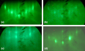
The sample preparation was checked by RHEED. In Fig. 1(a) can be seen the RHEED image taken from the cleaned and annealed Si(001) substrate, which shows sharp 2 × 1 reconstruction. Weak, so-called Kikuchi, lines are seen in the image, indicating the high quality of the Si(001) surface. After iron evaporation, in the case of 1.0-nm deposited Fe, the lines of RHEED image, originating from the substrate, totally disappeared, indicating that the iron film has polycrystalline character. After 60 min annealing at 500 °C according to the RHEED image, a new reconstructed surface developed, showing the epitaxial character of the formed iron silicide [Fig. 1(b)]. These lines are weak and diffuse as a consequence of the moderate quality of the surface, which was grown at relatively low temperature (500 °C). Higher temperature annealing produced sharper lines in the RHEED images. In Fig. 1(c) can be seen the RHEED image of 1-nm Fe annealed at 600 °C for 60 min, and in Fig. 1(d) 1-nm Fe annealed at 850 °C for 60 min.
RHEED images of (a) the cleaned and annealed Si(001) substrate, showing 2 × 1 reconstruction, which indicates the high quality of the surface. RHEED images of evaporated 1-nm Fe layer after annealing for 60 min on Si(001) at (b) 500 °C (c) 600 °C, and (d) 850 °C for iron silicide formation. The images show the epitaxial character of the formed iron silicide, but the weak and diffuse lines refer to a moderate quality surface.

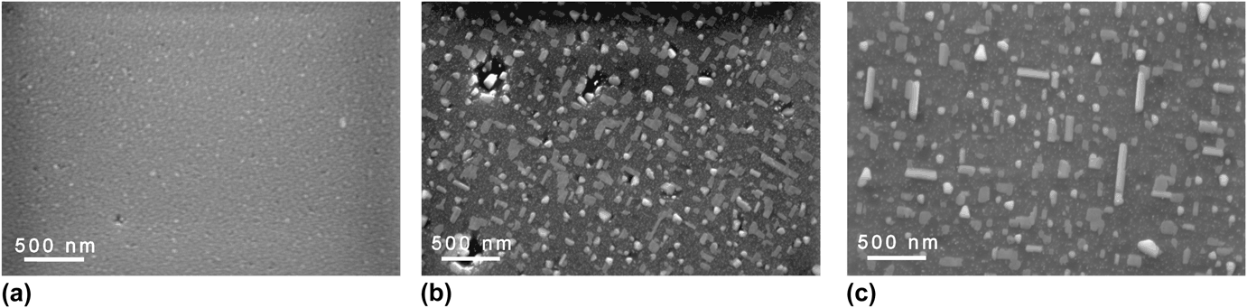
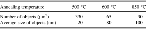
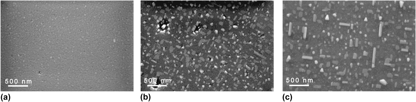
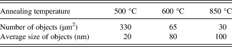
SEM images of the annealing temperature dependent formation of iron silicide nanostructures are presented in Figs. 2(a)–2(c). The nominal “film thickness” was 1.0 nm for each sample, and the heat treatments were carried out at (a) 500 °C, (b) 600 °C, and (c) 850 °C for 60 min. As can be seen, all the samples show aggregated iron silicide nanostructures, but at different scales. The size and the distribution of the islands depend on the temperature. The average dimension of the islands found was between 20 and 100 nm. The density of islands decreased with increasing temperature of annealing, while the size of the nanostructures increased. The average number of nanostructures/µm2, and the average size of the objects are listed in Table I for the three annealing temperatures. In the case of lower annealing temperature (500 °C), the characteristic shapes of nanostructures were dome-like and randomly shaped. Higher temperature annealing yielded triangular, rectangular, and elongated iron silicide nanostructures, which appear in square directions to each other in the plane of the surface, oriented epitaxially to the Si(001) substrate.
SEM images of 1.0 nm iron annealed on Si(001) for 60 min at (a) 500 °C, (b) 600 °C, (c) 850 °C.
The average number/µm2 and average size of the nanostructures versus annealing temperature.
The detailed transmission electron microscopy phase analysis of iron silicide nanostructures was presented in our previous paper.Reference Vouroutzis, Zorba, Dimitriadis, Paraskevopoulos, Dózsa and Molnár27 Where, the nanostructures contain three different iron disilicide phases, i.e., orthorhombic β-FeSi2, cubic γ-FeSi2, and tetragonal α-FeSi2. The epitaxy relationships, as deduced from the diffraction pattern are: (a) [14 $\bar 2$] β-FeSi2//[001] Si, (b) [001] γ-FeSi2//[001] Si and [100] γ-FeSi2//[100] Si, (c) [
 $\bar 12\bar 1$] α-FeSi2//(001) Si and [111] α-FeSi2//<111> Si and [
 $\bar 1$01] α-FeSi2//<
 $\bar 4$80> Si and <110> α-FeSi2//(001) Si and c axis of α-FeSi2 is 22° of the <1
 $\bar 1$0> Si direction.Reference Vouroutzis, Zorba, Dimitriadis, Paraskevopoulos, Dózsa and Molnár27 The ratio of the phases depends on the time and temperature of the annealing.
This ordering is a direct consequence of Ostwald ripening phenomena, where the bigger islands grow further at the cost of the smaller ones. In post-deposition conditions, where no additional material is being deposited, Ostwald ripening is considered the primary coarsening mechanism for island growth.Reference Zinke-Allmang28 The role of the Ostwald ripening is reinforced by the existence of depleted regions near the bigger objects.
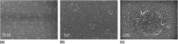
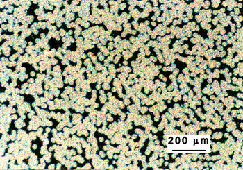
SEM images of the sample annealed at 600 °C for 60 min show interesting surface arrangements at lower magnification. On the surface of the sample, randomly distributed circles with 4–6 µm diameter are seen [Fig. 3(a)]. With the enhancement of the magnification of the microscope [Figs. 3(b) and 3(c)], the inner structure of the circles is more visible. The contours of the circles consist of nanostructures, which are locally bigger than their neighbors, and around them there are depleted regions. The question arises what is the reason for the above special geometric arrangement. To answer the above question an additional, thick iron silicide sample was prepared from 30 nm iron, which was annealed at 600 °C for 5 min. The optical microscopy image taken from this sample is shown in Fig. 4. The surface shows dense, randomly distributed, mainly coalesced circular objects with about 20 µm diameter in the background. This geometry is typical for nucleation-controlled type silicide solid-phase reactions.Reference d'Heurle9 The FeSi to FeSi2 phase transformation is a nucleation-controlled process.Reference Dimitriadis and Werner26 The nucleation-controlled solid-phase reactions produce rough surfaces as a consequence of the coalescence of growing nuclei from different spots. The question may arise, whether these circles refer the nucleation-controlled character of the reaction, or are artifacts. If the circular objects would be artifacts of improper cleaning or any other surface effects, they should have the same size in different samples. But, in the case of 1 nm Fe, their size is in the 4 µm range [Figs. 3(a)–3(c)], and for 30 nm Fe films, their size is around 20 µm (Fig. 4), and Ref. Reference Dimitriadis and Werner26 shows ∼50 µm size circular objects from the 150-nm Fe film. The size of the objects correlates the initial Fe film thickness. That is why they are not a consequence of an accidental interface contamination but caused by the nucleation-controlled growth mode of iron disilicide.
SEM images of 1.0 nm iron annealed at 600 °C for 60 min at enhancing [(a) toward (c)] magnifications. The circular objects show the nucleation-controlled growth of iron silicide at nanoscale.

Optical microscopy image of thin film type (30 nm evaporated Fe annealed at 600 °C for 5 min) iron silicide sample showing typical nucleation-controlled growth.

Apparently, the only possible explanation of the circles displayed in the sample with nanostructures [Figs. 3(a)–3(c)] is a nucleation controlled–type phase formation at the nanoscale too. It is interesting that the nanoobjects inside and at the contours of the circles show the same orientation as the other ones outside. The simultaneous effect of nucleation-controlled growth mode and epitaxial growth resulted a peculiar arrangement and morphology of the iron silicide nanostructures.
It is worth mentioning that the epitaxial growth in solid-phase thin film reactions basically differs from conventional deposit-substrate epitaxy. Initially, the polycrystalline layer reacts with the substrate, and the reaction products may grow epitaxially on the substrate. During the growth, the original (average) interface is shifted toward the substrate because some of the original substrate material has been consumed to grow the silicide layer. In fact, epitaxial silicides form within the substrate not on the substrate. This phenomenon has no special consequences in conventional planar thin film geometry, but in the case of nanostructures, it has a spectacular impact. Sometimes, the nanostructures are sunk into the substrate.Reference Chen, Chen and Chen29 Nowadays, this kind of epitaxy is called endotaxy (oriented growth within a substrate),Reference Mahato, Das, Juluri, Batabyal, Roy, Satyam and Dev30 but this is only a subcategory of epitaxy.
IV. CONCLUSIONS
Iron silicide nanoislands were grown by conventional Fe evaporation and subsequent annealing on the Si(001) substrate. The size distribution and shape of the formed islands depended on the annealing temperatures. In the case of lower annealing temperature, the characteristic shapes of nanostructures were dome-like and randomly shaped. Higher temperature annealing resulted in triangular, rectangular, and elongated iron silicide nanoobjects, which are oriented in square directions. In some cases, the nanoparticles were arranged in circles. This might be the direct consequence of a nucleation-controlled type of local transition of iron monosilicide to the iron disilicide phase at nanoscale.
ACKNOWLEDGMENT
The author thanks Zofia Vertesy for SEM images. The support of OTKA Grant No. 81998 is acknowledged.