Introduction
The recurrent laryngeal nerve (RLN) may be injured as a result of a variety of events, including iatrogenic surgical injury, neoplasm or neurological disease. Injury to the RLN results in vocal fold paresis or paralysis. Poor or absent movement of one or both vocal folds causes difficulties with voice, deglutition or breathing.Reference Eisele and Eisele1, Reference Zeitels, Casiano, Gardner, Hogikyan, Koufman and Rosen2 Current surgical options for a paralyzed vocal fold include medialisation and reinnervation procedures. However, these do not restore physiological movement to the paralyzed fold; they are geometric solutions for a dynamic neurological problem. Clearly, an intervention which restores physiological movement would be ideal and would have important therapeutic implications.
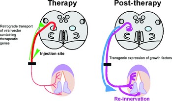
The introduction of therapeutic gene products to the central nervous system (CNS) represents a promising therapeutic approach to both peripheral and central nervous system pathology, including trauma and neurodegenerative disease.Reference Kang, Kaplitt and Loewy3, Reference Ghadge, Roos, Kang, Wollmann, Fishman and Kalynych4 Insulin-like growth factor I has neurotrophic and protective effects in the larynx.Reference Rubin, Mobley, Hogikyan, Bell, Sullivan and Boulis5 Viral vectors containing therapeutic genes for neurotrophic factors can be delivered to the CNS via remote injection.Reference Boulis, Turner, Dice, Bhatia and Feldman6–Reference Boulis, Bhatia, Anlar, Brindle and Feldman9 Previous research has shown that an adeno-associated virus vector, remotely injected into the RLN, travels in a retrograde fashion to the rat brainstem,Reference Rubin, Hogikyan, Sullivan, Boulis and Feldman10 and that injection of an adenoviral vector into a crushed RLN added no additional injury compared with crush alone.Reference Shiotani, O'Malley, Coleman and Flint11 A proposed novel therapeutic approach to RLN injury, by the introduction of growth factor genes to the brainstem via remote injection of viral vectors, is outlined in Figure 1. In order to study the effects of this approach, a quantifiable and reproducible injury model must first be defined.
Proposed viral gene therapy vector model for reinnervation of the injured recurrent laryngeal nerve.

The objective of this project was to develop and characterise an injury model for the study of potential viral gene therapy to treat RLN injury.
Materials and methods
Subjects
The study was undertaken in 20 adult, male, Sprague–Dawley rats (300 g weight).
Experimental design
A prospective, randomised study was undertaken. Rats were randomly assigned to one of five groups, and sacrificed at weekly intervals between one and five weeks in order to determine outcome measurements.
Outcome measures
The degree of reinnervation or denervation was quantified by measuring the percentage of nerve to motor endplate contact. This was defined as the number of motor endplates contacting nerves, divided by the total number of motor endplates.
Details of animal surgery
Anaesthesia and direct laryngoscopy
Rats were anaesthetised with 50 mg/kg intraperitoneal pentobarbital. Direct laryngoscopy was performed transorally with loupe magnification. Vocal fold mobility was documented. The animals were intubated with a 16 gauge angiocatheter and ventilated with a Harvard rat ventilator. Supplemental oxygen and 2 per cent isofluorane were used to maintain general anaesthesia during surgery.
Surgery
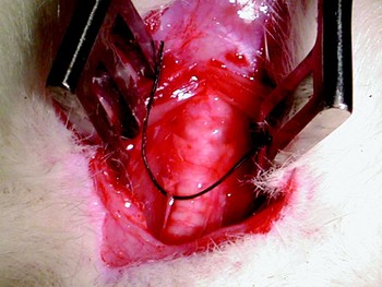
The neck was shaved and Betadine® applied. A vertical skin incision was made between the angle of the mandible and the sternal notch. The platysma and strap muscles were divided, revealing the thyroid gland, larynx and trachea. The RLN was identified with loupe magnification, within the tracheoesophageal groove (see Figure 2).
Animal surgery: exposure of the recurrent laryngeal nerve (RLN). The right RLN is indicated by black thread.

The right RLN was crushed with jeweler's forceps, with enough force to bring the tips into apposition, for a duration of exactly 30 seconds, at the level of the sixth tracheal ring, as previously described by Rubin et al. Reference Rubin, Mobley, Hogikyan, Bell, Sullivan and Boulis5 A multi-layer closure was performed after adequate haemostasis was obtained with bipolar cautery. Rats were extubated while they were breathing spontaneously. Vocal fold function was reassessed by direct laryngoscopy to confirm complete paralysis of the right vocal fold. All surgical procedures and laryngoscopies were performed by the same investigator (KF).
Sacrifice
At the specified time intervals, rats were sacrificed using an overdose of pentobarbital (200 mg/kg intraperitoneally) and intracardiac perfusion of paraldehyde. Rigor mortis was observed in order to ensure that the animal would not revive. This method was consistent with the recommendations of the panel of euthanasia of the American Veterinary Medical Association.
Tissue harvest
The larynx was removed, cryoprotected in phosphate buffer containing 20 per cent sucrose, and frozen in isopentane. Larynges were sectioned in 15 µm axial sections (resulting in longitudinal sections of the thyroarytenoid muscles).
Tissue staining
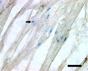
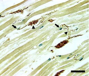
Motor endplates were stained for acetylcholinesterase, and neurofilament was stained using standard immunohistochemical methods previously described (see Figures 3 and 4).
Denervated thyroarytenoid muscle, one week post-crush injury. Motor endplates stain blue (acetylcholinesterase; arrow). Neurofilament–endplate contact = 11.6 per cent. Scale bar = 100 µm.

Reinnervated thyroarytenoid muscle, three weeks post-crush injury. Motor endplates stain blue (acetylcholinesterase; arrows). Neurofilaments stain brown (arrowheads). Neurofilament–endplate contact = 86.8 per cent. Scale bar = 100 µm.

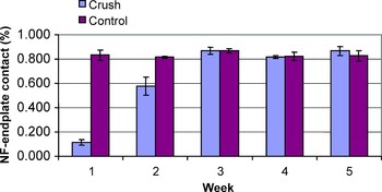
Percentage neurofilament to endplate contact as a function of time following crush injury. NF = neurofilament; control = non-injured side; error bars = standard deviation

Outcome measures
The degree of denervation or reinnervation was quantified by measuring the percentage of nerve to motor endplate contact. This was defined as the number of motor endplates contacting nerves, divided by the total number of motor endplates, per high power field. A blinded investigator determined the nerve–endplate contact percentage in the thyroarytenoid muscles under light microscopy, by manually counting the number of motor endplates contacting nerves and then dividing by the total number of motor endplates. Every seventh laryngeal section was evaluated. An average of 89 motor endplates were evaluated per rat. Analysis of variance and standard t-tests were used to compare the mean percentage of endplates contacted by neurofilament. Alpha <0.05 was considered significant.
Results and analysis
Results are summarised in Figures 3 to 5. As expected, the control (non-crushed) side exhibited normal vocal fold mobility on laryngoscopy, and the mean nerve–endplate contact percentage was high (86.8 per cent) and remained unchanged over time. In contrast, the crushed side exhibited complete vocal fold paralysis immediately after injury, and the mean nerve–endplate contact percentage was low (11.6 per cent) at one week and gradually increased with time (being 53.9 per cent at two weeks and 88.6 per cent at three weeks). Clinical recovery of vocal fold mobility mirrored these objective findings, and movement was normal by three weeks. Furthermore, the nerve–endplate contact percentage remained high thereafter (being 81.7 per cent at four weeks and 86.6 per cent at five weeks). The difference between results at week one and week three was statistically significant (p < 0.01), both for different time groups and for the injured side vs control side within individual animals.
Discussion
The optimal goal for surgical rehabilitation of recurrent laryngeal nerve injury is re-establishment of physiological function. Although current surgical techniques for treatment of RLN injury can ameliorate symptoms, they are not capable of producing physiological movement of the vocal fold. Transgenic expression of therapeutic factors, using viral gene therapy, may prove to be a novel method for realising the potential of laryngeal reinnervation. There are multiple candidate genes for treatment of peripheral nerve injury, including insulin-like growth factor I.Reference Shiotani, O'Malley, Coleman and Flint11
Before exploring the therapeutic effects of gene therapy, however, a reliable and predictable model of injury must be established. Preliminary results have suggested that crush injury will provide such a model, as it consistently produces axonotmesis.Reference Rubin, Hogikyan, Sullivan, Boulis and Feldman10 This model also seems likely to provide an intact neural conduit to allow retrograde transport of the viral vector to the brain stem. Thus, the broad, long-term objective of this study was to establish an injury model with which to investigate gene therapy as a therapeutic strategy for RLN injury.
The results of this study demonstrate that the anatomical integrity of neuromuscular connections can be reliably quantified by determining the nerve–endplate contact percentage in laryngeal muscle. Crush injury was shown to result initially in an unequivocal, dramatic decrease in nerve–endplate contact percentage, as an objective measure of denervation. Data at one week correlate well with those of an earlier study in which only one week post-crush data were obtained.Reference Rubin, Mobley, Hogikyan, Bell, Sullivan and Boulis5 Furthermore, following this injury, there was a gradual, predictable sequence of reinnervation, as evidenced by a progressive increase in nerve–endplate contact percentage. The time course of injury and recovery was fully characterised in the current study. The functional integrity of the laryngeal neuromuscular connections was determined through laryngoscopic observation of vocal fold mobility, and this functional variable correlated well with the histological measure. Thus, characterisation of this model provides an experimental system in which the effects of therapeutic interventions could readily be studied. Ameloriation of initial denervation and/or enhanced rates of reinnervation and functional recovery would be the goals of such studies.
We acknowledge that crush injury seems crude and difficult to control, but emphasise that in this study, the force applied (i.e. until the tips of the jeweler's forceps came into apposition) and the duration of crush (i.e. 30 seconds), were, in fact, well controlled and reproducible. The variability in our results was small (see standard deviations in Figure 3), which further supports the consistency and reproducible nature of the crush injury. Furthermore, previous studies have investigated the effects of nerve crush in other models.Reference Bridge, Ball, Mackinnon, Nakao, Brandt and Hunter12–Reference Park, Kim and Kim14 Bridge and associates demonstrated that crush injury to the sciatic nerve produced a consistent injury of axonotmesis (Sunderland second degree) evident at two weeks and recovered by four weeks.Reference Bridge, Ball, Mackinnon, Nakao, Brandt and Hunter12 Most importantly, they used a variety of methods to crush the nerve, and demonstrated no significant difference in the degree of injury amongst the various crush techniques. Mu and Yang studied crush injury to the canine RLN. They reported electromyography (EMG) evidence of complete denervation one week after crush injury, by loss of motor unit potentials and by involuntary fibrillation potentials. Motor unit potentials returned by four weeks, and at two months the EMG appeared normal. This suggested complete reinnervation of the intrinsic musculature of the canine larynx within eight weeks.Reference Mu and Yang15 Van Lith-Bijl and associates also examined the effect of crush injury to the RLN in cats. They demonstrated evidence of reinnervation by two and a half weeks post-crush, by laryngoscopy and EMG.Reference van Lith-Bijl, Mahieu, Stolk, Tonnaer, Groenhout and Konings16 These studies support the concept that crush injury is a consistent, reproducible injury which is followed by a predictable sequence of reinnervation. However, we feel that EMG might not be an ideal outcome measure, because it is (Reference Eisele and Eisele1) more technically difficult in smaller animals such as the rat, and (Reference Zeitels, Casiano, Gardner, Hogikyan, Koufman and Rosen2) considerably more subjective than assessment of nerve–endplate contact percentage.
It is important to note that this injury model does have limitations. Crush injury is not representative of the more severe type of neural insult often encountered clinically. Nerve transection is a more severe injury, and one with even greater need for improved therapy, but it is less optimal for initial proof-of-principle studies exploring viral gene therapy. A transection model introduces a significant variable, primary neurorrhaphy, making the experimental system more prone to variability that could confound interpretation of results. We acknowledge that if gene therapy is shown to accelerate reinnervation or limit denervation in the setting of crush injury, then further studies would be warranted, involving a more severe type of neural injury.
A potential limitation of this model is the near-complete degree of reinnervation within three weeks. However, we see this as an exciting opportunity. Current evidence supports our contention that the effects of gene therapy can be studied within this relatively short time period, for several reasons.
Firstly, recombinant adeno-associated viral vector (rAAV) carrying the gene for green fluorescent protein (GFP) has been shown to travel to the rat brainstem via remote injection in the intact RLN by three weeks.Reference Rubin, Hogikyan, Sullivan, Boulis and Feldman10 The same group then went on to demonstrate that adenoviral vector (containing no transgene) delivered to the crushed RLN, in a rat model, travels retrograde to the brainstem within one week. This is evidenced by the presence of virus in brainstem sections (using fluorescent in situ hybridisation) in animals sacrificed one week after injection.Reference Rubin, Mobley, Hogikyan, Bell, Sullivan and Boulis5
Secondly, in a different model, the vagus nerve was avulsed from the skull base, and adenoviral glial cell line-derived neurotrophic factor (GDNF) was injected into the jugular foramen. Animals sacrificed at four days expressed B-galactosidase activity in the nucleus ambiguous.Reference Saito, Shiotani, Watabe, Moro, Fukuda and Ogawa17
Thirdly, in a transection model, a muscle-specific vector injected into the thyroarytenoid muscle resulted in gene expression as well as a significant increase in nerve–endplate contact, four weeks post-injection.Reference Shiotani, O'Malley, Coleman and Flint11 In this study, no animals were sacrificed prior to four weeks, so the exact kinetics of gene expression remain unknown, but must be within a four week time-frame in the transection model (i.e. completely denervated muscle endplates), which is more rigorous than a crush model (i.e. incompletely denervated muscle endplates).
Therefore, we can infer from this evidence that, in the crush model, gene expression at the level of the brainstem should occur within four to seven days of injection, and that the subsequent downstream effects on nerve–endplate contact should occur well within four weeks. If no effects are seen in the proposed model by three weeks, efficacy of gene therapy cannot be ruled out. However, if positive effects are seen in the proposed model by three weeks, this finding would be quite compelling.
We wish to clarify that it was the purpose of this paper to define, create and quantify an injury model that could be used for gene therapy studies. The results of the model suggest that earlier time points should be investigated when designing these studies. Although the model in this paper examined the effects of nerve crush at weekly intervals, it is clear that the model could and should be applied to earlier time points (i.e. within a two-week time period). This model demonstrates a proof-of-principle, and should be used as a hypothesis-generating springboard for future studies.
We propose that crush injury is a good choice for an experimental gene therapy model, for the following reasons. Firstly, crush injury yields a predictable sequence of denervation followed by reinnervation. Secondly, this can be quantified by determining the nerve–endplate contact percentage, using light microscopy, and the functional consequences can be observed via laryngoscopy. Thirdly, crush preserves the neural conduit necessary for retrograde axonal transport of a viral vector and for ingrowth of recovering neural elements.
• This study aimed to develop and characterise an experimental model of recurrent laryngeal nerve (RLN) injury for the study of viral gene therapy, using the rat as an animal model
• Crush injury to the RLN resulted in a dramatic, measurable decrease in nerve-to-endplate contact percentage
• Spontaneous recovery was observed by three weeks post-injury
• This experimental model will be used to investigate the potential therapeutic role of viral gene therapy for the treatment of RLN injury
To date, the most compelling evidence supporting our experimental model comes from a recently published Japanese study.Reference Araki, Shiotani, Watabe, Saito, Moro and Ogawa18 This paper described a similar crushed RLN model, and demonstrated improved neurofunctional recovery following adenoviral glial cell line derived neurotrophic factor gene transfer. Remote injection of an adenoviral vector encoding glial cell line derived neurotrophic factor, following crush injury, resulted in positive identification of gene expression at the brainstem, as well as improved motor nerve conduction velocity, larger axonal diameter, improved remyelination and improved clinical recovery of vocal fold movement. Furthermore, this neurofunctional recovery was evident by two weeks.
Conclusion
Crush injury to the recurrent laryngeal nerve results in a dramatic, measurable decrease in nerve-to-endplate contact percentage. This decrease is reliably quantifiable. Spontaneous recovery is observed by three weeks post-injury. This experimental model will be used to investigate the potential therapeutic role of viral gene therapy for the treatment of RLN injury.
Acknowledgements
This work was supported by a grant from the National Institutes of Health (NS38849) and the Program for Understanding Neurological Diseases (PFUND).