The Amplatzer muscular ventricular septal defect device had been used for closure of the native right ventricular outflow tract in the setting of single-ventricle post-Glenn shunt, which prevents the forward flow from the systemic ventricle to the pulmonary arteries.Reference Thanopoulos, Georgakopulos and Tsaousis 1 This technique in the closure of the native right ventricular outflow tract in biventricular repair has not been reported.
Case report
We report a case of a 13-year-old girl (weight 64 kg, height 148 cm) who underwent tetralogy of Fallot repair at the age of 3 years. Owing to abnormal coronary artery branch crossing the right ventricular outflow tract, a 22 mm Hancock valved conduit was utilised to bypass the right ventricular outflow tract obstruction, leaving the native outflow tract patent. At 9 years of age, the patient showed signs and symptoms of conduit dysfunction due to severe stenosis and moderate regurgitation and underwent percutaneous Melody valve implant. After 4 years, the native outflow tract regurgitation increased from mild to severe.
Pre-procedure evaluation
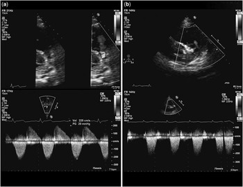
Transthoracic echocardiography showed moderate right ventricular dilatation, mild stenosis at the Melody valve, and a peak Doppler gradient of 20 mmHg with trivial regurgitation. There was severe regurgitation through the native right ventricular outflow tract (Fig 1a).
Parasternal short-axis view, two-dimensional echocardiogram with continuous wave Doppler colour flow imaging. ( a ) Severe regurgitation across the native right ventricular outflow tract. The corresponding continuous wave Doppler demonstrates the severity of the regurgitation. ( b ) The native right ventricular outflow tract after closure with an Amplatzer muscular ventricular septal defect device with trivial regurgitation from the Melody valve and none from the native right ventricular outflow tract are shown. The device is in good position. The corresponding continuous wave Doppler shows no regurgitation and no change in the stenosis of the Melody valve.

To determine the regurgitant volume from the native outflow tract, she underwent cardiac magnetic resonance imaging, which showed that the Melody valve and its stent are in a stable position, with mild narrowing at the proximal portion. There was trivial incompetence with an estimated regurgitation fraction of 3%. The native pulmonary valve was thickened and stenotic. Flow acceleration was evident on cine-images with moderate to severe regurgitation – regurgitant fraction 29%. The right ventricle was moderately dilated and hypertrophied with low normal systolic function; the right ventricular end-diastolic volume was 185 ml, right ventricular end-systolic volume was 97 ml, and right ventricular ejection fraction was 48%.
Despite the fact that the cardiac magnetic resonance imaging data showed only borderline indications to intervene, the colour echo Doppler suggested severe native right ventricular outflow tract regurgitation. This fact added to the patient showing easy fatigability and the right ventricle being dilated, and we thought that reducing the rate of regurgitation from the native right ventricular outflow tract would possibly reduce the probability of right ventricular dysfunction.
Procedure
Cardiac catheterisation procedure was performed under general anaesthesia.
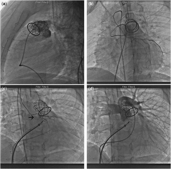
The right and left femoral veins were accessed using 7 Fr sheath. With the help of a 7 Fr Berman angiographic catheter, haemodynamic measurements were obtained. Right ventricular angiography showed forward flow through the Melody valve and the native right ventricular outflow tract. Cineangiography in the main pulmonary artery showed moderately severe regurgitation across the native right ventricular outflow tract with trivial incompetence of the Melody valve (Fig 2a).
Still-frame cineangiogram. ( a ) Lateral view of the right ventricular outflow tract with moderately severe regurgitation of the native right ventricular outflow tract. ( b ) Simultaneous balloon sizing and coronary angiogram. ( c ) The device in the right ventricular outflow tract with simultaneous coronary angiogram. ( d ) Pulmonary angiogram through the Melody valve showing a competent Melody valve, and no evidence of right ventricular outflow tract regurgitation.

A 7 Fr wedge balloon catheter was advanced through the native right ventricular outflow tract to the right pulmonary artery. Then, a Lunderquist exchange guide wire (Cook Medical, Bloomington, Indiana, United States of America) was passed to the right pulmonary artery. The balloon wedge catheter was exchanged with AGA sizing balloon 24 mm (AGA Medical Corporation, Golden Valley, Minnesota, United States of America) to estimate the diameter of the native right ventricular outflow tract, and at the same time simultaneously coronary angiography was obtained (Fig 2b). The waist of the balloon was measured to be 14 mm at the narrowest diameter. Thereafter, the sizing balloon was removed and a 10 Fr Amplatzer sheath was advanced through the native right ventricular outflow tract to the main pulmonary artery. A size 16 mm Amplatzer muscular occluder device (AGA Medical Corporation) was advanced under fluoroscopic guidance and positioned into the native right ventricular outflow tract, making sure that the two discs of the device were straddling the narrowest area of the native right ventricular outflow tract. Repeat coronary angiography showed that the implanted device did not impinge on the coronary artery (Fig 2c). The device was kept attached to the cable for 20 minutes. During this period, we assured that there was neither haemodynamic instability nor signs of ischaemia. A repeated pulmonary artery angiography showed that the device was in good position and the waist straddling the native right ventricular outflow tract with trivial angiographic evidence of regurgitation (Fig 2d). Thus, the device was released and maintained in its position.
Post procedure
The patient was discharged the next day on low-dose aspirin. On follow-up, transthoracic echocardiography showed the device in good position and no evidence of regurgitation across the native right ventricular outflow tract accepting trivial regurgitation and unchanged gradient across the Melody valve (Fig 1b). This result remained unchanged till last follow-up 18 months post procedure.
Limitation of the study
We acknowledge that the duration of follow-up is short and that long-term follow-up is warranted.
Discussion
During the past 12 years, percutaneous pulmonary valve implantation was established as a valid alternative to surgical replacement of failed conduit repair in the right ventricle outflow tract.Reference Bonhoeffer, Boudjemline and Saliba 2 , Reference Lurz, Nordmeyer and Giardini 3 The indication was expanded to involve the right ventricular outflow patch.Reference Momenah, El Oakley and Al Najashi 4 – Reference Bouzaz, Kilner and Gatzoulis 6
Occasionally, the association of tetralogy of Fallot with abnormal coronary arteries might preclude a repair involving transannular patch. In this situation, often a conduit is used to bypass the right ventricular outflow tract.Reference Dandolu, Baldwin and Norwood 7 Some surgeons leave the native right ventricular outflow tract patent, to prevent low cardiac output if inadvertent obstruction to the new conduit occurred. In addition, this may allow implanting a smaller conduit. However, when there is a need to exchange the conduit, the native right ventricular outflow tract may need to be addressed if there is severe regurgitation.Reference Tchervenko, Peeletier and Shum-Tim 8 For cases of a well-functioning conduit valve implant, in the presence of a regurgitant native right ventricular outflow tract, there are different management options. Aside from surgical intervention, the option of implanting a second pulmonary valve in the native outflow tract could be contemplated. We did not choose this as it is technically more challenging. We presumed that it might cause inadvertent compression of the abnormal coronary arteries. Furthermore, it would have been extremely difficult to retrieve the implantable second valve once in place if ischaemia develops. Thus, a surgical operation would have been required on an urgent basis. We chose instead to close the native right ventricular outflow tract utilising a muscular ventricular septal defect device, as it was an easier and safer alternative. As it has been demonstrated during the procedure, this option allowed testing the coronary artery by angiography, after positioning the device, and thus it could have been retrieved easily if needed.
Conclusions
Our case demonstrates the effective use of a muscular ventricular septal defect device to close the native right ventricular outflow tract in the setting of tetralogy of Fallot following surgical repair with a conduit. We believe that this procedure is a valid alternative to either surgical approach or implanting a Melody valve in the position of the native right ventricular outflow tract. Long-term results of this procedure need to be observed.
Acknowledgement
We are thankful to our non-invasive echo lab staff including Dr Abdul Rehman Almoukirish, Mr Mohammed Alonazi and Miss Fareeda Al Hazmi for excellent images. We are thankful to cath lab staff Mr Aymen N Alnasir and Miss Vivienne Vella.
Financial Support
The research received no specific grant from any funding agency, commercial or not for profit sector.
Conflicts of Interest
None.