Case summary
Fallot’s Pentalogy is a cyanotic congenital heart malformation that includes an atrial septal defect, a ventricular septal defect, subvalvular or valvular pulmonary stenosis, right ventricular hypertrophy, and an overriding aorta. Hypoplasia or atresia of the pulmonary arteries may further increase right-to-left shunting. Major systemic-to-pulmonary collaterals (MAPCA) may provide supplementary blood flow to the lungs in this case.
Dynamic CT angiography is a novel imaging modality in patients with complex cardiac malformations. It allows for detailed visualisation of cardiac anatomy as well as for haemodynamic assessment because of its high spatial and temporal resolution. We present a dynamic CT angiography (four-dimensional CT SOMATOM Force; Siemens, Forchheim, Germany) of a 55-year-old man with Fallot’s Pentalogy (Supplementary videos 1 and 2). The four-dimensional CT angiography was performed using the shuttle-mode technique in order to cover the whole chest. In order to reduce radiation dose, 10 dynamic acquisitions were performed with a interscan delay of 1.5 second using a low tube voltage setting of 70 kVp together with automated tube current modulation. The total radiation dose of the scan was 12.5 mSv, which compares with a retrospectively gated cardiac CT acquisition performed with 120 kVp.
The patient’s initial treatment consisted of a Blalock shunt at age 5. Closure of the atrial septal defect and the ventricular septal defect, resection of the subpulmonary stenosis, cross-section of the pulmonary valve, and implantation of a transannular patch were performed at age 8. A pseudoaneurysm was diagnosed and resected in adulthood together with closure of a major aortopulmonary collateral to the left pulmonary artery. An implantable cardioverter-defibrillator was implanted at the age of 47 because of episodes of sustained ventricular tachycardia.
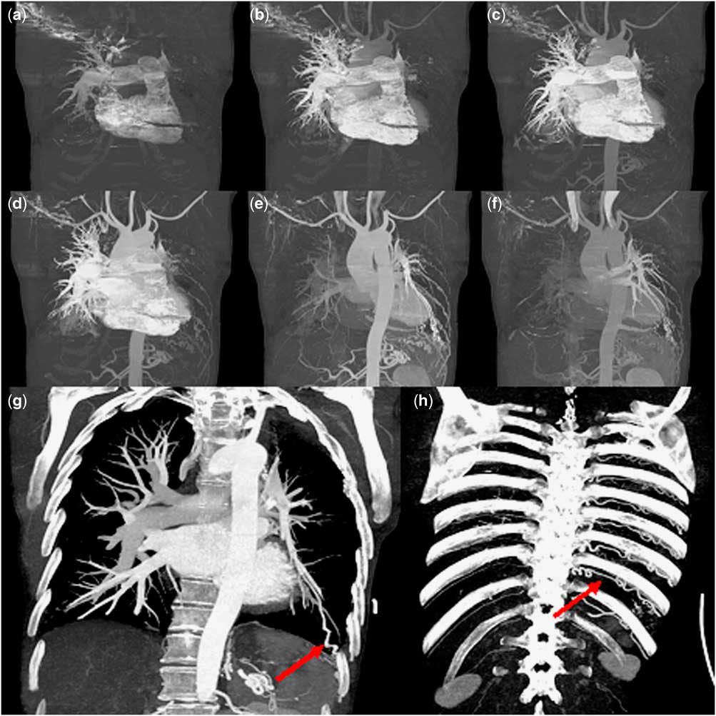
The current presentation of the patient was due to ventricular tachycardia. Dynamic CT angiography (Fig 1) showed subtotal occlusion of the left pulmonary artery with mixed perfusion of the left lung (Fig 1a–f) by a prominent infra-diaphragmatic major aortopulmonary collateral (Fig 1g) as well as multiple collaterals from the unilateral dilated intercostal arteries (Fig 1h).
Dynamic CT angiography demonstrates retrograde perfusion of the left lung ( a – f ) by a prominent infra-diaphragmatic major aortopulmonary collateral ( g ; arrow) as well as multiple collaterals from the unilateral, dilated intercostal arteries ( h ; arrow).

This case demonstrates that dynamic CT angiography is a useful second-line alterative to MRI in non-paediatric patients with a clinical need for functional perfusion analysis and a contraindication for MRI.
Acknowledgements
None.
Financial Support
This research received no specific grant from any funding agency or from commercial or not-for-profit sectors.
Conflict of Interest
None.
Supplementary material
To view supplementary material for this article, please visit https://doi.org/10.1017/S1047951117000610