1. Introduction
Structure-based drug design (SBDD) involves the use of three-dimensional (3D) structural data to advance lead identification and subsequent optimization for drug discovery. The exponential growth of the Protein Data Bank (PDB) and improvement in homology modeling techniques makes SBDD applicable to an ever-growing number of pharmaceutically relevant targets with an available 3D structure. SBDD studies are generally centered upon at least one of the following three goals: prediction of binding modes, prediction of binding affinities and prediction of novel binding partners. Depending on these goals and available data, SBDD can involve different approaches, which are often separated into docking techniques and de novo design. Docking is implemented to predict the binding mode and affinity of small molecules. When docking is applied to a large compound database, it is referred to as virtual screening (VS) and often centered on the prediction of new ligands with high affinity for a target molecule to enrich the compound set for experimental testing. De novo design is performed with the intent of predicting new compounds in novel chemical space. Fragment-mapping techniques that use functional groups to probe binding sites are often applied for this type of approach. Over the past 20 years, these SBDD studies have produced viable leads, enabling the development of successful clinical drugs and leading to the extensive implementation of SBDD in medicinal chemistry research (Jorgensen, Reference Jorgensen2004; Taft et al. Reference Taft, Da Silva and Da Silva2008; Talele et al. Reference Talele, Khedkar and Rigby2010).
X-ray crystallography and NMR studies have clearly demonstrated conformational differences between many receptors’ holo (bound) and apo (unbound) states. Sampling ligand conformations is straightforward; most SBDD protocols now include ligand flexibility (with on-the-fly sampling being superior to a rigid set of pre-generated conformations), yet this is insufficient for the most accurate results. Despite data demonstrating the influence of protein flexibility on ligand binding, most SBDD efforts still rely upon a static receptor structure because of the resources required to account for its many degrees of freedom. Although a few proteins can have their binding potential represented with a single, fixed conformation, for most systems the information presented by a rigid structure is simply inadequate.
In a comparative study of 10 docking programs and 37 scoring functions, no single method outperformed the others when performing docking of diverse compounds to a set of eight rigid proteins (Warren et al. Reference Warren, Andrews, Capelli, Clarke, LaLonde, Lambert, Lindvall, Nevins, Semus, Senger, Tedesco, Wall, Woolven, Peishoff and Head2006). The scoring functions were unable to accurately predict binding affinity or relatively rank the compounds. Ginalski and co-workers evaluated the binding predictions and scoring results for 1300 protein–ligand complexes from PDBbind 2007 with Surflex, LigandFit, Glide, GOLD, FlexX, eHiTS, and AutoDock (Plewczynski et al. Reference Plewczynski, Lazniewski, Augustyniak and Ginalski2011). The authors found that the programs achieved a mean RMSDTop-Score that ranged from 2.77 Å (GOLD) to 4.37 Å (FlexX) for the docked poses, but those top scoring poses did not typically match the best RMSD pose (matching occurred in 40–60% of cases), and none of the scoring functions were able to achieve a reasonable correlation between the best pose score and the experimental activity (Spearman correlation ranged from 0.07 (LigandFit) to 0.39 (eHiTS)).
Several recent articles and reviews have been published, which discuss the deficiencies of existing scoring functions for docking (Englebienne & Moitessier, Reference Englebienne and Moitessier2009; Huang et al. Reference Huang, Grinter and Zou2010; Leach et al. Reference Leach, Shoichet and Peishoff2006; Moitessier et al. Reference Moitessier, Englebienne, Lee, Lawandi and Corbeil2008; Mukherjee et al. Reference Mukherjee, Balius and Rizzo2010; Warren et al. Reference Warren, Andrews, Capelli, Clarke, LaLonde, Lambert, Lindvall, Nevins, Semus, Senger, Tedesco, Wall, Woolven, Peishoff and Head2006). Importantly, Englebienne and Moitessier's careful study of 209 protein–ligand complexes and 18 scoring functions demonstrated that the accuracy of certain scoring functions was negatively impacted by protein flexibility and solvation. Mukherjee et al. (Reference Mukherjee, Balius and Rizzo2010) evaluated the relationship between success, sampling failure, and scoring failure in static, fixed anchor, and anchor-and-grow docking outcomes with DOCK. They showed that scoring failures increased as sampling errors decreased, and scoring failures appeared to peak at an RMSD between 1.5 and 2.0 Å. Klebe and co-workers hypothesized that there is an intimate link between docking and scoring, postulating that correct prediction of accurate binding geometries will simultaneously solve the scoring problem (Klebe, Reference Klebe2006; Velec et al. Reference Velec, Gohlke and Klebe2005; Zentgraf et al. Reference Zentgraf, Steuber, Koch, La Motta, Sartini, Sotriffer and Klebe2007). Therefore, while we acknowledge the limitations of current scoring functions, we focus on the variety of techniques for incorporating protein flexibility in SBDD.
A number of reviews have included some consideration of the impact of protein flexibility on drug discovery (Alonso et al. Reference Alonso, Bliznyuk and Gready2006; Bottegoni, Reference Bottegoni2011; Cozzini et al. Reference Cozzini, Kellogg, Spyrakis, Abraham, Costantino, Emerson, Fanelli, Gohlke, Kuhn, Morris, Orozco, Pertinhez, Rizzi and Sotriffer2008; Durrant & McCammon, Reference Durrant and McCammon2010; Fuentes et al. Reference Fuentes, Dastidar, Madhumalar and Verma2011; Henzler & Rarey, Reference Henzler and Rarey2010; Klebe, Reference Klebe2006; Leach et al. Reference Leach, Shoichet and Peishoff2006; Rao et al. Reference Rao, Subramanian and Sharma2009; Sotriffer, Reference Sotriffer2011; Sousa et al. Reference Sousa, Fernandes and Ramos2006; Teague, Reference Teague2003; Teodoro & Kavraki, Reference Teodoro and Kavraki2003; Totrov & Abagyan, Reference Totrov and Abagyan2008). Many of these reviews concentrated on only a subset of protein flexibility methods or emphasized a particular technique. The individual roles of sampling protein flexibility and accurate scoring upon docking success and failure remain unclear. Without a standard for reporting methodological outcomes or a consistent set of test cases, it is difficult to tease apart the relative influence of conformational searching versus interaction scoring. Furthermore, it may be difficult, if not impossible, to judge the extent to which conformational sampling appropriately models the true potential energy landscape. Here, we try to address these difficult questions through the examination of a variety of techniques that account for flexibility in protein–ligand binding, not only studies where the primary focus is on docking but also studies that concentrate on identifying potential binding sites with small molecules or fragments.
2. Protein flexibility
2.1 Conformational variability
Our understanding of receptor–ligand binding has significantly advanced from the original lock-and-key model proposed by Fischer (Reference Fischer1890). Early experimental studies showed that the act of ligand binding influences the protein conformation, referred to as conformational induction or induced fit (Koshland, Reference Koshland1958). Another model of ligand binding is conformational selection, wherein the ligand chooses a binding partner from among available states in the conformational ensemble, thereby shifting the population distribution (Berger et al. Reference Berger, Weber-Bornhauser, Eggenberger, Hanes, Pluckthun and Bosshard1999; Foote & Milstein, Reference Foote and Milstein1994; Kumar et al. Reference Kumar, Ma, Tsai, Sinha and Nussinov2000; Monod et al. Reference Monod, Wyman and Changeux1965; Tsai et al. Reference Tsai, Kumar, Ma and Nussinov1999a, Reference Tsai, Ma and Nussinovb, Reference Tsai, Ma, Sham, Kumar and Nussinov2001). Recently, several papers have been published examining the evidence for whether receptor binding occurs because of induced fit or conformational selection. Sullivan & Holyoak (Reference Sullivan and Holyoak2008) studied the kinetics of phosphoenolpyruvate carboxykinase and concluded that induced fit and conformational selection were not mutually exclusive, rather they were complementary avenues for binding. Weikl & von Deuster (Reference Weikl and von Deuster2009) developed equations of binding kinetics using four states, the dominant apo and holo protein conformations without a bound ligand and those same conformations with a ligand bound, to describe the protein–ligand complex. They used the equilibrium constants from this model to distinguish between the induced and selected fit. Hammes et al. (Reference Hammes, Chang and Oas2009) examined the binding pathways for dihydrofolate reductase (DHFR) and flavodoxin and determined that a mixed binding mechanism was most likely. They noted that the relative importance of induced fit or conformational selection for a particular case could be analyzed by comparing the flux through the reaction path of ligand-binding and conformational change. Both mechanisms of binding will produce the same result; it is important only that some mechanism of receptor conformational change be incorporated in docking simulations. This is particularly useful in SBDD because it implies that computationally inexpensive methods that include protein flexibility should correctly predict binding modes.
2.2 Allostery
Protein–ligand flexibility upon binding is crucial for proper understanding of allosteric regulation. Nussinov and co-workers postulated that most proteins exist in an ensemble of states, and thus most proteins have the potential for allostery (Gunasekaran et al. Reference Gunasekaran, Ma and Nussinov2004). Based on the experimental literature, they demonstrated that the binding of an allosteric ligand shifted the population of conformational substates, thereby influencing the ability of other ligands to bind an alternate site.
Drug resistance is a growing problem that calls for new approaches to drug therapy. Exploring allosteric control in protein targets provides exciting opportunities for finding new modes of action and hence, overcoming emergent resistance, and developing cocktails of drugs to improve treatment.
3. Protein–ligand binding
3.1 The cross-docking problem
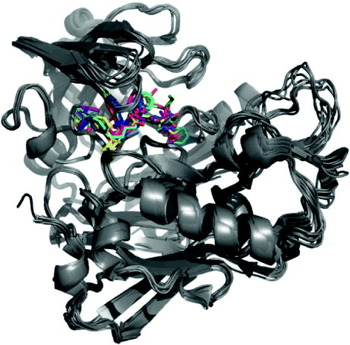
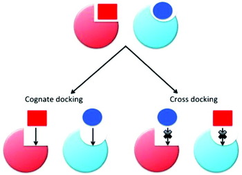
Research into protein flexibility and allostery has lent support to the importance of representing multiple states in binding studies (Fig. 1). Mobley & Dill (Reference Mobley and Dill2009) noted that binding free energy (ΔG bind) and entropy are influenced by the shape and width of the entire conformational landscape, rather than a single rigid pose. Murray et al. (Reference Murray, Baxter and Frenkel1999) examined approaches for using rigid receptors in docking studies. When the authors attempted to dock a known ligand into a protein structure solved in the presence of a different ligand (referred to as cross-docking), they found that the active site was biased toward to the native ligand (Fig. 2). A variety of differences in the surface of the binding site were identified for the same protein solved with different ligands or in the absence of a ligand. Movement was observed in the backbone, side chain (both dependent and independent of the backbone Cα), and active site metals. As a consequence, active sites were biased toward a particular ligand type, negatively impacting docking efforts. The resultant misdocking could not be overcome without accounting for critical conformational shifts. Najmanovich et al. (Reference Najmanovich, Kuttner, Sobolev and Edelman2000) examined the percentage of residues that actually undergo a change upon ligand binding, based on two non-redundant datasets containing paired holo- and apo-protein structures from the PDB. No significant correlation was found between backbone movement and side-chain flexibility, but Lys, Arg, Gln, and Met were identified as the residues most likely to demonstrate side-chain rearrangement upon ligand binding. Najmanovich et al. (Reference Najmanovich, Kuttner, Sobolev and Edelman2000) showed that 60–70% of the binding site undergoes some changes in the side-chain orientation. Taken together, these studies clearly illustrated that the lack of protein flexibility in typical SBDD efforts severely limits the identification of true ligands and accurate docked poses.
The conformational variation in holo- and apo structures of beta-secretase 1 illustrates structural differences frequently seen across multiple crystal structures of the same protein.

The same protein crystallized with different ligands. This simple two ligand-one protein set demonstrates the influence of structure on the shape of the binding pocket.

An underlying assumption in rigid docking efforts is that the complexed state is the lowest free energy state. However, the ligand-bound state is not always the lowest energy state for either the protein or the ligand. Smith et al. (Reference Smith, Redfield, Smith, Dobson, Clore, Gronenborn, Walter, Naganbushan and Wlodawer1994) were first to examine conformational similarity, they compared the four available structures of interleukin-4, two from NMR and two from crystallography. They found that differences in the experimental methods clearly affected the structure, and that the two NMR structures were more similar to the crystal structures than to each other. Further, they noted that the exposed regions of the surface, particularly the highly flexible loops, showed the largest conformational difference among the structures. Eyal et al. (Reference Eyal, Gerzon, Potapov, Edelman and Sobolev2005) evaluated a set of 659 structure pairs from the PDB and showed that experimental variation in protein structures certainly exists, as a result of refinement, crystal packing, and/or crystallization conditions. Chopra et al. (Reference Chopra, Summa and Levitt2008) employed Molecular Dynamics (MD) on a set of 75 proteins to explore the role of solvent in structural determination and concluded that solvent effects have an enormous influence on the final refined protein structure.
3.2 Sources of structural information
Use of a set of similar conformations only generates a finite amount of information on potential binding partners. Additional receptor space should be explored, especially in scenarios where alternate binding modes are of interest. X-ray crystallography, NMR structures, homology models, normal mode analysis (NMA), and molecular mechanics simulations are all potential sources of structural diversity. Advances in technology have allowed for significant increases in available structural information for a vast number of receptor targets. The PDB now contains over 76 000 structures (as of 9/27/11), up from 13 605 structures in 2000 (Berman et al. Reference Berman, Bhat, Bourne, Feng, Gilliland, Weissig and Westbrook2000). Several research groups, as well as thousands of computer users, have dedicated their computer power to distributed structure prediction of proteins with Folding@HOME, POEM@HOME, and Rosetta@HOME. Martin-Renom et al. (Reference Marti-Renom, Stuart, Fiser, Sanchez, Melo and Sali2000) estimated that almost one-third of all known protein sequences can have their structure predicted via homology modeling efforts. Schafferhans & Klebe (Reference Schafferhans and Klebe2001) found that accuracy increases when the results of several different homology modeling programs are combined. While predictive models are not as accurate as structures solved by x-ray crystallography or NMR, they enable the study of targets that are difficult to determine experimentally. No consensus has been reached on the best source of data for including structural flexibility in SBDD, and a number of studies clearly demonstrate the difficulty and variability in assessing appropriate conformations (Bolstad & Anderson, Reference Bolstad and Anderson2008; Damm & Carlson, Reference Damm and Carlson2007; Laughton et al. Reference Laughton, Orozco and Vranken2009; Philippopoulos & Lim, Reference Philippopoulos and Lim1999; Yang et al. Reference Yang, Eyal, Chennubhotla, Jee, Gronenborn and Bahar2007). Bolstad and Anderson (Reference Bolstad and Anderson2008) found that their docking protocol correctly placed all forty tested LcDHFR ligands into the crystal structure, while the scoring function correctly ranked only 80% of the ligands. Including an NMR structure, homology model, or an ensemble of structures increased the number of incorrectly placed ligands while decreasing or maintaining the correct ranking of affinities. The authors found a similar trend when docking 19 ligands to hDHFR. This suggested that both sampling and scoring failures might lead to inaccuracies within a study. Sampling failures may occur without careful selection of flexibility information, but scoring may also negatively impact the results if the function cannot correctly classify binding poses.
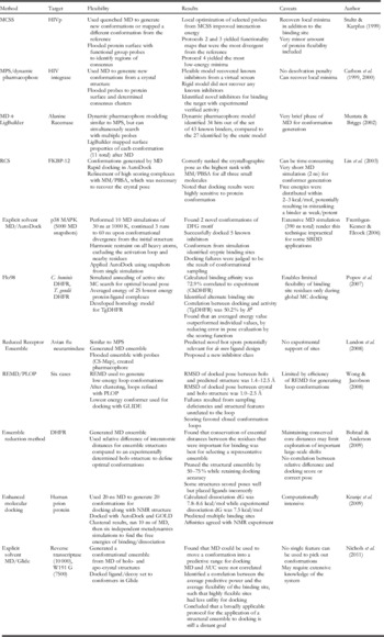
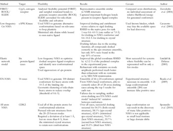
The following sections are organized based on different techniques for including protein flexibility in docking and de novo design (Table 1). Soft docking allows limited discovery of new conformation space while docking with flexible side-chain samples observed conformational space. More recent techniques incorporate ensembles of structures to allow for a greater measure of native flexibility or to reveal new conformations. By relying on several protein conformations, new chemical space can be explored, allosteric sites can be discovered, and more accurate binding conformations can be generated. The accepted metric for a well-docked pose is ⩽2.0 Å RMSD from the experimentally determined structure. Protein flexibility can be incorporated into a binding-site model before docking is initiated or it can be allowed post-docking through refinement of the bound complex. Docking can be performed against an average structure or ensemble, a set of receptor conformations (multiple receptor conformations (MRC)), or with conformations generated ‘on the fly’ (induced-fit docking (IFD)).
Techniques for incorporating full protein flexibility into docking approaches

Experimental methods for SBDD inherently include protein–ligand flexibility. However, lead development and design that is based solely on the experimental data for ligand binding can be time-consuming and prohibitively expensive since every lead optimization step must be tested. Fragment-based drug design (FBDD) has enabled the application of experimental methods to searching the interaction potential along the protein surface with small organic probes. The use of fragment-based studies in SBDD enables the identification of ligands with high affinity for targets that have been traditionally difficult to characterize. These insights have greatly influenced computational approaches to protein flexibility, where fragment mapping can be applied to flexible receptors with functional group probes to find binding hot spots and identify specific interaction potentials. The use of molecular dynamics and/or probe minimizations to search for important binding sites with small probes has been frequently applied in SBDD projects because the results enable the development of a pharmacophore model for use in virtual screening studies.
4. Early protein–ligand flexibility in docking
4.1 Soft docking
It was not until the mid-1990s that researchers began to tackle the problem of receptor degrees of freedom in SBDD. The original technique involved soft docking, which accounted for minimal backbone movement, and side-chain flexibility through attenuation of the Lennard–Jones repulsion term between the rigid protein and ligand (Jiang & Kim, Reference Jiang and Kim1991). This allowed for some penetration between the ligand and protein, and was followed by a rigid-body minimization. Soft docking increased the number of high scoring hits compared to the use of a single structure for docking. A study of T4 lysozyme and aldose reductase showed that soft docking was superior to docking to a static structure (Ferrari et al. Reference Ferrari, Wei, Costantino and Shoichet2004). However, the authors also found that serial docking to a structural ensemble of rigid receptors identified six novel compounds targeting aldose reductase that soft docking had ranked poorly. Of these six compounds, four were experimentally verified and one had a low micromolar IC50 value. Ferrari et al. conjectured that the differences in ranking were due to the differences in the scoring function applied for soft versus serial docking.
Soft docking is a fast and easy method for including some protein flexibility into docking studies, and as a result is it included as a step in many of the methods presented below.
4.2 Relaxation methods
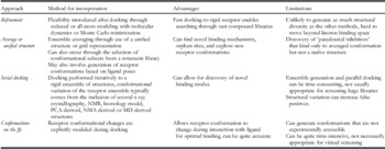
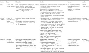
Refinement of the docked complex is another simple approach that adjusts for protein flexibility by modeling induced-fit effects. The incorporation of protein flexibility on the ‘back end’ can only be done when the docking technique was based on an all-atom structure; it cannot be performed on a grid, or in any other situation where the protein is not explicitly represented. Monte Carlo (MC) or MD simulations are a popular choice for refinement because they enable the optimization of docked poses, investigation of solvent effects, examination of the kinetic stability, and prediction of ΔG bind from physics-based scoring functions. Refinement is frequently performed as a final step in many of the docking approaches discussed here; Table 2 lists protein–ligand docking methods that limit the inclusion of full flexibility to the refinement stage (Mizutani et al. Reference Mizutani, Takamatsu, Ichinose, Nakamura and Itai2006; Pencheva et al. Reference Pencheva, Lagorce, Pajeva, Villoutreix and Miteva2008; Taylor et al. Reference Taylor, Jewsbury and Essex2003).
Studies including full receptor flexibility after docking (post-docking refinement)

4.3 Docking with flexible side chains
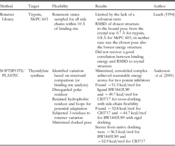
Early efforts at on-the-fly docking focused on the side chains, with the use of a rotamer library based on backbone dihedral angles to describe protein flexibility. In 1993, rotamer libraries were first used to predict side-chain conformations while studying the protein folding problem (Dunbrack & Karplus, Reference Dunbrack and Karplus1993). In 1994, the use of side-chain rotamer libraries was extended to protein–ligand docking with the examination of trypsin-benzamidine and antibody McPC 603-phosphocholine binding through a modified version of AMBER 4.0 (Leach). The study restricted the protein backbone completely while the side chains within 10 Å of the ligand were allowed for sampling a set of discrete rotameric states. Leach found that the presence of the ligand revealed additional accessible conformational states by modulating the protein's potential energy surface. Similar discrete sampling methods for modeling side-chain flexibility in docking include SOFTSPOTS/PLASTIC (Anderson et al. Reference Anderson, O'Neil, Surti and Stroud2001), as presented in Table 3a.
Docking studies that included side-chain flexibility through rotamer libraries. Caveats for each method are similar in that flexibility of the side chains alone has limited success in describing receptor motion for most binding events and the implementation of a rotamer library limits the conformational space that can be sampled

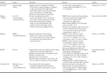
Side-chain motion upon ligand binding has also been approached through continuum sampling methods. Alberts et al. (Reference Alberts, Todorov and Dean2005a) found that random movement of side chains was preferable to a rotamer library for successful docking results. Techniques that apply some form of continuum sampling to side-chain motion are listed in Table 3b, including AutoDock 4.0 (Morris et al. Reference Morris, Huey, Lindstrom, Sanner, Belew, Goodsell and Olson2009), Dynasite/GOLD (Kallblad & Dean, Reference Kallblad and Dean2003), FlexX (Rarey et al. Reference Rarey, Kramer, Lengauer and Klebe1996), ICM/MC (Abagyan et al. Reference Abagyan, Totrov and Kuznetsov1994), Mining Minima (Kairys & Gilson, Reference Kairys and Gilson2002), Skelgen (Alberts et al. Reference Alberts, Todorov and Dean2005a, Reference Alberts, Todorov, Kallblad and Deanb), and SLIDE (Schnecke & Kuhn, Reference Schnecke and Kuhn2000; Zavodszky et al. Reference Zavodszky, Sanschagrin, Korde and Kuhn2002; Zavodszky & Kuhn, Reference Zavodszky and Kuhn2005). A rotamer library can be limited by the information it contains, while continuum sampling may locate unrealistic conformational states.
Docking studies where selected side-chain flexibility was accounted for through continuum sampling. This approach to flexibility at the side-chain positions may explore new space but cannot describe large-scale motion

Allowing side-chain flexibility is less resource intensive than full flexibility methods and it enabled some conformational variability through the exploration of low-energy orientations of side chains. However, incorporating side-chain flexibility has failed in cases of proteins with large-scale hinge or loop rearrangements or even backbone-dependent movement of side chains, neither of which can be taken into account by discrete or continuum sampling.
5. Induced fit docking
5.1 Conformational generation on the fly
Significant progress has been made since the initial implementation of flexible side chains in docking. Sampling motion of the side chains on the fly increases the potential energy space but can still overlook global conformational shifts. Allowing for conformational changes during docking can be a highly accurate technique for modeling bound poses of protein–ligand complexes. IFD is important because it can allow the docking simulation to search new conformational space; however, this sampling of receptor and ligand degrees of freedom is also quite computationally intensive, which limits its application in large-scale virtual screening studies.
The first on-the-fly method to expand beyond conformational sampling of side chains without relying upon a composite representation was based on DOCK 4.0 and ITScore: the ensemble docking algorithm (Huang & Zou, Reference Huang and Zou2007). The authors included the conformational ensemble as an adjustable variable in the IFD algorithm as opposed to performing serial docking across an entire ensemble. In addition to the usual six degrees of freedom for ligand flexibility, the authors added a protein conformation term, which allowed the protocol to simultaneously optimize the ligand conformation and choose the best protein structure for binding. In order to initially orient the ligand, a reference conformation comprising the largest possible binding site was generated from the combination of existing conformations. Ten separate protein ensembles were used for development and were obtained from the set by Cavasotto & Abagyan (Reference Cavasotto and Abagyan2004), the set by Claussen et al. (Reference Claussen, Buning, Rarey and Lengauer2001), and two HIV-1 protease (HIVp) ensembles. Huang and Zou achieved an average success rate of 92% based on both pose and energy score, where the energy score was required to be within 1.0 kcal/mol of the native score or better and the five highest-ranked poses were included using a threshold for success of 2.5 Å RMSD from the experimental pose. They noted that docking accuracy based on binding pose alone was insufficient, as demonstrated by the influence of the binding energy upon results with rigid docking. Their ensemble method ran for a similar amount of time as one rigid docking calculation and was 12.9 times faster than serial docking (data averaged over ten systems, with an average of 10.5 structures).
RosettaLigand was introduced as an extension of the protein–protein docking program, RosettaDock (Meiler & Baker, Reference Meiler and Baker2006). In this paper, side chains were repacked during docking through the implementation of a rotamer library, while the vdW repulsion term was softened and ligand conformations were perturbed with MC. Their benchmark set included 100 complexes for native docking and 20 for cross-docking. They found a success rate (RMSD <2.0 Å) of 80% for native docking and 70% for cross-docking; the authors noted that their RMSD metric accounted for hydrogen atoms and side chains in the binding site, thus capturing small changes in conformation overlooked by other methods. RosettaLigand was updated to include backbone sampling through a gradient-based minimization of the torsion angles for both protein and ligand as the final step in their docking procedure (Davis & Baker, Reference Davis and Baker2009; Davis et al. Reference Davis, Raha, Head and Baker2009). Although RosettaLigand has generated good results, with protein flexibility included, it requires 40–80 processor hours to generate a bound pose for the receptor–ligand complex and therefore is too time-consuming for use with large compound libraries.
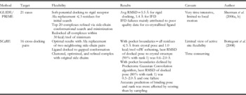
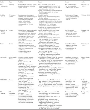
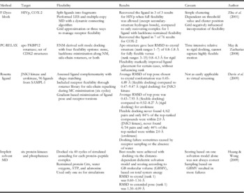
Other IFD methods for including protein flexibility include modeling flexibility for a limited number of receptor residues, Table 4a: GLIDE/PRIME (Sherman et al. Reference Sherman, Beard and Farid2006a, Reference Sherman, Day, Jacobson, Friesner and Faridb), SCaRE (Bottegoni et al. Reference Bottegoni, Kufareva, Totrov and Abagyan2008); discrete sampling, Table 4b: FlexX-Ensemble (Claussen et al. Reference Claussen, Buning, Rarey and Lengauer2001), a modified DOCK protocol (Wei et al. Reference Wei, Weaver, Ferrari, Matthews and Shoichet2004), Fleksy (Nabuurs et al. Reference Nabuurs, Wagener and De Vlieg2007), FLIPDock (Zhao & Sanner, Reference Zhao and Sanner2007, Reference Zhao and Sanner2008), FITTED (Corbeil et al. Reference Corbeil, Englebienne and Moitessier2007, Reference Corbeil, Englebienne, Yannopoulos, Chan, Das, Bilimoria, L'Heureux and Moitessier2008; Corbeil & Moitessier, Reference Corbeil and Moitessier2009), 4D Docking (Bottegoni et al. Reference Bottegoni, Kufareva, Totrov and Abagyan2009); and continuum sampling, Table 4c: F-DycoBlock (Zhu et al. Reference Zhu, Fan, Liu and Shi2001), PC-RELAX (Zacharias, Reference Zacharias2004, Reference Zacharias2008), RosettaLigand (Davis & Baker, Reference Davis and Baker2009), and implicit MD (Huang & Wong, Reference Huang and Wong2009).
Docking studies that modeled 2–3 residues to represent receptor flexibility during the performance of IFD

Docking studies that applied discrete sampling of flexibility through panels of receptor structures during the performance of IFD

Docking studies that applied molecular mechanics or minimization to incorporate receptor flexibility during IFD

6. Ensemble docking
Ensemble docking differs from IFD in that protein flexibility is accounted for prior to the actual docking. Although the studies by Huang and Zou as well as Bottegoni et al. used a pre-existing ensemble of conformations, they sampled those conformations on the fly, and so they are included above in the IFD section. Two different types of methods exist for representing receptor flexibility during docking: grid-based ensembles and structure-based ensembles. Frequently, alternate protein conformations are represented on a two-body potential grid, enabling fast, inexpensive docking simulations. Flexibility can also be incorporated into binding predictions through the sequential docking to structures in a conformational ensemble or docking to an averaged/united receptor structure. Due to the time required for initial ensemble generation and repeated docking runs to a set of static structures, sequential docking is typically the most computationally intensive approach. However, it avoids the discovery of receptor–ligand complexes that are physically impossible (paradoxical ligands), which are sometimes seen in the results from docking to an average structure or grid.
6.1 Grid-based ensembles
The first docking method to use a composite grid was performed through DOCK3.5 in order to evaluate the impact of representing conformational variability as a structural ensemble on binding pose results (Knegtel et al. Reference Knegtel, Kuntz and Oshiro1997). With HIVp, ras p21, retinol binding protein, and uteroglobin as their test cases, the authors compared the capacity of standard grids to energy- and geometry-weighted average grids to reproduce known binding poses and affinities. They found that docking scores were sensitive to the grid spacing and threshold parameters used to define the composite grid. The performance of the geometry-weighted grid was less dependent on the protein conformations that were available than the energy-weighted, to the extent that the geometry-weighted grid placed known ligands for HIVp within the top 21%, while the energy-weighted grid placed them in the top 33%. This occurred because the geometry-weighted grid ignored the repulsive potentials between flexible atoms, while the energy-weighted grid simply smoothed the repulsive potential, which can result in the retrieval of paradoxical inhibitors.
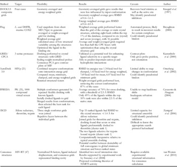
The incorporation of protein flexibility through docking to an interaction grid that represents the receptor ensemble is a common approach for SBDD, and additional methods are presented in Table 5, including those based on AutoDock 3.0 (Osterberg et al. Reference Osterberg, Morris, Sanner, Olson and Goodsell2002), Flog (Broughton, Reference Broughton2000), GRID/CPCA (Kastenholz et al. Reference Kastenholz, Pastor, Cruciani, Haaksma and Fox2000), IFREDA (Cavasotto & Abagyan, Reference Cavasotto and Abagyan2004), ISCD (Zentgraf et al. Reference Zentgraf, Fokkens and Sotriffer2006), and sets of consensus structures (van Westen et al. Reference van Westen, Wegner, Bender, Ijzerman and van Vlijmen2010).
Studies including receptor flexibility through grid-based ensemble docking

6.2 Structure-based ensembles
The multiple copy simultaneous search (MCSS) methodology was the earliest computational approach for mapping binding sites with functional group probes (Miranker & Karplus, Reference Miranker and Karplus1991). The authors simultaneously minimized or quenched the probes by MD to the binding site of the hemagglutinin protein to identify favorable minima and found some correspondences between the positions of the minima and the functional groups on the ligand, sialic acid. While the authors noted that their method could account for a limited amount of side-chain flexibility by including multiple copies of the side chains, the published work was performed against a rigid structure.
Stultz and Karplus (Reference Stultz and Karplus1999) explored the influence of protein flexibility on the results from MCSS where five different protocols for placing two functional group probes, methanol and methyl ammonium, were used to search the binding surface of HIVp. MCSS calculations were performed for 1: a minimized crystal structure, 2: a conformation generated from quenched MD of the initial structure, 3: an unrestrained structure, 4: the output of 1 subjected to quenched MD with functional groups restrained and multiple side-chain copies, and 5: a different rigid crystal structure of HIVp. The authors found that their results with protocols 4 and 5 yielded more favorable interaction energies than the reference protocol 1. Protocol 4 gave valuable information for the ligand design, while the different low-energy minima from protocol 5 supported the idea of performing MCSS against an ensemble of structures and comparing the results; therefore, the authors suggested a combination of 4 and 5. However, one of the limitations of MCSS and other ensemble methods is that they do not account for entropy or solvation during surface mapping, which hampers discrimination between druggable and irrelevant minima. Furthermore, as Schubert & Stultz (Reference Schubert and Stultz2009) point out in their review of MCSS, functionality maps are difficult to combine across different structures of a protein.
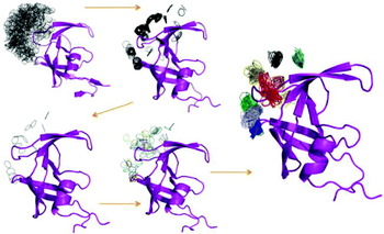
An original technique for structure-based ensemble docking was introduced by Carlson et al. (Reference Carlson, Masukawa, Rubins, Bushman, Jorgensen, Lins, Briggs and McCammon2000) as the dynamic pharmacophore model (Carlson et al. Reference Carlson, Masukawa and McCammon1999), later termed the multiple protein structure (MPS) method. Two pharmacophores for HIV integrase were generated, a static model based on a rigid crystal structure and a dynamic model based on MD snapshots initiated from the same crystal structure. The MPS method mapped the dynamic binding surface of the protein with common functional groups by running a series of multi-unit search for interacting conformers (MUSIC) simulations (Fig. 3), wherein hundreds of probes were used to flood the protein surface and then minimized by MC sampling. During MUSIC simulations, all of the probe–probe interactions were ignored, which allowed probes to interact with the rugged binding potential of the protein and cluster into hot-spot positions along the protein surface. These cluster sites were used to develop a pharmacophore for virtual screening, and where the rigid model was unable to locate any of the test case inhibitors, the dynamic pharmacophore model not only identified known inhibitors, but it also predicted new inhibitors that were confirmed by experiment. The MPS method is one of the few ensemble-based methods that has been successfully used to find novel leads with demonstrated activity (Bowman et al. Reference Bowman, Nikolovska-Coleska, Zhong, Wang and Carlson2007; Damm et al. Reference Damm, Ung, Quintero, Gestwicki and Carlson2008).
MUSIC simulations of benzene probes (gray) to the surface of an HIVp monomer (purple). The initial stages are universal to mapping methods, but the combination of the structures in the fourth and final frames are unique to the MPS method. Five hundred probes were flooded onto the protein surface, minimized, and clustered together. Parent probes were identified for each monomer conformation, the HIVp monomers were aligned through a weighted-RMSD function, and then the parent probes were clustered together and retained when at least half of the protein conformations contained a probe at that site. This resulted in low-energy probe clusters (colored red through purple in the final frame) of benzene that could be combined with ethane and methanol clusters to develop a pharmacophore model.

McCammon and co-workers developed the relaxed complex scheme (RCS) as a technique for incorporating receptor flexibility prior to docking (Lin et al. Reference Lin, Perryman, Schames and McCammon2002, Reference Lin, Perryman, Schames and McCammon2003; McCammon, Reference McCammon2005). Initially the method was developed to generate conformational ensembles of the apo state through MD, which were then used in AutoDock to screen compound mini-libraries and score the receptor–ligand complexes. The 2003 study by Lin et al. examined FKBP-12 by RCS and implemented a final refinement step with MM/PBSA (Kollman et al. Reference Kollman, Massova, Reyes, Kuhn, Huo, Chong, Lee, Lee, Duan, Wang, Donini, Cieplak, Srinivasan, Case and Cheatham2000) to yield final results. Further updates to this method have included extension to virtual screening and reduction of the conformational ensemble to a representative configurational set (Amaro et al. Reference Amaro, Baron and McCammon2008). RCS was successfully used to identify cryptic binding pockets and/or lead inhibitors of HIVp (Perryman et al. Reference Perryman, Lin and McCammon2006), avian influenza neuraminidase (Cheng et al. Reference Cheng, Amaro, Xu, Li, Arzberger and McCammon2008), acetylcholine binding protein (Babakhani et al. Reference Babakhani, Talley, Taylor and McCammon2009), DNA polymerase β (Barakat & Tuszynski, Reference Barakat and Tuszynski2010), MDM2/MDMX (Barakat et al. Reference Barakat, Mane, Friesen and Tuszynski2010), and cruzain (Durrant et al. Reference Durrant, Keranen, Wilson and McCammon2010). Cheng et al. noted that conformational sampling markedly influenced the re-ranking of compounds based on interactions gained or lost compared to the crystal structure. The authors noted that the most dominant structures did not have the lowest binding energy, but given the limitations of MD, they believed that the dominant clusters were good representatives of the ensemble. Babakhani et al. suggested that both the scoring function and the conformational variability contributed to several failures in reproducing known binding modes.
To determine the best process for developing a structural ensemble, Rueda et al. (Reference Rueda, Bottegoni and Abagyan2010) used ICM to examine the same set of 1068 conformations from 99 pharmaceutically relevant proteins that was used in the 4D docking study. The authors judged performance in virtual screening based on the area under the curve (AUC) of a ROC plot and found that holo conformations yielded improved AUC values compared to apo conformations. It was interesting that the authors found that ensembles of holo plus apo conformations did not significantly improve results compared to holo-only ensembles. In cases without a holo structure, the authors recommended the use of an apo structure with the largest pocket volume. The authors proposed the use of a ligand-guided approach to find the optimal protein conformation, but stated that docking to an ensemble would be an acceptable substitute in the absence of ligand activity data. Several groups have found that there tends to be a single receptor conformation that grants the best performance in docking studies (Barril & Morley, Reference Barril and Morley2005; Rueda et al. Reference Rueda, Bottegoni and Abagyan2010).
NMA has been frequently used as a tool for examining protein flexibility relevant to ligand binding (Cavasotto et al. Reference Cavasotto, Kovacs and Abagyan2005; Keseru & Kolossvary, Reference Keseru and Kolossvary2001; May & Zacharias, Reference May and Zacharias2005, Reference May and Zacharias2008; May et al. Reference May, Sieker and Zacharias2008; Zacharias, Reference Zacharias2004; Zacharias & Sklenar, Reference Zacharias and Sklenar1999; Zavodszky & Kuhn, Reference Zavodszky and Kuhn2005). It is important to note that low-frequency modes identify large-scale motions, while high-frequency modes identify small-scale motion. Rueda et al. (Reference Rueda, Bottegoni and Abagyan2009) showed that they could use NMA on elastic network models for all heavy atoms of the receptor in order to derive an ensemble of protein conformations and improve results from cross-docking in ICM. Fourteen proteins, each with two structures and their cognate ligands, were used as the benchmark test set. Optimal results involved the generation of 100 different conformations from NMA on all heavy atoms within 10 Å of the ligand-binding site. Larger conformational ensembles, such as those with 200 members, negatively affected false positive rates. Another NMA-based method for conformational selection was based on cyclin dependent kinase-2 (CDK-2), because it had a sufficient amount of available experimental data on ligand binding for validation of the NMA protocol (Sperandio et al. Reference Sperandio, Mouawad, Pinto, Villoutreix, Perahia and Miteva2010). Unlike most other NMA-based methods, Sperandio et al. included all of the protein atoms in the calculation in order to better represent conformational changes and found that too many protein conformations negatively impacted docking results by recovering additional false positives. They suggested addressing this by developing scoring protocols specifically tailored to the use of a conformational ensemble.
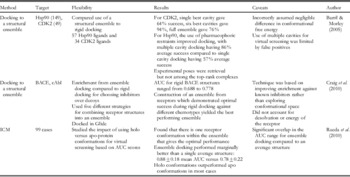
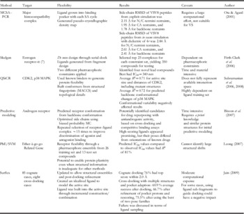
Of the variety of techniques that have been developed to improve binding predictions for protein–ligand complexes, the use of a conformational ensemble is perhaps the most common approach. Representative cases and novel methods based on structure-based ensemble docking are listed in Tables 6a–6d. Crystal structure and NMR-based ensemble methods include docking to a large set of conformations (Barril & Morley, Reference Barril and Morley2005; Craig et al. Reference Craig, Essex and Spiegel2010), and ICM (Rueda et al. Reference Rueda, Bottegoni and Abagyan2010), as listed in Table 6a. Procedures that rely upon molecular dynamics to provide receptor flexibility include MCSS (Stultz & Karplus, Reference Stultz and Karplus1999), MPS (Carlson et al. Reference Carlson, Masukawa and McCammon1999, Reference Carlson, Masukawa, Rubins, Bushman, Jorgensen, Lins, Briggs and McCammon2000), MD+LigBuilder (Mustata & Briggs, Reference Mustata and Briggs2002; Mustata et al. Reference Mustata, Soares and Briggs2003), RCS (Lin et al. Reference Lin, Perryman, Schames and McCammon2003), MD+AutoDock (Frembgen-Kesner & Elcock, Reference Frembgen-Kesner and Elcock2006), Flo98 (Popov et al. Reference Popov, Yee and Anderson2007), a reduced receptor ensemble (Landon et al. Reference Landon, Amaro, Baron, Ngan, Ozonoff, McCammon and Vajda2008), REMD/PLOP (Wong & Jacobson, Reference Wong and Jacobson2008), the ensemble reduction method (Bolstad & Anderson, Reference Bolstad and Anderson2009), enhanced molecular docking (Kranjc et al. Reference Kranjc, Bongarzone, Rossetti, Biarnes, Cavalli, Bolognesi, Roberti, Legname and Carloni2009) and MD+Glide (Nichols et al. Reference Nichols, Baron, Ivetac and McCammon2011), as listed in Table 6b. Ensembles have also been generated for docking through NMA, as demonstrated by FIRST/ROCK/SLIDE (Zavodszky et al. Reference Zavodszky, Lei, Thorpe, Day and Kuhn2004), low-frequency NMA (Cavasotto et al. Reference Cavasotto, Kovacs and Abagyan2005), ENM (Lindahl & Delarue, Reference Lindahl and Delarue2005), EN–NMA (Rueda et al. Reference Rueda, Bottegoni and Abagyan2009), and all atom NMA (Sperandio et al. Reference Sperandio, Mouawad, Pinto, Villoutreix, Perahia and Miteva2010), as listed in Table 6c. Finally, ligand-induced flexibility has also been used as another source for conformers, as shown in Table 6d and applied by MCSA-PCR (Ota & Agard, Reference Ota and Agard2001), Skelgen (Firth-Clark et al. Reference Firth-Clark, Willems, Williams and Harris2006), QSiCR (Subramanian et al. Reference Subramanian, Sharma and B-Rao2006, Reference Subramanian, Sharma and B-Rao2008), PhE/SVM (Leong, Reference Leong2007; Leong & Chen, Reference Leong and Chen2008; Leong et al. Reference Leong, Chen, Chen and Chen2009), structure prediction (Bisson et al. Reference Bisson, Cheltsov, Bruey-Sedano, Lin, Chen, Goldberger, May, Christopoulos, Dalton, Sexton, Zhang and Abagyan2007), and Surflex (Jain, Reference Jain2003, Reference Jain2007, Reference Jain2009). Although listed in Table 5 due to its incorporation of protein flexibility within an interaction grid, IFREDA also relies upon ligand-induced conformations for contributing flexibility.
Studies including full receptor flexibility through collection of a crystal or NMR structural ensemble

Studies including full receptor flexibility through a structural ensemble previously generated by MD

Studies including full receptor flexibility through a normal mode analysis, or other non-MD-based conformational generation, prior to docking

Studies including full receptor flexibility through ligand-induced flexibility

7. De novo approaches through fragment-based drug design
Several interesting experimental methods that naturally account for protein flexibility have greatly influenced computational SBDD. The burgeoning field of FBDD has allowed for the identification of low-molecular weight binders, which allow for the identification of difficult binding sites (Erlanson et al. Reference Erlanson, McDowell and O'Brien2004). First introduced by the multiple solvent crystal structure (MSCS) (Mattos & Ringe, Reference Mattos and Ringe1996) and SAR-by-NMR (Shuker et al. Reference Shuker, Hajduk, Meadows and Fesik1996) techniques, FBDD can be performed via NMR or x-ray crystallography, and provides a different set of hits from high-throughput screening (HTS) experiments. Screening proteins with a fragment collection both verifies the druggability of a target system and identifies fragments that can be linked and optimized to develop a viable lead compound (Wiesmann et al. Reference Wiesmann, Barr, Kung, Zhu, Erlanson, Shen, Fahr, Zhong, Taylor, Randal, McDowell and Hansen2004). Importantly, hits from FBDD have been experimentally validated and identified novel compounds shown to work in the clinic (Howard et al. Reference Howard, Berdini, Boulstridge, Carr, Cross, Curry, Devine, Early, Fazal, Gill, Heathcote, Maman, Matthews, McMenamin, Navarro, O'Brien, O'Reilly, Rees, Reule, Tisi, Williams, Vinkovic and Wyatt2009; Murray & Rees, Reference Murray and Rees2009).
The MSCS technique was originally introduced as a crystallography tool for the characterization of the binding potential of pancreatic elastase. Other groups have also used MSCS to probe ligand interactions in thermolysin (English et al. Reference English, Done, Caves, Groom and Hubbard1999, Reference English, Groom and Hubbard2001), subtilisin (Fitzpatrick et al. Reference Fitzpatrick, Steinmetz, Ringe and Klibanov1993; Schmitke et al. Reference Schmitke, Stern and Klibanov1997; Reference Schmitke, Stern and Klibanov1998), RNase A (Fedorov et al. Reference Fedorov, Joseph-McCarthy, Fedorov, Sirakova, Graf and Almo1996; Dechene et al. Reference Dechene, Wink, Smith, Swartz and Mattos2009), p53 core (Ho et al. Reference Ho, Luo, Zhao, Chai, Fitzgerald and Marmorstein2006), lysozyme (Wang et al. Reference Wang, Zhu, Huang, Qian, Shao, Jia and Tang1998), and H-Ras (Buhrman et al. Reference Buhrman, de Serrano and Mattos2003). During crystallization, the water and organic solvent acted as probes of binding affinity because the organic solvent, which was chosen to represent a common functional group, displaces bound waters only at locations with favorable affinity for the particular interaction type presented by the protein. MSCS can be performed with a variety of probes, thus enabling the results from the protein crystallography experiment to be superimposed in order to identify regions of consensus binding.
When Joseph-McCarthy et al. (Reference Joseph-McCarthy, Fedorov and Almo1996) compared their results from studies with MCSS to results from MSCS, they found that it was important for traditional MCSS to consider occupancy at a first site in order to accurately predict occupancy at a second binding site. English et al. (Reference English, Groom and Hubbard2001) compared the use of MSCS, GRID, and MCSS through the exploration of the binding surface of thermolysin with acetone, acetonitrile, isopropanol, and phenol acting as functional group probes. The authors found that in comparison with MSCS, the MCSS, and GRID methods overestimated electrostatic interactions, retrieved an overabundance of local energy minima, and could not provide detailed descriptions of the interactions between the protein and probes.
Although both SAR-by-NMR and MSCS allow for a larger amount of protein flexibility then current methods for FBDD do, they are also both material and time-intensive. Furthermore, as discussed by English et al. (Reference English, Groom and Hubbard2001), MSCS can be quite slow and probe choice is limited by the fragility of protein crystals. Computational solvent mapping is a complementary approach to experimental fragment-binding studies; however, most computational approaches do not account for either the impact of protein flexibility or proper solvation effects, which can lead to poor mapping results. Over the past two years, several groups have developed new methodology for including flexibility and solvent competition in computational fragment mapping. While the results of any fragment-based study can be translated into a consensus pharmacophore model and used in virtual screening applications, the primary objective of MSCS and computational solvent mapping has been the correct identification of potential binding sites.
Locus Pharmaceuticals developed a method based on a grand-canonical (GC) MC simulation for pharmacophore development (Clark et al. Reference Clark, Guarnieri, Shkurko and Wiseman2006, Reference Clark, Meshkat and Wiseman2009; Moore, Reference Moore2005). Both studies from Clark et al. were performed against a static structure. Their later study concentrated on deriving accurate ΔG bind, while their earlier study used ten simulated annealing runs of GC-MC to explore the protein surface of thermolysin with 2 probes and of T4 lysozyme with 14 probes (2006). Based on the simulated annealing runs, GB/SA was used to predict binding affinities and Clark et al. found that their results retrieved some of the hot spots observed in the published MSCS data. Moore discussed the use of torsion-space dynamics to generate protein flexibility in combination with general outcomes from the use of a 500-member fragment library to search target surfaces with GC-MC; however, no specific details were given.
Vadja and co-workers developed a fast algorithm for searching protein surfaces with small organic probes that was based on a fast Fourier Transform correlation approach (FTMAP) (Brenke et al. Reference Brenke, Kozakov, Chuang, Beglov, Hall, Landon, Mattos and Vajda2009). In a manner akin to MPS, billions of solvent probes are minimized to the protein surface and then clustered based on a simple greedy algorithm. The probe clusters demonstrate the position and orientation of proposed hot spots along the target, where the largest cluster is defined as the maximal hot-spot location; the second largest is the second most-important site; and etc. Although FTMAP is another method for computational solvent mapping that does not inherently include protein flexibility, receptor dynamics could be modeled through serial searches over multiple conformations.
A novel method for probe mapping incorporated solvent competition and protein flexibility through MD (Seco et al. Reference Seco, Luque and Barril2009). Based on MSCS, the authors used isopropyl alcohol (IPA) and water together to perform a single MD simulation over 16 ns. The use of binary solvent MD inherently accounted for solvation effects and protein flexibility. This method was applied to three proteins with experimental MSCS results and five pharmaceutically relevant receptors that have not been studied by MSCS; simulation probe occupancy was used to differentiate binding sites and predict ΔG bind. This method was much more computationally demanding than other computational solvent-mapping techniques such as FTMAP (Brenke et al. Reference Brenke, Kozakov, Chuang, Beglov, Hall, Landon, Mattos and Vajda2009), but may be more competent at distinguishing between druggable and non-druggable sites because of the use of flexibility data. Yang & Wang (Reference Yang and Wang2010) used this same solvent-mapping technique to study hot-spot mapping against thermolysin together with a double-decoupling method, which provided a more rigorous calculation of ΔG bind at potential hot spots. It is important to note that the analysis of probe occupancy to locate binding sites necessarily assumes that Boltzmann sampling occurred during simulation; therefore, careful selection of the trajectory length required for convergence is exceedingly important but frequently neglected in method development.
Guvench & MacKerell (Reference Guvench and MacKerell2009) published a similar method, Site Identification by Ligand Competitive Saturation (SILCS), where small molecule probes were used to examine the binding surface. Their technique used all-atom MD simulations of protein in a box of propane, benzene, and water as the explicit solvent probes. Ten independent simulations over 5 ns were generated for analysis and the mapping results were represented on a 1 Å grid of volume occupancy. SILCS was capable of reproducing known binding interactions for the test case, BCL-6 oncoprotein, and a follow-up study further validated the approach on seven proteins from five different protein families (Raman et al. Reference Raman, Yu, Guvench and Mackerell2011). In their follow-up study, MacKerell and co-workers extended the simulation time to 10 runs of 20 ns each and scored their occupancy results based on a normalized calculation of the observed:expected occupancy, which they termed grid free energy (GFE). When compared to the positions of known ligands, their GFE was able to select for the crystallographic pose of the bound ligand in several protein families. However, in the only available figure of an entire protein surface, SILCS is clearly shown to preferentially map many irrelevant minima before identifying the binding site.
We have developed a computational method that achieves hot-spot mapping results that are similar to available experimental data; Mixed-solvent Molecular Dynamics (MixMD). Our MPS method (Carlson et al. Reference Carlson, Masukawa, Rubins, Bushman, Jorgensen, Lins, Briggs and McCammon2000) for pharmacophore development demonstrated success in mapping protein systems for drug design (Bowman et al. Reference Bowman, Nikolovska-Coleska, Zhong, Wang and Carlson2007; Damm et al. Reference Damm, Ung, Quintero, Gestwicki and Carlson2008) and MixMD complements MPS while simultaneously allowing protein flexibility and probe competition with water. MixMD was inspired by the MSCS technique but incorporates more explicit conformational sampling. Since MSCS results identified specific electron density that can be attributed to the placement of organic solvent along the protein surface, we specifically compare grids of solvent occupancy from simulation to that of experimental electron density. Using many, short MD simulations of protein in 50% weight/weight mixtures of acetonitrile and water, we successfully validated MixMD based on the available MSCS structures (Lexa & Carlson, Reference Lexa and Carlson2011). Similar results were obtained with isopropanol (unpublished data).
8. Limitations of structure-based drug design
Most published methods for flexible protein–ligand docking are based on limited sets for benchmarking results. The use of a large test set for methods development is crucial for demonstrating performance across a range of different targets. Furthermore, docking to a flexible protein is more resource and time-intensive than docking to a rigid receptor and in many cases ensemble docking is unrealistic for screening huge compound libraries. Through the careful development of methods for docking and scoring, the computational requirements for accurate simulation may be sufficiently lessened to allow for more widespread implementation of flexibility.
The use of multiple conformations for docking is limited in that several protein conformations may decrease the selectivity of lead compounds by increasing the false positive rate. Using combinations of features from different conformations may also lead to the creation of a ligand with high affinity for an average receptor structure that is not experimentally accessible, a so-called ‘paradoxical inhibitor’. As a result, there is the potential for introduction of bias through user intervention in many of these methods for flexible docking.
Due to the rugged landscape of most proteins, not every conformation that is included in a low-energy ensemble will adequately represent its true binding potential. This has led many recent studies to dedicate their focus to identifying the optimal method for selection of only the relevant protein conformations. For this process of conformer selection and weighting to succeed, it is crucial that the internal energy of the individual protein conformations be included in the scoring process (though this is more difficult to properly calculate). Furthermore, it is necessary that the scoring functions used in fully flexible procedures be as accurate as possible so as to provide the most physically realistic results.
Within the field of SBDD, it remains unclear whether sampling or scoring is the limiting factor in our ability to correctly predict binding modes and affinities. Hampering our ability to address the clear need for improved methods is an unfortunate lack of careful documentation in the literature covering docking or scoring failures for a given study. Careful examination of the techniques presented in this review yielded 30 papers with enough detail to allow assessment of the possible factors resulting in inaccurate results. Of these papers, 14 leaned toward scoring being the dominant factor, 11 toward sampling, four were evenly split, and one toward poor crystallographic density. This depicts the unfortunate scenario where both sampling and scoring can negatively impact success in the field. The results from several papers convincingly argue that the combination of sampling and scoring can be applied to predict better complexes than those identified without flexibility. Beyond applying RMSD metrics to experimental structures, there is no extensive evidence for whether the current sampling approaches model changes in conformation accurately; while some results successfully predict known experimental poses, other results conflict with, or have not been corroborated by, experimental data. It is important that consistent data and analysis be presented in the future so that these sources of error in SBDD can be better understood.
Experimental FBDD approaches such as SAR-by-NMR and MSCS are time-consuming, costly, and dependent upon the receptor size and its potential for crystallization. On the other hand, computational FBDD approaches can be just as time-consuming and are heavily dependent on proper parameterization and analysis. The trend toward estimating binding affinity from computational solvent grids has shown only partial success when compared with experimental affinities. Computational FBDD shows great promise as a tool for including flexibility and probe-water competition explicitly in a ‘docking’ experiment; however, implementation of these methods requires careful consideration of the underlying chemistry to ensure reliable results.
9. Future directions
Considerable progress in the field of SBDD is still required before achieving the ultimate goal: accurately predicting the native state conformation of novel receptor–ligand complexes. Since there is a strong dependency upon the test set for both docking and scoring functions, the use of the high-quality test sets is essential for method development and the sets from Murray and co-workers have given researchers a resource for building docking and scoring functions that are applicable across a wide range of protein targets (Hartshorn et al. Reference Hartshorn, Verdonk, Chessari, Brewerton, Mooij, Mortenson and Murray2007; Verdonk et al. Reference Verdonk, Mortenson, Hall, Hartshorn and Murray2008). Some of the congeneric series in the CSAR-NRC set developed in our group are also very valuable to this effort (Dunbar et al. Reference Dunbar, Smith, Yang, Ung, Lexa, Khazanov, Stuckey, Wang and Carlson2011; Smith et al. Reference Smith, Dunbar, Ung, Esposito, Yang, Wang and Carlson2011). As technology for structure determination advances, the structures of many more pharmaceutically relevant receptors can be solved. Additionally, as structural information grows, so too does our ability to develop reliable homology models that closely mimic the native conformation of the protein; thus allowing the continued exploration of new targets for drug discovery. However, the successful use of homology models in SBDD is obviously wedded to accuracy in methods for full flexibility in docking and scoring.
Computational methods for SBDD aim to use docking and de novo methods to shorten the period of time required for lead discovery and optimization. As we improve our ability to represent protein flexibility in the presence of potential binders, structurally diverse binding sites can be better characterized in order to improve predictions of cross-reactive or promiscuous ligands. Additionally, allowing conformational variability in SBDD can assist particular goals in lead optimization, such as broad-spectrum treatments or improved selectivity for a specific homologue.
Unfortunately, the timeline for SBDD programs in industry requires that results be quickly obtained in order to contribute to lead development. Techniques for incorporating protein flexibility are quite exciting, but they often increase computation time as well as the number of hits. For fully flexible methodology to become more widely applicable, scoring functions must be devised that can identify the optimal conformers for the receptor and decrease false positive hits. Although many VS papers still measure accuracy and success in terms of recovery rather than experimentally validated leads, scientific journals are beginning to require experimental evidence to support VS success. Hopefully, this trend will inspire the development of more accurate scoring functions.
Acknowledgements
This work has been supported by the National Institutes of Health (GM65372). KWL acknowledges Rackham Graduate School, the Pharmacological Sciences Training Program (GM07767), and the American Foundation for Pharmaceutical Education for funding.