I. INTRODUCTION
Recent advances in the field of inorganic oxides (e.g., titanium oxide) have led to consideration of these materials for use in diverse applications such as medicine, energy conversion, electronics, and decontamination. The size and morphology of the oxide structures affect the ability of these oxides to perform these varied functions.Reference Shao, Xu, Huang, Zhang and Ma1–Reference Wu, Yu, Liu and Xu11 Current industrial methods for forming inorganic oxides involve the use of organic solvents, corrosive pH, and/or high temperature and pressure. Though effective at producing the oxide, they provide limited control over morphology.Reference Chen, Yi, Brennan and Brook12 In contrast, nature has the ability to form intricate and unique structures of silicon dioxide (silica) in an aqueous environment, at neutral pH and low temperature. For example, the diatom, a single-celled organism, produces intricately designed exoskeletons largely made out of silica, with each subspecies possessing its own unique structure. Mechanistic studies have revealed that the diatom uses small peptides, modified with a polyamine, to form these silica structures. The peptide (silaffin) and the polyamine act as polycationic agents, forming Lewis acid/base complexes with silicic acid. The silicic acid is rapidly hydrolyzed within the complexes, followed by condensing to the silica. Exoskeleton morphology can be controlled in part by varying the composition of the peptide sequence and the chain length of the polyamine.Reference Kroger, Deutzmann, Bergsdorf and Sumper13,Reference Kroger, Deutzmann and Sumper14 In laboratory experiments, both the silaffin peptide and polyamines alone have been shown to produce silica.Reference Kroger, Deutzmann and Sumper14–Reference Menzel, Horstmann, Behrens, Barnreuther, Krueger and Jahns16 Using the mechanistic knowledge obtained from studying the diatom, several compounds have been identified as potential bioinspired agentsReference Dickerson, Sandhage and Naik17 for silica precipitation.
Block co-polymers, small biomolecules, and polyamines have all demonstrated the ability to form silica in vitro using a mechanism similar to nature, though with greater morphological variability.Reference Lopez, Gautier, Livage and Coradin18–Reference Bernecker, Wieneke, Riedel, Seibt, Geyer and Steinem23 Further research demonstrated that many of these bioinspired silica precipitating agents have the ability to form other inorganic oxides, including gallium oxide, titanium dioxide (titania), and zinc oxide.Reference Roth, Zhou, Yang and Morse21,Reference Kroger, Dickerson, Ahmad, Cai, Haluska, Sandhage, Poulsen and Sheppard24,Reference Chen and Rosi25 As in the formation of silica, these agents act as Lewis bases, forming complexes with the precursor that fuse and aggregate to precipitate the inorganic oxide. The reactivity of the metal oxide precursor plays a role in the precipitation, affecting the morphology of the resulting precipitate.Reference Brutchey and Morse26
For example, though many of the agents mentioned above showed reactivity towards titanium complexes to form titania, much of the morphological control was lost, creating smooth coatings instead of discrete particles. One notable exception, the R5 peptide, is derived from naturally occurring silaffin. The R5 peptide structure consists of the same 19-mer repeat unit found in silaffin, but without the oligo-amine post-translational modifications created by the diatom. This peptide produced titania with a particulate morphology; this inspired research groups to test other natural substances, such as enzymes and small proteins, for reactivity.Reference Luckarift, Dickerson, Sandhage and Spain20,Reference Cole and Valentine27,Reference Fang, Pousen, Dickerson, Cai, Jones, Naik, Kroger and Sandhage28 Though the basic chemical mechanism for titania formation is similar to other biomimetically formed materials, structural factors unique to the R5 peptide must be driving this level of morphological control.
Previous studies, inspired by the silaffin peptide, have investigated aspects of primary structure in biological materials to provide some insight into structure-function relationships. Contributions of individual amino acids to the precipitation capability of silica have been elucidated,Reference Belton, Paine, Patwardhan and Perry29 but this may not translate directly to other inorganic oxides. In addition, each amino acid in a peptide will have local interactions (e.g., electrostatic, van der Waals forces) that will affect these contributions. Studies that precipitate nonbiologically derived oxides have used reaction conditions that can produce mixtures of inorganic materials, complicating the structure-function analysis and morphology of the precipitate.Reference Dickerson, Jones, Cai, Ahmad, Naik, Kroger and Sandhage30 Therefore, it is necessary to explore individual amino acid effects on precipitation through structural modification of the R5 peptide, under conditions that will have minimal impact on the precipitation.
To assess the impact of primary structural changes on metal oxide precipitation and morphology, modified R5 peptides were surveyed against a titania precursor for reactivity. Herein is reported the results of this study, in which the R5 peptide primary structure was modified through systematic amino acid deletions and substitutions. There are a number of functional side chains on the R5 peptide, such as hydroxyl, amine, guanidine, and carboxyl, which could impact the titania precipitation. Systematic changes in the primary structure provide insight into the peptide components that direct the precipitation and impart morphology to the titania.
II. EXPERIMENTAL SECTION
A. Materials
Trizma® hydrochloride (Tris·HCl), titanium(IV)bis-(ammonium lactate)-dihydroxide (TBALD), L-lysine, Lys–Lys dihydrochloride (2-Lys), Lys–Lys–Lys (3-Lys), Lys–Lys–Lys–Lys (4-Lys), and Lys–Lys–Lys–Lys–Lys (5-Lys) were purchased from Sigma Aldrich (St. Louis, MO) and used as received. Tiron® reagent was purchased from Fisher Scientific. Designed peptide arrays were synthetically produced by New England Peptide, LLC (Gardner, MA). Centrifugation was done with a Spectrafuge 16M from Labnet international (Edison, NJ). All scanning electron microscopy (SEM) and energy dispersive x-ray spectroscopy (EDS) data were obtained on an EVO 60 SEM, equipped with an EDAX Genesis EDS X-ray Detector from Zeiss (Peabody, MA). X-ray diffraction (XRD) data were obtained on an AXS D8 Advance XRD spectrometer from Bruker (Billerica, MA).
B. Methods
Peptide arrays were synthesized by New England Peptide as powders cleaved from the synthesis supports (beads). Polymers and peptide powders were dissolved in deionized water at a concentration of 100 mg/mL. In a typical reaction, a 15 µL aliquot of a peptide or polymer solution was added to 45 µL of 25 mM Trizma buffer or 25 mM sodium phosphate buffer (pH 7.2) in one well of a 96-well polystyrene microtiter plate (VWR #29442-396). After equilibrating for 10 min, a 15 µL aliquot of TBALD (50% wt/vol) was added to each well. The microtiter plate was covered and allowed to incubate for 24 h at room temperature with mild agitation. Any precipitates were collected via centrifugation at 3000 rpm and washed with water. This wash process was repeated three more times, and then the precipitates were allowed to air dry overnight.
C. Powder analysis
Dried precipitate powder was loaded onto SEM studies using carbon tape and visualized at 1000–50,000× magnification. Confirmation of the precipitate composition was obtained using EDS at 10,000× magnification. Crystal structure information was obtained by placing 1–2 mg of powder into a 1.0 mm quartz tube (Charles Supper), obtaining a room temperature spectrum in the XRD, and then heating samples to 600 °C in an oven for 3 h before irradiating again to observe changes in the XRD spectrum.Reference Choi, Tan, Jang, Um, Yoo and Choe31,Reference Zhang, Yang, Zhang, Lu and Qi32
D. Quantification procedure
The initial volume of the R5 precipitation (20 mg/mL R5 and 10% TBALD) was kept constant until the last hour of the reaction, when 75 µL of 25 mM Tris HCl, pH 7.2 was added to double the volume. The volume was doubled for quantification only. The microplates were spun down for 30 min at 4 °C, and 20 µL of supernatant were removed from each well three times. The supernatant solutions were added to 180 µL of 5 mM Tiron® reagent (4,5-dihydroxy-1,3-benzenedisulfonic acid, disodium salt monohydrate) prepared with 1 M sodium acetate buffer, pH 4.7. Solutions in the wells were left to equilibrate for one hour at room temperature before absorbance values were measured at 380 nm using an EON microplate reader (BioTek U.S., Winooski, Vermont). Necessary dilutions were made if the absorbance values were too high and calculations were performed to determine how much TiIV (TBALD) had reacted by comparing to a calibration curve. Average values are reported after four independent quantification reactions were performed. The remaining solids were washed three times with water and dried overnight for SEM imaging.
IV. RESULTS AND DISCUSSION
The silaffin peptide has many functional amino acids that could be important for both precipitation and morphological control (e.g., particles, tubes, rods, sheets). To identify the most important primary structural components, the peptide was modified to localize effects due to these changes. In nature, the lysine residues in the silaffin peptide have been post-translationally modified with oligo-amines, which have been left off, not only for synthetic simplicity, but more importantly to focus in on primary structural contributions of the peptide (R5) to the precipitation. The importance of basicity to precipitation initiation has been illustrated by the use of materials such as spermine, poly-L-lysine, and ethanolamine,Reference Tomczak, Glawe, Drummy, Lawrence, Stone, Perry, Pochan, Deming and Naik19,Reference Roth, Zhou, Yang and Morse21,Reference Cole and Valentine27 which led initially to a focus on modifications that would adjust the number of basic residues in the R5 peptide. However, initiation of the precipitation and morphological control of that precipitation could be dictated by different residues; hence, the need to explore sequential truncations and their effect on the precipitate.
Using the R5 peptide as a model compound, strategic modifications were designed to determine the relative importance of both individual amino acids and regions within the peptide structure for controlling the formation of a nonbiological oxide, titania. Modifications included truncations of 2–12 amino acids from both the N- and C-terminal ends, amino acid additions within the sequence, and complete substitution of select functional groups.
A. Titania precipitation from truncated R5 peptide in Trizma buffer
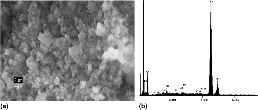
The addition of TBALD to the R5 peptide solution readily forms a white precipitate, which was analyzed by SEM/EDS for morphology and elemental composition. EDS confirms the presence of titanium in the precipitate (Fig. 1). Although R5 is known to precipitate a form of titania, the conditions we used differed from previous studies; therefore, positive titanium identification in EDS supported further exploration of the material. Examination of bulk precipitate produced using our conditions at room temperature (RT) generated a XRD spectrum with no detectable features, indicating the material to be amorphous [Fig. 2(a)]. To confirm that the amorphous material is a form of titania, the precipitate prepared at RT was heated to 600 °C to improve its crystallinity and remove entrapped proteinaceous material. An XRD spectrum was then generated with the heated sample, and peaks appeared that can be attributed to crystalline anatase form of titania [Fig. 2(b)], as illustrated previously.Reference Choi, Tan, Jang, Um, Yoo and Choe31–Reference Filocamo, Stote, Ziegler and Gibson33
(a) SEM of titania precipitate formed in 25 mM Tris buffer by R5 peptide at 10,000× magnification, and (b) corresponding EDS showing strong Ti Kα and Kβ peaks.

(a) XRD of titania generated at room temperature. (b) XRD of titania precipitated by R5 peptide heated to 600 °C. The red lines mark the location of anatase peaks.
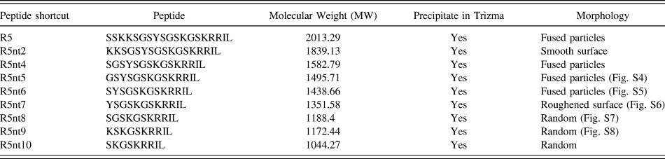
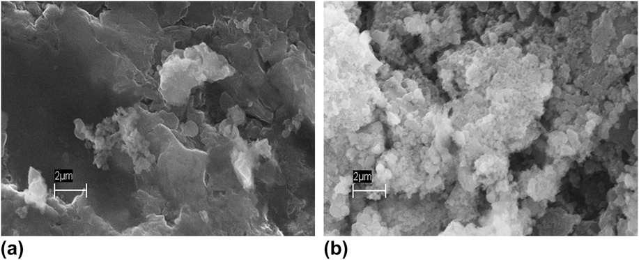
Using SEM to evaluate morphology, the R5 peptide precipitate indicates a level of control not demonstrated by most biomimetic polymers. The particulate nature of the precipitate from the R5 peptide is in stark contrast to the smooth, film-like nature of titania precipitates generated in previous studies.Reference Dickerson, Sandhage and Naik17,Reference Kroger, Dickerson, Ahmad, Cai, Haluska, Sandhage, Poulsen and Sheppard24 Furthermore, truncations of up to 10 amino acids from the N-terminus of the R5 peptide do not inhibit the ability of the peptide to precipitate, although the amount of precipitate is visually decreased as the length of the peptide decreases (Table I). More specifically, removal of the first 2 serine residues (Table I: R5nt2) results in a complete loss of morphological control, creating a smooth surface [Fig. 3(a)]. However, further truncation of the 2 lysines leaves a serine as the N-terminal residue (Table I: R5nt4, Fig. S1), and the morphological control returns [Fig. 3(b)]. Peptides that have eight or more amino acids removed from the N-terminus, formed precipitates that have a random structure, and this did not change with subsequent truncations. It appears that at some point the size of the peptide limits the types of morphology that the precipitate can form.
Truncated R5 peptides from the N-terminus and their precipitation capability.
SEM images (10,000× magnification) of precipitates formed in 25 mM Tris buffer using R5 peptide with (a) 2 serines truncated from N-terminus, and (b) 2 serines and 2 lysines truncated from the N-terminus.
When the R5 peptide was truncated from the C-terminal end, results were more pronounced. Removal of the isoleucine–leucine residues (Table II: R5ct1 and R5ct2) has no effect on the precipitation ability or the morphology, which was expected due to the lack of reactive groups on the side chains. However, further removal of the 2 arginine residues (Table II: R5ct4) resulted in a visible significant reduction in the amount of precipitate formed. Additional removal of the lysine residue closest to the C-terminus (R5ct5) rendered the resulting peptide incapable of precipitating titania. No further subtraction from the C-terminal side restored the precipitating capability in Trizma buffer. Due to the relatively low purity of truncated R5 peptides in the microtiter array (Tables I and II), quantification of titania would be difficult to assess. However, quantification of titania precipitates formed from purified R5 peptide (synthesized separately from the microtiter array) was accomplished using a colorimetric technique,Reference Puddu, Slocik, Naik and Perry34 and the average value of 1.850 mol TiO2/mol R5 was obtained. We would expect that value to be the upper bound of precipitation generated using the procedure described above.
Truncated R5 peptides from the C-terminus and their precipitation capability.
In fact, a pattern emerged that suggests that a critical number of basic residues are needed for precipitation to initiate. This number is partially dependent on the relative basicity of the residue. Further modifications of the R5 peptide confirmed that reliable precipitation at significant levels visually can occur when (a) three lysines and at least one arginine are present, (b) four lysines and more than two serines are present or (c) two arginines and two lysines are present. Though serine is not a basic residue, previous research has shown that hydroxyl side chains do contribute, albeit limited, to the precipitation capability.Reference Choi, Tan, Jang, Um, Yoo and Choe31 Truncating amino acids from both the C- and N-terminal ends simultaneously affected the precipitation only after the number of reactive elements dropped below one of these critical combinations (results not shown).
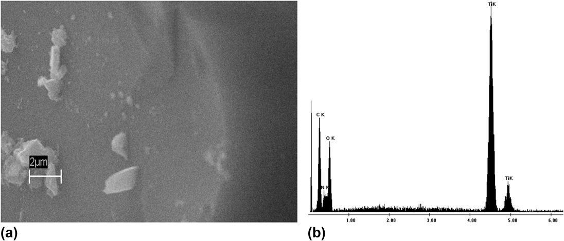
Small lysine-containing peptides were tested to support this observation. Compounds consisting of 2–5 lysine residues were tested under identical conditions for the ability to precipitate titania. After 24 h, only the 5-lysine residue peptide precipitated titania, forming as fused sheets with no discernable structure (Fig. 4). The 5-mer is highly charged, so we would not expect this molecule to have significant intermolecular interactions prior to precipitation. Therefore, the ability to precipitate must be localized on each molecule. The fact that only the 5-mer precipitated titania supports the theory that a minimal number of reactive groups (e.g., amines) is needed to facilitate titania precipitation. The “local concentration” effect is based on both the structure of the reactive group, and its Lewis basicity, which is supported by previous research that looked at the effect of local concentration and basicity on precipitating ability.Reference Filocamo, Stote, Ziegler and Gibson33,Reference Kharlampieva, Slocik, Singamaneni, Poulsen, Kroger, Naik and Tsukruk35,Reference Ahn, Park and Lee36
(a) SEM of titania precipitate formed in 25 mM Tris buffer by KKKKK at 10,000× magnification, and (b) corresponding EDS showing strong Ti Kα and Kβ peaks.
B. Titania precipitation from substituted R5 peptides in Trizma buffer
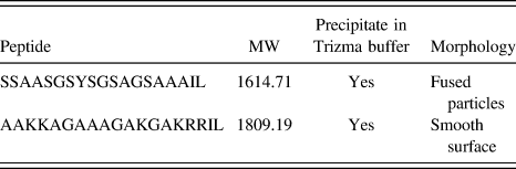
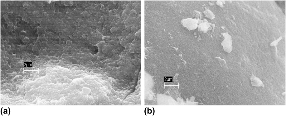
Based upon the rules that have been identified, amino acids with hydroxyl or basic side chains were selectively substituted. The substitutions were designed to replace all of the amino acids containing hydroxyl side chains (serines), or all the basic side chains (lysines and arginines) with alanine, which has a nonreactive methyl side chain (Table III). Both substituted R5 peptides precipitated titania (Fig. 5), though at a reduced amount compared with the unmodified R5 peptide. The ability to precipitate titania with the serine-substituted peptide was expected, since the amines are the primary nucleating elements. However, even the lysine/arginine-modified peptide produced a very small amount of titania. From previous studies, hydroxyl side chains have been shown to precipitate titania at a slower rate than nitrogen-containing side chains.Reference Brutchey and Morse26,Reference Filocamo, Stote, Ziegler and Gibson33 Since the R5 peptide has seven serines residues separated by 0 to 2 amino acids there should be sufficient hydroxyl groups, in relatively close proximity to each other, for the initiation of titania formation. The lysine/arginine-substituted peptide yielded precipitates composed of fused particles while the serine-substituted peptide with the amine side chain intact precipitated as a smooth surface [Figs. 5(a) and 5(b)]. These results support the concept that the basic residues are the main driver for precipitation, but the hydroxyl residues are the primary contributors to morphological control.
R5 peptide with substitutions of either all amines or all hydroxyls.
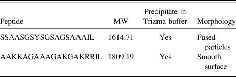
SEM images (10,000× magnification) of precipitates formed in 25 mM Tris buffer using R5 peptides (a) with alanine substitutions for lysine and arginine, and (b) with alanine substitutions for serine.
C. Titania precipitation from truncated silaffin R5 peptide in phosphate buffer.
Reaction conditions can have a profound impact on precipitation results and morphology.Reference Nguyen5,Reference Li, Chen, Ma, Zhang, An and Huang8,Reference Dickerson, Jones, Cai, Ahmad, Naik, Kroger and Sandhage30 Much of the research performed to date on biomimetic silaffin precipitations has been performed in phosphate buffer. Silica precipitations in phosphate buffer show no evidence of phosphorus being incorporated into the oxide matrix. However, precipitations involving titania precursors, using phosphate buffers as the medium, are not exclusively titania, but are either in whole or in part titanium phosphate.Reference Cole and Valentine27,Reference Kharlampieva, Slocik, Singamaneni, Poulsen, Kroger, Naik and Tsukruk35 The presence of titanium phosphate implies that the phosphate is not a passive player when used as the medium for titania precipitation. This more active role was tested with the same truncated R5 peptides and lysine compounds reacted in Trizma buffer.
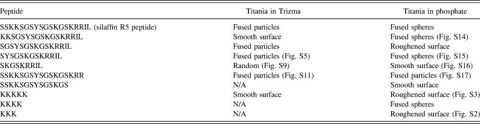
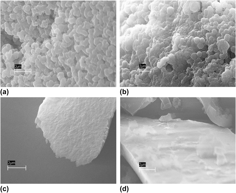
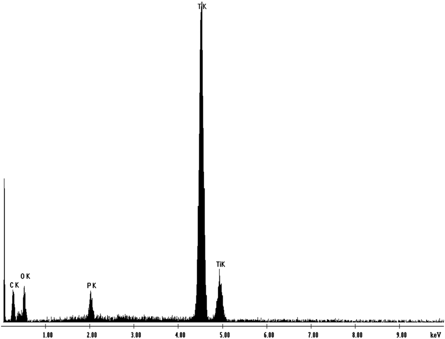
All peptides active in Trizma buffer were also able to precipitate in phosphate. In addition, all of the peptides with 3 or 4 amino acids containing basic side chains formed a precipitate, which differs from the results seen in Trizma (Table IV). A lower local concentration of reactive groups is needed, in the presence of phosphate, to produce a precipitate. This was confirmed using the 2–5 lysine residue compounds as precipitating agents; 3-Lys, 4-Lys [Fig. 6(a)], and 5-Lys were successful at forming a precipitate in phosphate buffer, whereas only 5-Lys was active in Trizma. EDS data confirm the presence of titanium, as well as a significant amount of phosphorus, in the precipitates formed (Fig. 7). The extensive washing procedure prior to imaging would remove residual buffer salts, indicating that the precipitate has phosphorus incorporated into the structure. Therefore, in phosphate buffer, this process produces at best a co-precipitate of titania and titanium phosphate. In fact, as observed by Puddu et al., TBALD will precipitate material containing phosphorus without any peptide initiator in the presence of a phosphate buffer at concentrations >100 mM.Reference Puddu, Slocik, Naik and Perry34 In contrast, Trizma buffer concentrations up to 500 mM produced no precipitate after 24 h. The results indicate that buffer composition can be an active participant in the precipitation reaction.
Comparison of precipitation capabilities in Trizma and phosphate buffers.
SEM images of precipitates (10,000× magnification) formed in 25 mM sodium phosphate buffer using the following peptides: (a) 4-Lys, (b) R5 peptide (SSKKSGSYSGSKGSKRRIL) (c) SGSYSGSKGSKRRIL, (d) SSKKSGSYSGSKGS.
EDS showing strong Ti Kα and Kβ peaks and P Kα (@ 2.00 keV) from titania precipitate generated using 5-Lys in phosphate buffer.
Morphological control in these reactions varied, depending on both the number of basic groups available and their presence or absence at the N- and C-terminal ends of the peptide. When basic residues were present at the termini, fused particles of sizes up to 1 µm were formed [Fig. 6(b)], consistent with structures reported in the literature.Reference Puddu, Slocik, Naik and Perry34,Reference Kharlampieva, Slocik, Singamaneni, Poulsen, Kroger, Naik and Tsukruk35,Reference Sewell and Wright37 However, deletion of the lysine residues at the N-terminus or basic residues at the C-terminus resulted in significant loss of morphological control [Figs. 6(c) and 6(d)]. The importance of amino acids with basic side chains in the morphological control of titanium phosphate is in stark contrast with titania precipitates in Trizma buffer, which were influenced more by the amino acids with hydroxyl side chains. Therefore when studying the formation of nonbiological materials using biomimetic processes, it will be vital to understand that not only how a precipitate is formed, but also how that precipitation is influenced by reaction conditions.
V. CONCLUSION
Using the R5 peptide as a model compound, we investigated the effect of primary structure on the ability of bioinspired materials to precipitate nonbiological oxides. Precipitation of titania using modified R5 peptides with systematic truncations and substitutions has shown that basic- and hydroxyl side chains have a prominent role to play in the reaction. The basic side chains are vital to initiating the reaction, with a minimum local concentration of basic residues necessary to sustain the precipitation. The hydroxyl residues appear to be necessary to impart morphological structure to the precipitates. In addition, we have demonstrated that reaction buffer can influence the composition and morphology of biomimetically-formed precipitates. The use of Trizma or phosphate buffer in the precipitation reaction changed both the capability to precipitate, and the morphology. Analysis of the precipitate revealed a high presence of phosphate in the precipitates matrix formed in the phosphate buffer, suggesting the creation of a very different material in different buffers.
These concepts provide a template from which new, simple peptides that primarily consist of lysine and serine residues can be designed that precipitate titania with controlled morphology. Small peptide sequences containing only lysine and serine residues have already shown promising results in the formation of titania with defined structures. In a broader sense, they illustrate the sophistication and adaptation inherent in the design and organization of natural materials. Although originally designed for silica formation, these peptidic structures are amenable to building other oxides from precursors of similar reactivity. Control over material formation at low energy costs are hallmarks of biological processes, and a fundamental understanding of these procedures is important to harness biomimetics for the next generation of materials.
Supplementary Material
To view supplementary material for this article, please visit http://dx.doi.org/10.1557/jmr.2016.165.
SEM images taken at 10,000× magnification representing each morphology identified: roughened surface, smooth surface, fused particles, random, and fused spheres.
ACKNOWLEDGMENTS
We thank the Defense Threat Reduction Agency for supporting this research, and Mr. Dave Ziegler for assistance with XRD measurements. We gratefully acknowledge Dr. Charlene Mello for helpful discussions regarding this work.