Introduction
Major depressive (MDD) and bipolar disorder (BD) are disabling diseases because of their cognitive impairments, which negatively impact daily functioning, recovery and treatment efficacy (Dunkin et al. Reference Dunkin, Leuchter, Cook, Kasl-Godley, Abrams and Rosenberg-Thompson2000; Jaeger et al. Reference Jaeger, Berns, Uzelac and Davis-Conway2006; Goodwin et al. Reference Goodwin, Martínez-Arán, Glahn and Vieta2008b ; Mur et al. Reference Mur, Portella, Martínez-Arán, Pifarre and Vieta2009; Beblo et al. Reference Beblo, Sinnamon and Baune2011; Baune et al. Reference Baune, Li and Beblo2013; Daniel et al. Reference Daniel, Montali, Gerra, Innamorati, Girardi, Pompili and Amore2013; McIntyre et al. Reference McIntyre, Cha, Soczynska, Woldeyohannes, Gallaugher, Kudlow, Alsuwaidan and Baskaran2013). These impairments are present during depressive and manic episodes, and may persist, albeit probably less severely, during remission (Quraishi & Frangou, Reference Quraishi and Frangou2002; Martínez-Arán et al. Reference Martínez-Arán, Vieta, Reinares, Colom, Torrent, Sánchez-Moreno, Benabarre, Goikolea, Comes and Salamero2004; Clark et al. Reference Clark, Sarna and Goodwin2005; Bearden et al. Reference Bearden, Glahn, Monkul, Barrett, Najt, Villarreal and Soares2006; Gruber et al. Reference Gruber, Rathgeber, Bräunig and Gauggel2007; Mur et al. Reference Mur, Portella, Martínez-Arán, Pifarré and Vieta2008; Beblo et al. Reference Beblo, Sinnamon and Baune2011; Xu et al. Reference Xu, Lin, Rao, Dang, Ouyang, Guo, Ma and Chen2012; Wekking et al. Reference Wekking, Bockting, Koeter and Schene2012; Bourne et al. Reference Bourne, Aydemir, Balanze-Marinez, Bora, Brissos, Cavanagh, Clark, Cubukcuoglu, Videira Dias, Dittmann, Ferrier, Fleck, Frangou, Gallagher, Jones, Kieseppa, Martínez-Arán, Melle, Moore, Mur, Pfennig, Raust, Senturk, Simonse, Smith, Bio, Soeiro-de-Souza, Stoddart, Sundet, Szoke, Thompson, Torrent, Zalla, Craddock, Andreassen, Leboyer, Vieta, Bauer, Worhunsky, Tzagarakis, Rogers, Geddes and Goodwin2013; Lim et al. Reference Lim, Baldessarini, Vieta, Yucel, Bora, Sim and Lim2013). BD subjects tend to show more of these impairments than MDD subjects (Wolfe et al. Reference Wolfe, Granholm, Butters, Saunders and Janowsky1987; Borkowska & Rybakowski, Reference Borkowska and Rybakowski2001; Smith et al. Reference Smith, Muir and Blackwood2006; Gildengers et al. Reference Gildengers, Butters, Chisholm, Anderson, Begley, Holm, Rogers, Reynolds and Mulsant2012; Xu et al. Reference Xu, Lin, Rao, Dang, Ouyang, Guo, Ma and Chen2012). However, the nature of these neurocognitive deficits is controversial, due to the use of the variety of neuropsychological tests (Yatham et al. Reference Yatham, Torres, Malhi, Frangou, Glahn, Bearden, Burdick, Martínez-Arán, Dittmann, Goldberg, Ozerdem, Aydemir and Chengappa2010) and the heterogeneity of clinical samples, e.g. regarding medication use (Beblo et al. Reference Beblo, Sinnamon and Baune2011). More knowledge about cognitive impairments would improve our understanding of the pathophysiology underlying mood disorders and may help to differentiate MDD and BD. This is important, since MDD and BD are difficult to distinguish clinically, especially during depression or remission (Altshuler et al. Reference Altshuler, Post, Mikalauskas, Rosoff, Leverich and Ackerman1995; Ghaemi et al. Reference Ghaemi, Boiman and Goodwin2000, Reference Ghaemi, Rosenquist, Ko, Baldassano, Kontos and Baldessarini2004; Hirschfeld et al. Reference Hirschfeld, Lewis and Vornik2003; Goodwin et al. Reference Goodwin, Anderson, Arango, Bowden, Henry, Mitchell, Nolen, Vieta and Wittchen2008a ; Perlis et al. Reference Perlis, Ostacher, Goldberg, Miklowitz, Friedman, Calabrese, Thase and Sachs2010). However, whether MDD and BD show similar profiles of cognitive deficits is still debated, as both comparable (Sweeney et al. Reference Sweeney, Kmiec and Kupfer2000; Bearden et al. Reference Bearden, Glahn, Monkul, Barrett, Najt, Villarreal and Soares2006) and different profiles (Taylor Tavares et al. Reference Taylor Tavares, Clark, Cannon, Erickson, Drevets and Sahakian2007) have been reported.
Executive dysfunction is a common feature in mood disorders (Quraishi & Frangou Reference Quraishi and Frangou2002; Beblo et al. Reference Beblo, Sinnamon and Baune2011; Snyder, Reference Snyder2013). Executive functioning comprises several higher-order cognitive processes to control and regulate lower-level cognitive processes and goal-directed behaviour (Smith & Jonides, Reference Smith and Jonides1999; Miyake et al. Reference Miyake, Friedman, Emerson, Witzki, Howerter and Wager2000; Funahashi, Reference Funahashi2001; Alvarez & Emory, Reference Alvarez and Emory2006). One of these processes is planning (Paelecke-Habermann et al. Reference Paelecke-Habermann, Pohl and Leplow2005; Kulkarni et al. Reference Kulkarni, Jain, Janardhan Reddy, Kumar and Kandavel2010), referring to the ability to organize cognitive behaviour in time and space, in order to achieve a goal through a series of intermediate steps (Owen, Reference Owen1997). One test to examine planning is the Tower of London (ToL; Shallice, Reference Shallice1982), a well-validated functional magnetic resonance imaging (fMRI) paradigm, known to activate an extensive fronto-parieto-thalamic-striatal network (Lazeron et al. Reference Lazeron, Rombouts, Machielsen, Scheltens, Witter, Uylings and Barkhof2000; Van den Heuvel et al. Reference Van den Heuvel, Groenewegen, Barkhof, Lazeron, van Dyck and Veltman2003; Unterrainer & Owen, Reference Unterrainer and Owen2006; Wagner et al. Reference Wagner, Koch, Reichenbach, Sauer and Schlösser2006), in which the prefrontal cortex is assumed to be critical for actual planning (Unterrainer & Owen, Reference Unterrainer and Owen2006). The parietal cortex is thought to be involved in visuospatial aspects of the task, whereas interactions between the prefrontal cortex and basal ganglia may be involved in the selection and switching of behavioural components during planning (Van den Heuvel et al. Reference Van den Heuvel, Groenewegen, Barkhof, Lazeron, van Dyck and Veltman2003). Planning thus draws on several subprocesses such as attention, working memory and inhibition (Unterrainer & Owen, Reference Unterrainer and Owen2006). For all of these subprocesses there is evidence of deficits in MDD and BD, both on a behavioural and neuronal level, albeit sometimes contradictory. For example, MDD and BD subjects have shown poorer performance on various visuospatial tasks (e.g. learning, working memory and reasoning; see e.g. Borkowska & Rybakowski, Reference Borkowska and Rybakowski2001; Quraishi & Frangou, Reference Quraishi and Frangou2002; Porter et al. Reference Porter, Gallagher, Thompson and Young2003; Tiihonen et al. Reference Tiihonen, Haukka, Henriksson, Cannon, Kieseppä, Laaksonen, Sinivuo and Lönnqvist2005; Snyder, Reference Snyder2013). Compromised working memory in MDD and BD has frequently been reported (see e.g. Sweeney et al. Reference Sweeney, Kmiec and Kupfer2000; Borkowska & Rybakowski, Reference Borkowska and Rybakowski2001; Weiland-Fiedler et al. Reference Weiland-Fiedler, Erickson, Waldeck, Luckenbaugh, Pike, Bonne, Charney and Neumeister2004; Taylor Tavares et al. Reference Taylor Tavares, Clark, Cannon, Erickson, Drevets and Sahakian2007; Kaneda, Reference Kaneda2009; Wekking et al. Reference Wekking, Bockting, Koeter and Schene2012; Bourne et al. Reference Bourne, Aydemir, Balanze-Marinez, Bora, Brissos, Cavanagh, Clark, Cubukcuoglu, Videira Dias, Dittmann, Ferrier, Fleck, Frangou, Gallagher, Jones, Kieseppa, Martínez-Arán, Melle, Moore, Mur, Pfennig, Raust, Senturk, Simonse, Smith, Bio, Soeiro-de-Souza, Stoddart, Sundet, Szoke, Thompson, Torrent, Zalla, Craddock, Andreassen, Leboyer, Vieta, Bauer, Worhunsky, Tzagarakis, Rogers, Geddes and Goodwin2013) paralleled by abnormally increased lateral prefrontal, parietal and anterior cingulate cortex (ACC) activity (Adler et al. Reference Adler, Holland, Schmithorst, Tuchfarber and Strakowski2004; Harvey et al. Reference Harvey, Fossati, Pochon, Levy, Lebastard, Lehéricy, Allilaire and Dubois2005; Rose et al. Reference Rose, Simonotto, Spencer and Ebmeier2006; Matsuo et al. Reference Matsuo, Glahn, Peluso, Hatch, Monkul, Najt, Sanches, Zamarripa, Li, Lancaster, Fox, Gao and Soares2007; Deckersbach et al. Reference Deckersbach, Rauch, Buhlmann, Ostacher, Beucke, Nierenberg, Sachs and Dougherty2008; Fitzgerald et al. Reference Fitzgerald, Srithiran, Benitez, Daskalakis, Oxley, Kulkarni and Egan2008; Frangou et al. Reference Frangou, Kington, Raymont and Shergill2008; Marquand et al. Reference Marquand, Mourão-Miranda, Brammer, Cleare and Fu2008; Pu et al. Reference Pu, Yamada, Yokoyama, Matsumura, Kobayashi, Sasaki, Mitani, Adachi, Kaneko and Nakagome2011; Jogia et al. Reference Jogia, Dima, Kumari and Frangou2012; Korgaonkar et al. Reference Korgaonkar, Grieve, Etkin, Koslow and Williams2013). Impaired inhibition (Stordal et al. Reference Stordal, Lundervold, Egeland, Mykletun, Asbjørnsen, Landrø, Roness, Rund, Sundet, Oedegaard and Lund2004; Larson et al. Reference Larson, Shear, Krikorian, Welge and Strakowski2005; Stefanopoulou et al. Reference Stefanopoulou, Manoharan, Landau, Geddes, Goodwin and Frangou2009; Gotlib & Joormann, Reference Gotlib and Joormann2010; Bora et al. Reference Bora, Harrison, Yücel and Pantelis2013) has also been associated with abnormal prefrontal and ACC activity (Blumberg et al. Reference Blumberg, Leung, Skudlarski, Lacadie, Fredericks, Harris, Charney, Gore, Krystal and Peterson2003; Gruber et al. Reference Gruber, Rogowska and Yurgelun-Todd2004; Strakowski et al. Reference Strakowski, Adler, Holland, Mills, DelBello and Eliassen2005; Wagner et al. Reference Wagner, Koch, Reichenbach, Sauer and Schlösser2006; Schlösser et al. Reference Schlösser, Wagner, Koch, Dahnke, Reichenbach and Sauer2008; Hajek et al. Reference Hajek, Alda, Hajek and Ivanoff2013; Korgaonkar et al. Reference Korgaonkar, Grieve, Etkin, Koslow and Williams2013; Welander-Vatn et al. Reference Welander-Vatn, Jensen, Otnaess, Agartz, Server, Melle and Andreassen2013). Nevertheless, there are indications that the ToL assesses predominantly planning abilities per se (Unterrainer & Owen, Reference Unterrainer and Owen2006). Previous mood disorder studies using the ToL have mainly been conducted in depressed MDD subjects. These studies demonstrated increased dorsolateral prefrontal cortical (DLPFC) and parietal activity (Fitzgerald et al. Reference Fitzgerald, Srithiran, Benitez, Daskalakis, Oxley, Kulkarni and Egan2008; Van Tol et al. Reference Van Tol, Van der Wee, Demenescu, Nielen, Aleman, Renken, Van Buchem, Zitman and Veltman2011), associated with either decreased (Fitzgerald et al. Reference Fitzgerald, Srithiran, Benitez, Daskalakis, Oxley, Kulkarni and Egan2008) or normal (Van Tol et al. Reference Van Tol, Van der Wee, Demenescu, Nielen, Aleman, Renken, Van Buchem, Zitman and Veltman2011) task performance. In contrast, two small PET and SPECT studies (N = 6 and N = 9 MDD, respectively) found decreased activity paralleled by decreased performance (Elliott et al. Reference Elliott, Baker, Rogers, O'Leary, Paykel, Frith, Dolan and Sahakian1997; Goethals et al. Reference Goethals, Audenaert, Jacobs, Van de Wiele, Ham, Pyck, Vandierendonck, Van Heeringen and Dierckx2005). One study investigating remitted MDD subjects showed no differences v. healthy controls (HC) (Van Tol et al. Reference Van Tol, Van der Wee, Demenescu, Nielen, Aleman, Renken, Van Buchem, Zitman and Veltman2011). In BD, ToL neuroimaging studies are not available; therefore it is unknown whether any planning deficits in BD are related to similar neural substrates as in MDD, or whether planning abnormalities in BD are more severe during depression than remission, similar to MDD (Van Tol et al. Reference Van Tol, Van der Wee, Demenescu, Nielen, Aleman, Renken, Van Buchem, Zitman and Veltman2011). Finally, in all studies of neurocognitive functioning in mood disorders using the ToL psychotropic medication was allowed, which may affect cognitive functioning on neural (Bell et al. Reference Bell, Willson, Wilman, Dave and Silverstone2005; Silverstone et al. Reference Silverstone, Bell, Willson, Dave and Wilman2005; Rose et al. Reference Rose, Simonotto, Spencer and Ebmeier2006; Völlm et al. Reference Völlm, Richardson, McKie, Elliott, Deakin and Anderson2006; Haldane et al. Reference Haldane, Jogia, Cobb, Kozuch, Kumari and Frangou2008; Wingen et al. Reference Wingen, Kuypers, Van de Ven, Formisano and Ramaekers2008) and neuropsychological (Savitz et al. Reference Savitz, Solms and Ramesar2005; Baune & Renger, Reference Baune and Renger2014) levels.
We therefore investigated the neural correlates of planning in medication-free depressed and remitted MDD and BD subjects, using the ToL. We hypothesized there would be an effect of the presence of mood disorder, with both MDD and BD subjects showing frontoparietal abnormalities (hyper-or hypoactivity) associated with similar or decreased behavioural performance compared to HC; an effect of state, with greater abnormalities in depressed v. remitted subjects; and an effect of mood disorder subtype, with greater abnormalities in BD v. MDD subjects.
Materials and method
Subjects
After approval by the local Medical Ethical Committee and written informed consent, we collected ToL fMRI data of 41 MDD subjects [21 remitted (MDDr), 20 depressed (MDDd)], 34 BD-I/II subjects (24 BDr, 10 BDd), matched for age, gender, education and depression severity, and 35 HC. These subjects were part of a sample described previously (Rive et al. Reference Rive, Mocking, Koeter, van Wingen, De Wit, Van den Heuvel, Veltman, Ruhé and Schene2015). Diagnoses were assessed using the Structured Clinical Interview for DSM-IV Axis I Disorders (SCID-I), conducted by a SCID-trained psychiatrist or psychiatry resident. In case of doubt, a second opinion was provided by a second trained psychiatrist.
Inclusion criteria for MDD and BD-I/II subjects were: age 18–60 years; age at first mood episode ⩽40 years; a history of at least two major depressive episodes (MDEs); current MDE or remission [i.e. not fulfilling the criteria of MDE or (hypo)manic episode]; and illness duration of ⩾5 years since the first episode (in order to reduce the likelihood of including late MDD-to-BD converters in the MDD group). Exclusion criteria for MDD and BD subjects were: current (hypo)mania; a current comorbid Axis I disorder (including any current substance use disorder; assessed by SCID-I) except for anxiety disorders; a diagnosis of cluster B personality disorder [assessed by SCID-II, if suspected based on (hetero)anamnestic information]; current use of antidepressants, anticonvulsants, mood stabilizers or antipsychotics (stopped at least 1 month before scanning); electroconvulsive therapy within 2 months before scanning. Incidental benzodiazepine use was allowed, but was stopped at least 1 day before scanning. In addition, MDD subjects were excluded when they had a history of (hypo)manic derailment after antidepressant use or a first-degree family history of bipolar disorder (in order to prevent inclusion of MDD subjects with possible BD traits). BD subjects were excluded if (hypo)manic episodes solely appeared during the use of antidepressants, again to ensure inclusion of ‘true’ BD subjects; and in case of a Young Mania Rating Scale score >8, to prevent inclusion of BD subjects with (subclinical) (hypo)manic symptoms.
Inclusion criterion for HC was: age 18–60 years. Exclusion criteria for HC were: presence of a lifetime psychiatric diagnosis (Axis I); use of any psychopharmacological agent; first-degree relatives with a history of a psychiatric diagnosis (Axis I).
Furthermore, for all subjects contra-indications for MRI scanning were: a history of head trauma or neurological disease; severe general physical illness; claustrophobia or implanted metal objects.
Demographic and clinical measurements
We used the Dutch version of the New Adult Reading Test (Schmand et al. Reference Schmand, Bakker, Saan and Louman1991), to provide an estimate of premorbid intelligence level; the Hamilton Depression Rating Scale (HAMD; Hamilton Reference Hamilton1960, Reference Hamilton1967) to assess depression severity; and the Bipolarity Index to assess the level of bipolarity based on lifetime characteristics (Sachs, Reference Sachs2004).
Data acquisition
Functional and structural MRI data were acquired on a 3.0-T MRI scanner (Philips Intera, Philips Medical Systems, The Netherlands) with body coil excitation and an 8-channel SENSE head coil. For the blood oxygen level dependent (BOLD) functional scans the following parameters were used: echo time 30 ms, repetition time 2300 ms, flip angle = 80°, matrix 96 × 96, 40 ascending slices, no gap, 2.29 × 2.29 × 3 mm voxels. For anatomical co-registration purposes, a 7-min T1-weighted structural image was acquired (echo time 4.6 ms, repetition time 9.6 ms, flip angle = 8°, matrix 256 × 256, 182 slices, 1 × 1 × 1.2 mm voxels).
fMRI paradigm
We used a pseudo-randomized, self-paced parametric version of the ToL (abbreviated version of the paradigm of Van den Heuvel et al. Reference Van den Heuvel, Groenewegen, Barkhof, Lazeron, van Dyck and Veltman2003). From a starting configuration of coloured beads, subjects were asked to work out the minimal number of steps required to achieve a target configuration (Supplementary Fig. S1). There were five different task loads, ranging from 1 to 5 steps. In the baseline condition, subjects were asked to count the yellow and blue beads. Directly after responding, the next trial was presented; maximum response time was 1 min. The total duration of the task was 12 min.
Data analysis
Behavioural data
We used IBM SPSS Statistics v. 20 (IBM Corp., USA) for all behavioural analyses. With the non-parametric Jockheere–Terpstra test we assessed whether in each group (HC, MDDr, BDr, MDDd, BDd) task performance as measured by response time and accuracy (i.e. percentage correct trials) decreased with increasing task load. This test was used because the assumption of normality was violated and transformation did not suffice.
To assess whether group moderated the relationship between outcomes (response times, accuracies) and task load, we used separate linear mixed effects regression models for each outcome. We used group, task load and the group × task load interaction term as independent variables, in addition to age and IQ (to correct for their confounding effects). To assess whether group moderated overall mean task performance (irrespective of task load), mean response times and accuracies across all planning tasks were also investigated with separate linear mixed effects regression model for each outcome. We used group as categorical independent variable, and age, IQ, and baseline response times/accuracies as continuous independent variables (to correct for their confounding effects).
We tested the following planned contrasts: HC v. patients, remitted v. depressed patients, MDDr v. BDr and MDDd v. BDd. The significance threshold was set at p < 0.0125 (i.e. Bonferroni-corrected, four comparisons). We used bootstrapping with 20 000 bootstrap samples to account for violation of the normality assumption of the dependent variable.
fMRI data
Statistical Parametric Mapping (SPM8, http://www.fil.ion.ucl.ac.uk/spm/software/SPM8) was used for analysis of fMRI data. The following pre-processing steps were applied: slice timing; realignment; co-registration of functional and structural data; segmentation; spatial normalization to standard stereotactic space using a template from the Montreal Neurological Institute and resampling to 3 mm isotropic voxels; smoothing of data with an 8 mm full-width-at-half-maximum Gaussian kernel.
At first level, data were analysed according to the general linear model, comprising (i) separate regressors for trials of each task load and baseline in order to calculate the mean BOLD response during mean overall task performance (task > baseline); (ii) one regressor for all except baseline trials with an additional first-order parametric modulator for task load, in order to assess the relationship between BOLD response and linearly increasing task load. In both models, movement regressors were added to correct for subject motion. With these models, two aspects of brain activity in response to the task were analysed: (i) mean BOLD response elicited by the task irrespective of task load (task > baseline) and (ii) the linear relationship between BOLD response and task load (linear task load) (Van den Heuvel et al. Reference Van den Heuvel, Groenewegen, Barkhof, Lazeron, van Dyck and Veltman2003; Van Tol et al. Reference Van Tol, Van der Wee, Demenescu, Nielen, Aleman, Renken, Van Buchem, Zitman and Veltman2011). To remove high-frequency noise, a 128-Hz high-pass filter was applied.
Main task effects
To assess main task effects across subjects, we performed two one-sample t tests using within-subject contrast images task > baseline and linear task load.
Between-group comparisons
To assess group differences in overall task effects and effects of increasing task load, each within-subject contrast image (task > baseline, linear task load) was entered in a second-level random-effects analysis (ANCOVA, groups: HC, MDDr, BDr, MDDd, BDd; covariates: IQ, age). Since we a-priori hypothesized abnormalities would be greater in depressed v. remitted and in BD v. MDD subjects, we used the following planned contrasts: patients v. HC, remitted patients v. depressed patients, BDr v. MDDr and BDd v. MDDd, to follow-up the effects of group (F test) on BOLD activity. We chose to use planned contrasts to follow-up group effects in order to prevent chance capitalization by partitioning of experimental variance. In secondary analyses, when suggested by post-hoc plots of the group effects, we also tested for state × diagnosis interactions and linear trends over groups. Results were considered significant if surviving family-wise error (FWE) small volume correction (SVC) (p FWE_SVC) for a 16 mm sphere (i.e. twice the size of the smoothing kernel) around the peak voxel (Van Tol et al. Reference Van Tol, Van der Wee, Demenescu, Nielen, Aleman, Renken, Van Buchem, Zitman and Veltman2011). To ensure that any significant voxels were within anatomically plausible regions, we focused on seven specific regions of interest (RoIs): the left and right superior parietal gyrus, left and right inferior parietal gyrus, ACC (all Automated Anatomical Labelling atlas defined) and left and right DLPFC (BA 9/46) (WFU Pickatlas v. 2.4; Maldjian et al. Reference Maldjian, Laurienti, Burdette and Kraft2003, Reference Maldjian, Laurienti and Burdette2004). We applied Bonferroni correction to account for the number of ROIs, adjusted for the mean correlation (r = 0.18) between our ROIs, rendering a corrected threshold of p FWE_SVC < 0.01 (equivalent to p < 0.05 uncorrected for the number of ROIs) (http://www.quantitativeskills.com/sisa/calculations/bonfer.htm). Non-ROI results surviving whole-brain FWE correction of p FWE_WB < 0.05 are also reported.
Sensitivity and correlation analyses
Because of significant differences in the number of previous depressive episodes and prevalence of previous substance use disorder, analyses were repeated with these variables as additional covariates. Furthermore, we checked whether results would hold without adding age and IQ as covariates, since mean age and IQ did not differ between groups and were only added to rule out any effect of age and IQ on possible MDD v. BD differences, in line with previous ToL studies (De Ruiter et al. Reference De Ruiter, Reneman, Boogerd, Veltman, Van Dam, Nederveen, Boven and Schagen2011; Van Tol et al. Reference Van Tol, Van der Wee, Demenescu, Nielen, Aleman, Renken, Van Buchem, Zitman and Veltman2011). Effects of performance on task-related BOLD activity were assessed with correlation analyses.
Ethical standards
The authors assert that all procedures contributing to this work comply with the ethical standards of the relevant national and institutional committees on human experimentation and with the Helsinki Declaration of 1975, as revised in 2008.
Results
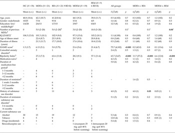
Demographics and clinical characteristics (Table 1)
Three subjects (1 BDr, 1 BDd, 1 MDDd) were excluded because of excessive movement or scanning artifacts. MDD (MDDr: N = 21; MDDd: N = 19), BD (BDr: N = 23; BDd: N = 9) and HCs (N = 35) were comparable regarding mean age, gender, education and IQ; MDD and BD subjects regarding prevalence of comorbid anxiety disorders. Mean HAMD scores differed between depressed and remitted subjects (p < 0.001), but not between MDD and BD within the remitted/depressed subgroups (p > 0.05). Although mean age of illness onset and illness duration did not differ between groups (p = 0.5 and p = 0.7, respectively), the median number of previous MDEs was higher in BDd than MDDd (p = 0.04). Furthermore, the proportion of subjects with a history of substance use disorder was greater in BDr than MDDr (p = 0.05, Table 1).
Demographic and clinical characteristics

BDd, Bipolar disorder, depressed state; BDr, BD, remitted state; DART, Dutch Adult Reading Test; HC, Healthy controls; MDDd, major depressive disorder, depressed state; MDDr, MDD, remitted state; MDE, major depressive episode.
Within the depressed and remitted groups, there were no significant differences between MDD and BD regarding demographic or clinical characteristics, except for the number of previous episodes.
a Low: primary education or preparatory middle-level applied education; middle: higher general continued education or middle-level applied education; high: preparatory scientific education, higher applied education, or scientific education.
b Missing values: BDr: 1.
c Kruskal–Wallis test comparing medians.
d >20 episodes are set on 20 episodes; reported are median and range.
e Missing values: MDDd: 1; BDr: 1.
f Hamilton Depression Rating Scale (17 items).
g Hypochondria, specific phobia, social phobia, panic disorder, obsessive-compulsive disorder.
h Missing values: MDDr: 1.
i Missing values: MDDd: 1.
j Missing values: MDDr: 4; BDr: 2; MDDd 3.
k Missing values: MDDr: 4; BDr: 2.
l Missing values: BDr: 2.
Behavioural results (Supplementary Fig. S2)
All subjects showed at least 85% accuracy on baseline trials and performed above chance level (range 63–95% accuracy) on the total number of trials. Thus, all subjects reliably engaged in the task and were included in the analyses. Overall, response times increased and accuracies decreased significantly with increasing task load (p < 0.001), without significant between-group differences (p > 0.0125).
fMRI results
Main task effects (Supplementary Fig. S3)
The task > baseline contrast was associated with activity in expected parieto-temporal and lateral/medial frontal regions, as well as in the precuneus, insula, caudate nucleus and pallidum (Supplementary Fig. S3A). Activity increased linearly with task load in similar areas (Supplementary Fig. S3B).
Between-group comparisons (Tables 2 and 3, Fig. 1)
Mean task performance (Tables 2a and 3a , Fig. 1a )
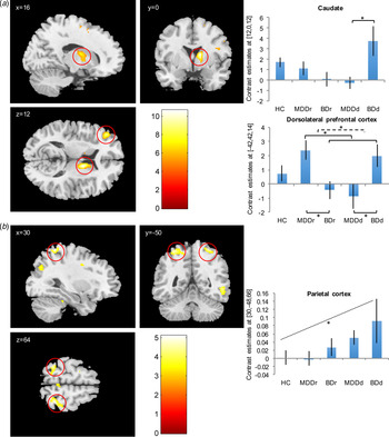
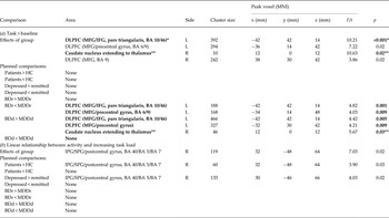
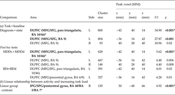
For task > baseline, the ANCOVA revealed significant effects of group in the left DLPFC (p FWE_SVC < 0.001) and caudate nucleus (p FWE_WB = 0.02); there was also trend-wise significant effects in another left-sided DLPFC cluster (p FWE_SVC = 0.02) and right DLPFC (p FWE_SVC = 0.02). Planned comparisons to investigate these group effects revealed increased DLPFC activity in MDDr v. BDr subjects (two left-sided clusters: p FWE_SVC = 0.001; p FWE_SVC = 0.009). BDd subjects also demonstrated increased activity compared to MDDd subjects in these clusters (p FWE_SVC = 0.005; p FWE_SVC = 0.009) and in the caudate (p FWE_WB = 0.03, Table 2 a). Post-hoc plotting revealed that the group effects of the three DLPFC clusters were driven by a state × diagnosis interaction in the patient group (Fig. 1 a). Secondary analysis (full factorial design) confirmed these state × diagnosis interactions [left DLPFC: p FWE_SVC = 0.001; right DLPFC: p FWE_SVC = 0.02 (trend); Table 3 a]. Post-hoc t tests (thresholded at p FWE_WB < 0.025/p FWE_SVC < 0.005, Bonferroni-corrected for two comparisons) showed significantly increased activity in MDDr compared to MDDd [three DLPFC clusters: left: p FWE_WB < 0.001; p FWE_SVC = 0.006 (trend); right: p FWE_SVC = 0.008 (trend)]. For additional results from the planned comparisons and secondary analysis outside group effects, see Supplementary Tables S1a, S2a).
Group effects for task > baseline and linear increasing task load. (a) For task v. baseline, there was a significant effect of group in the caudate and dorsolateral prefrontal cortex (DLPFC), driven by increased caudate activity in the BDd group [p = 0.02, whole-brain family-wise error (FWE) corrected] and, as revealed by secondary analysis based on post-hoc plotting of the group effects, a diagnosis × state interaction in the DLPFC (presented is one of the three DLPFC clusters; p < 0.001, small volume FWE corrected; also significant with whole-brain FWE correction). In turn, this diagnosis × state interaction was driven by increased MDDr v. MDDd and MDDr v. BDr activity (p < 0.001, small volume FWE corrected; MDDr v. MDDd also significant with whole-brain FWE correction) as well as increased BDd v. MDDd activity (p = 0.005, small volume FWE corrected). (b) For linearly increasing task load, there was a trendwise significant effect of group in the parietal cortex; secondary analysis based on post-hoc plotting this group effect revealed a significant linear trend of group (p < 0.001, small volume FWE corrected; also significant with whole-brain FWE correction). For visualization purposes, results are displayed at p < 0.001, uncorrected. *Indicates a significant result; –– indicates a difference between two means; ---- indicates an interaction effect; ++++ indicates a linear trend. Error bars represent 90% confidence intervals. BDd, bipolar disorder, depressed state; BDr, BP, remitted state; MDDd, major depressive disorder, depressed state; MDDr, MDD, remitted state.

Planned comparisons of activity related to task > baseline and task load
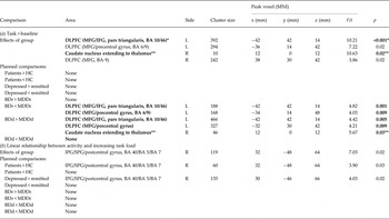
BA, Brodmann area; BDd, bipolar disorder, depressed state; BDr, BP, remitted state; DLPFC, dorsolateral prefrontal cortex; IFG, inferior frontal gyrus; IPG, inferior parietal gyrus; MDDd, major depressive disorder, depressed state; MDDr, MDD, remitted state; MFG, middle frontal gyrus; MNI, Montreal Neurological Institute; SFG, superior frontal gyrus; SPG, superior parietal gyrus.
Brain regions indicated in bold are significant results [for regions of interest (ROIs): p < 0.01 small volume (sphere r = 16) family wise error (FWE) corrected; or for non-ROIs: p < 0.05 whole-brain FWE corrected].
* Also significant at p < 0.05 whole-brain FWE corrected [dorsolateral prefrontal cortex (MFG/IFG, pars triangularis, BA 10/46), task > baseline, effect of group: cluster size k = 5, F = 10.21, p = 0.025).
** p < 0.05 whole-brain FWE corrected.
Secondary analysis based on post-hoc plots of effects of group for task > baseline (DLPFC) and task load (IPG/FPG)

BA, Brodmann area; BDd, bipolar disorder, depressed state; BDr, BP, remitted state; DLPFC, dorsolateral prefrontal cortex; IFG, inferior frontal gyrus; IPG, inferior parietal gyrus; MDDd, major depressive disorder, depressed state; MDDr, MDD, remitted state; MFG, middle frontal gyrus; SFG, superior frontal gyrus; SPG, superior parietal gyrus.
Brain regions indicated in bold are significant results [for regions of interest (ROIs): p < 0.01 for interaction or linear effects; p < 0.005 for post-hoc comparisons (Bonferroni-corrected for two post-hoc tests), both small volume (sphere r = 16) family-wise error (FWE) corrected; or, for non-ROIs, p < 0.05 for interaction or linear effects; p < 0.025 for post-hoc comparisons (Bonferroni-corrected for two post-hoc tests), both whole-brain FWE corrected].
* Also significant at p < 0.05 whole-brain FWE corrected [DLPFC (MFG/IFG, pars triangularis, BA 10/46) task > baseline, state × diagnosis: cluster size k = 28, F = 34.90, p = 0.007; DLPFC (MFG/IFG, pars triangularis, BA 10/46), task > baseline, MDDr > MDDd: k = 18, t = 5.62, p = 0.009; IPG/SPG (BA 40/BA 5/BA 7), linear trend over groups, linearly increasing task load: k = 4, t = 5.04, p = 0.033].
In summary, mean task performance was associated with a diagnosis × state interaction in the bilateral DLPFC, primarily driven by increased activity in MDDr v. both MDDd and BDr. Furthermore, BDd subjects showed greater left-sided DLPFC and caudate activity than MDDd.
Task load (Tables 2 b and 3 b, Fig. 1b )
For increasing task load, the ANCOVA revealed a borderline significant effect of group in the right parietal cortex (p FWE_SVC = 0.02; Table 2 b). Planned comparisons revealed trend-wise increased activity in the same region for patients > HC (p FWE_SVC = 0.03) and depressed > remitted patients (p FWE_SVC = 0.02). Post-hoc plotting of the parietal group effect suggested a linear trend over groups, i.e. MDDr demonstrated the lowest parietal activity, followed by BDr, MDDd and BDd (Fig. 1 b). Secondary analysis testing for this trend showed that this linear effect was significant (p FWE_WB < 0.001, Table 3 b). For additional results from the planned comparisons and secondary analysis outside group effects, see Supplementary Tables S1b and S2b).
Sensitivity and correlation analyses
Repeating the analysis adjusting for the number of previous episodes or previous substance use disorders did not change the pattern of results, nor did removing the regressors age and IQ (data available on request). There were no correlations between behavioural performance and BOLD activity.
Discussion
The present study is the first to investigate the neural correlates of planning comparing medication-free remitted and depressed BD and MDD. With increasing task load, post-hoc analyses indicated a stronger increase in parietal activity in more severely affected subjects (i.e. BD more than MDD, depressed more than remitted): secondary analysis revealed that HC and MDDr showed the least parietal activity increase, followed by MDDd, BDr and BDd.
Investigating MDD only (Van Tol et al. Reference Van Tol, Van der Wee, Demenescu, Nielen, Aleman, Renken, Van Buchem, Zitman and Veltman2011) also demonstrated a greater load-related activity increase in depressed v. remitted MDD and HC, albeit in the DLPFC. Although not found as ANCOVA group effect, our secondary analysis testing for the linear trend over groups revealed increasing activity in the same DLPFC cluster (Supplementary Table S2). Together, these results suggest that the more severely subjects are affected, the more activity is required to effectively engage in increasing levels of planning. Since the relationship between decreasing performance and increasing task load did not differ between groups, activity increases may be considered as compensatory. Previous research using the ToL indicated that the parietal cortex is involved in visuospatial attention (Newman et al. Reference Newman, Greco and Lee2009), whereas the DLPFC is involved in planning (Ruh et al. Reference Ruh, Rahm, Unterrainer, Weiller and Kaller2012). Thus, additional parietal and DLPFC activity may have been necessary to overcome attentional and/or planning difficulties, e.g. due to decreased neural efficiency or interference by an overactive emotion processing system, which requires control to maintain performance even in ‘cold’ cognitive tasks (Strakowski et al. Reference Strakowski, Adler, Holland, Mills and DelBello2004). Either way, our results indicate that this is particularly important in (depressed) BD subjects. Nevertheless, these results should be interpreted with caution given the fact that results of the primary analysis did not survive stringent multiple comparison correction.
To engage in the ToL irrespective of task load, MDDr subjects demonstrated increased bilateral DLPFC activity (different clusters than for increasing task load) compared to MDDd subjects. Again, this increased activity may have been compensatory to perform successfully. Increased activity in MDDr may thus be interpreted as reflecting an intact ability to use compensatory DLPFC resources, as opposed to MDDd subjects, who may not be capable of doing so due to their depressed state. Indeed, when inspecting behavioural data in a secondary analysis without correction for baseline performance, mean performance of MDDd subjects was slightly worse than of HC (accuracy: p = 0.09, Cohen's d = −0.2), whereas MDDr subjects showed no performance difference with HC (p = 0.5, d = 0.07). However, the MDDd v. HC difference was small and non-significant, possibly due to power issues, so replication in larger samples is needed.
BDd subjects showed increased left DLPFC and caudate activity compared to MDDd. Because performance was – unexpectedly – normal (accuracy, BDd v. HC: p = 0.3, d = 0.1), increased activity in these regions may have been compensatory in BDd as well. Indeed, in addition to the DLPFC, the caudate is known to be activated by visuospatial planning (Elliott, Reference Elliott2003; Van den Heuvel et al. Reference Van den Heuvel, Groenewegen, Barkhof, Lazeron, van Dyck and Veltman2003; Alvarez & Emory, Reference Alvarez and Emory2006), suggesting that BDd subjects require more compensatory activity. However, why BDd subjects – as opposed to MDDd subjects – could maintain normal performance remains unclear. As our BDd sample size was small, this may have limited the power to find significant behavioural performance differences. Second, abnormal DLPFC and caudate activity in BDd may be a reflection of the frontostriatal dysfunction implicated in the pathophysiology of mood disorders (Sheline, Reference Sheline2003; Savitz et al. Reference Savitz, Solms and Ramesar2005; Drevets et al. Reference Drevets, Price and Furey2008; Marchand & Yurgelun-Todd, Reference Marchand and Yurgelun-Todd2010; Bora et al. Reference Bora, Harrison, Davey, Yücel and Pantelis2012). Disturbances in this circuit (associated with motor, cognitive and emotional processes) are thought to precipitate affective, somatic and cognitive symptoms both in MDD and BD (Strakowski et al. Reference Strakowski, Adler and Delbello2002; Savitz et al. Reference Savitz, Solms and Ramesar2005; Bora et al. Reference Bora, Harrison, Davey, Yücel and Pantelis2012). Increased DLPFC and caudate activity in BDd compared to MDDd may reflect qualitatively different frontostriatal functioning in BD depression. The one study directly comparing frontostriatal functioning in relation to executive functioning (response inhibition) in MDD and BD, suggested similar dysregulation in both patient groups (Pompei et al. Reference Pompei, Dima, Rubia, Kumari and Frangou2011). However, all MDD subjects in this study were BD relatives; hence this dysregulation could have been a reflection of a bipolar trait, contrary to our sample. Moreover, indirect comparisons of MDD and BD indeed suggest disease-specific differences in frontostriatal functioning, especially during tasks involving motor activation, emotion and reward. Depression (and (hypo)mania) in BD has been associated with increased striatal activity (Blumberg et al. Reference Blumberg, Stern, Martinez, Ricketts, De Asis, White, Epstein, McBride, Eidelberg, Kocsis and Silbersweig2000; Marchand & Yurgelun-Todd, Reference Marchand and Yurgelun-Todd2010; Delvecchio et al. Reference Delvecchio, Fossati, Boyer, Brambilla, Falkai, Gruber, Hietala, Lawrie, Martinot, McIntosh, Meisenzahl and Frangou2012; Hajek et al. Reference Hajek, Alda, Hajek and Ivanoff2013); whereas in remitted BD, both increased (Wessa et al. Reference Wessa, Houenou, Paillère-Martinot, Berthoz, Artiges, Leboyer and Martinot2007; Marchand & Yurgelun-Todd, Reference Marchand and Yurgelun-Todd2010) and decreased (Marchand & Yurgelun-Todd, Reference Marchand and Yurgelun-Todd2010; Hajek et al. Reference Hajek, Alda, Hajek and Ivanoff2013) activity was reported. In depressed MDD, however, there are indications for striatal hypoactivity (Elliott et al. Reference Elliott, Baker, Rogers, O'Leary, Paykel, Frith, Dolan and Sahakian1997; Ernst et al. Reference Ernst, Nelson, McClure, Monk, Munson, Eshel, Zarahn, Leibenluft, Zametkin, Towbin, Blair, Charney and Pine2004; Forbes et al. Reference Forbes, Hariri, Martin, Silk, Moyles, Fisher, Brown, Ryan, Birmaher, Axelson and Dahl2009; Pizzagalli et al. Reference Pizzagalli, Holmes, Dillon, Goetz, Birk, Bogdan, Dougherty, Iosifescu, Rauch and Fava2009; Marchand & Yurgelun-Todd, Reference Marchand and Yurgelun-Todd2010; Smoski et al. Reference Smoski, Rittenberg and Dichter2011; Delvecchio et al. Reference Delvecchio, Fossati, Boyer, Brambilla, Falkai, Gruber, Hietala, Lawrie, Martinot, McIntosh, Meisenzahl and Frangou2012), which may normalize upon remission (Norbury et al. Reference Norbury, Selvaraj, Taylor, Harmer and Cowen2010). Furthermore, structural neuroimaging studies indicate that the caudate and putamen may be larger in BD than in MDD (Strakowski et al. Reference Strakowski, Adler and Delbello2002; Bonelli et al. Reference Bonelli, Kapfhammer, Pillay and Yurgelun-Todd2006; Kempton et al. Reference Kempton, Salvador, Munafo, Geddes, Simmons, Frangou, Williams, With and Disorder2011; Bora et al. Reference Bora, Harrison, Yücel and Pantelis2013). Our results corroborate these findings by showing that striatal activity during executive functioning (planning) is specifically increased in depressed BD subjects.
Strengths and limitations
A major strength of our study is the inclusion of medication-free subjects, excluding the possibility that current medication influenced the results. However, this may unwantedly have introduced a selection of less severe patients who could manage without psychotropic medication, at least for some period. On the other hand, if these MDD/BD samples were indeed less severe, this would probably only have reduced the likelihood of detecting differences between MDD and BD. Second, the inclusion of MDD subjects without a BD family history and an illness duration ⩾5 years reduced the chance of including latent BD subjects as MDD. Finally, with both depressed and remitted subjects, we could investigate the effects of diagnosis and mood state.
Some limitations warrant mentioning. First, the BDd sample size was small, due to the stringent inclusion criteria and an inclusion stop because of replacement of the MRI scanner; thus, results should be interpreted with caution. Second, we did not find any significant behavioural differences between patient groups and HC, which may be due to selection bias, i.e. our attempts to recruit drug-free patients. Other possible explanations are insufficient power, or a lack of sensitivity of the MRI version of the ToL to identify behavioural abnormalities (Snyder, Reference Snyder2013). Moreover, visuospatial planning is just one aspect of executive functioning and may not be the most sensitive indicator of executive dysfunction in MDD and BD. Thus, to examine behavioural differences in general executive functioning between unmedicated MDD, BD and HC, future research should test several aspects with a broader battery of cognitive tests. Nevertheless, our ToL paradigm proved sufficiently sensitive to demonstrate group differences on a neuronal level. Third, our cross-sectional design cannot distinguish whether differential neural activity patterns associated with visuospatial planning are the result of a preexisting vulnerability to develop MDD or BD, or the result of ‘scarring’ due to previous depressive and/or (hypo)manic episodes. Fourth, we included both BD-I and BD-II subjects. BD-II may be considered an intermediate between BD-I and MDD (bipolar spectrum); hence, if BD subtype would have affected our results, inclusion of BD-II subjects would likely have reduced the observed differences. This was indeed suggested by plots of DLPFC and parietal, but not caudate, activity (Supplementary Fig. S4A,B). Fifth, SVC was applied to a 16-mm sphere around the peak voxel, instead of correcting for the size of an anatomical ROI. However, since sample sizes were small to modest and anatomical ROIs would have been large (DLPFC, parietal cortex), this threshold reduces the risk of missing relevant results due to Type-II error. Importantly, most of the findings discussed were also significant at the stringent threshold of p < 0.05, whole-brain FWE corrected. Sixth, as a consequence of the use of planned contrasts to follow-up group effects, we could not compare the HC group with one of the patient groups. However, we were able to compare MDD and BD within each state (depressed or remitted), which was the main goal of our study.
Conclusion and recommendations
This study demonstrates that the executive function of planning is associated with aberrant neural activity in medication-free, recurrent MDD and BD. Results of our secondary analysis indicate that both BD and depressed MDD patients require additional parietal activity to engage in visuospatial planning at increasing load, with more abnormalities in more severely affected subjects (i.e. depressed more than remitted, and BD more than MDD). Frontostriatal activity during planning per se differed between depressed MDD and BD, which suggests differential pathophysiology implicated in MDD v. BD. This may provide biomarkers to aid in the differentiation between mood disorders. Therefore, further research in larger samples, focusing on prediction of diagnosis and behavioural outcomes is warranted.
Supplementary material
For supplementary material accompanying this paper visit http://dx.doi.org/10.1017/S0033291716000933.
Acknowledgements
Geeske van Rooijen, MD and Robert Klanderman helped with collecting the data; Lotte Dijkstra helped in conducting the pre-processing.
This work was supported by a grant from The Netherlands Organization for Health Research and Development (ZonMw), programme Mental Health, education of investigators in mental health (OOG; no. 100-002-034 to M.M.R.) and a grant from The Netherlands Organization of Scientific Research (NWO)/Netherlands Organization for Health Research and Development (ZonMw) (VENI-Grant; no. 016.126.059 to H.G.R.).
Declaration of Interest
None.