Introduction
Blood-dwelling flukes of the genus Schistosoma are the causative agent of schistosomiasis (Gryseels, Reference Gryseels2012). More than 200 million individuals are infected, 97% of whom are concentrated in Africa (Utzinger et al., Reference Utzinger, Raso, Brooker, De Savigny, Tanner, Ornbjerg, Singer and N'goran2009). Treatment with praziquantel is the core component of schistosomiasis control programs (WHO, 2011). There are worries that the heavy reliance on a single drug for schistosomiasis control may promote the selection and spread of drug-resistant parasites (Xiao et al., Reference Xiao, Mei and Jiao2011). Indeed, in different regions of endemicity, low cure rates have already been observed (Doenhoff et al., Reference Doenhoff, Cioli and Utzinger2008). An additional disadvantage of praziquantel is its much lower efficacy against young developing stages of schistosomiasis (Ingram et al., Reference Ingram, Ellis and Keiser2012; Keiser et al., Reference Keiser, Silue, Adiossan, N'Guessan, Monsan, Utzinger and N'Goran2014).
Recently, experimental studies have shown that the anti-malarial drug mefloquine exhibits potential effects against various stages of juvenile and adult worms in Schistosoma mansoni, when the drug is administered to infected mice at a single dose of 200 or 400 g/kg body weight (Fakahany et al., Reference Fakahany, Younis, El Hamshary, Fouad, Hassan and Ali2014).
Schistosomiasis is associated with liver injury and extensive liver fibrosis (Burke et al., Reference Burke, McManus, Ramm, Duke, Li, Jones and Gobert2010; Melek et al., Reference Melek, El-Sharkawy and Khaery2011). Granuloma formation is initiated by antigens secreted by the miracidium. The granulomas formed around the eggs impair blood flow in the liver and consequently induce portal hypertension. The trapping of S. mansoni eggs in the liver is coupled with the elevation of levels of liver serum markers (Setty et al., Reference Setty, Quereshi, Viswanath Swamy, Patil, Prakash, Prabhu and Gouda2007). High levels of the immunoglobulin IgG have been associated with periportal fibrosis and portal hypertension in patients with advanced schistosomiasis mansoni (Tawfeek et al., Reference Tawfeek, Alafifi and Azmy2003).
Drugs that are used to treat this disease are unable to reduce these pathological reactions. Therefore, the search for a new strategy capable of decreasing granuloma formation and fibrosis is quite relevant for treatment of schistosomiasis (Mata-Santos et al., Reference Mata-Santos, Lino, Rocha, Paiva, Castelo Branco and Pyrrho2010).
Silymarin, a standardized extract of the milk thistle (Silybum marianum) has been used for a long time as a traditional herbal remedy; it has been introduced as a ‘hepatoprotective’ agent in Europe and Asia (Jahan et al., Reference Jahan, Khan, Imran and Sair2015). Silymarin promotes protein synthesis that helps the regeneration of liver tissue. Moreover, it is a potent anti-inflammatory, immunomodulatory and anti-fibrogenic agent in the liver (Jeong et al., Reference Jeong, Lee, Jeong, Do, Yang, Yuan, Park, Kim and Jeong2005; Paiva & Pyrrho, Reference Paiva and Pyrrho2014). It was reported that silymarin did not affect the oviposition capacity of S. mansoni, yet treatment with it led to the reduction in both the area of hepatic granulomas and liver fibrosis (Mata-Santos et al., Reference Mata-Santos, Lino, Rocha, Paiva, Castelo Branco and Pyrrho2010). The co-administration of silymarin with praziquantel in S. mansoni infection decreased the granulomatous inflammatory reactions in acute and chronic stages of infection (El-Lakkany et al., Reference El-Lakkany, Hammam, El-Maadawy, Badawy, Ain-Shoka and Ebeid2012; Abdel-Hady et al., Reference Abdel-Hady, Abaza, Fathy, Badawey, Nada, El-Shafei and Abdel-Aziz2013).
The present study was conducted to test the anti-inflammatory/anti-fibrotic effects of silymarin, alone or combined with mefloquine, on acute schistosomiasis, by evaluating parasitological, histopathological, biochemical and immunological parameters.
Materials and methods
Experimental design and worm/egg recovery
Seventy male CD1 Swiss albino mice, each weighing 18–20 g, were used in this study. Mice were treated according to internationally valid guidelines at the Schistosome Biological Supply Program Unit of Theodor Bilharz Research Institute, Giza, Egypt. Mice were housed in a controlled temperature and light environment and were given water and a commercial pelleted diet. Mice were infected subcutaneously with 60 ± 10 S. mansoni cercariae (Peters & Warren, Reference Peters and Warren1969). Cercariae of S. mansoni were obtained from infected Biomphalaria alexandrina snails, which were reared and maintained at Schistosome Biological Supply Program, Theodor Bilharz Research Institute, Giza, Egypt.
Silymarin (Legalon®, purchased from Chemical Industries Development, Giza, Egypt under license from Madaus GmbH, Germany) was given orally in a dose of 140 mg/kg body weight, 3 days/week for 8 weeks, in the form of an aqueous suspension in 2% Cremophor El (Sigma Chemical Co., St. Louis, Missouri, USA). Mefloquine (F. Hoffmann-La Roche, Aesch-Basel, Switzerland) was suspended in 7% (v/v) Tween-80 and 3% (v/v) ethanol and was administered orally in a dose of 400 mg/kg body weight after 21 days from the beginning of the experiment. Mice were divided into seven groups, comprising healthy controls; uninfected mice treated with silymarin or mefloquine; infected, untreated mice; and infected mice treated with silymarin and/or mefloquine. All mouse groups were sacrificed 8 weeks post-infection (pi) and/or post-treatment and were subjected to the following investigations.
Hepatic portal and mesenteric veins were perfused for recovery of worms and subsequent counting (Duvall & DeWitt, Reference Duvall and DeWitt1967). Worm recovery (%) was calculated by the method of Tendler et al. (Reference Tendler, Pinto, Oliveira, Gebara and Katz1986) as follows: P= [(C–V)/C] × 100, where P= % of protection (worm recovery), C= mean number of parasites recovered from infected animals, V= mean number of parasites recovered from treated animals. The numbers of eggs/g tissue in livers and intestines were counted (Cheever & Anderson, Reference Cheever and Anderson1971). The percentages of eggs at different developmental stages were examined (Pellegrino et al., Reference Pellegrino, Oliveira, Faria and Cunha1962).
Histopathology and immunological protocols
Liver tissues were fixed in 10% buffered neutral formalin and processed to make paraffin blocks. The blocks were cut into 4-μm sections and stained with haematoxylin and eosin (Bancroft & Stevens, Reference Bancroft and Stevens1990). An ocular micrometer was used to establish the diameters of granulomas surrounding single mature schistosome eggs in liver sections (Mahmoud & Warren, Reference Mahmoud and Warren1974).
Blood was collected from each mouse, using heart acupuncture. Sera were separated, centrifuged at 2000 rpm for 15 min and stored in aliquots at − 20°C. Alanine aminotransferase (ALT) and aspartate aminotransferase (AST) were determined using commercial kits purchased from Bioadwic (Bioadwic El-Nasr pharmaceutical chemicals, Abou Zaabal, Cairo, Egypt) (Reitman & Frankel, Reference Reitman and Frankel1957). Alkaline phosphatase (ALP) was estimated using a commercial kit (BioMerieux, Marcy-L'Étoile, France) (Kind & King, Reference Kind and King1954). A spectrum kit (Spectrum diagnostics, Obour City, Cairo, Egypt) was used to determine gamma-glutamyl transferase (γ-GGT) (Szasz, Reference Szasz1974).
Determination of anti-schistosomal soluble egg antigen (anti-SEA) immunoglobulins (total IgG and IgM) in sera of mice was performed using an indirect enzyme-linked immunosorbent assay (ELISA) based on the method of Engvall & Perlmann (Reference Engvall and Perlmann1971) with few modifications, using a BioRad microplate reader (Model 3550, BioRad, Richmond, California, USA). Absorbance was measured at 492 nm.
Data analysis
One-way analysis of variance (ANOVA) followed by a post-hoc least significant difference (LSD) test for multiple comparisons was performed using the Statistical Package for the Social Sciences (SPSS, Chicago, Illinois, USA), version 16.0. Differences were considered statistically significant when P < 0.05. Data were expressed as mean ± standard error (SE).
Results
Egg and worm recovery
Silymarin administration alone produced minimal reduction in worm burden, egg counts/g tissue in liver and intestine, and the percentages of eggs at different developmental stages, when compared with the infected, untreated group. On the other hand, treatment with mefloquine alone or in combination with silymarin showed the highest percentage of reduction in worm load, egg counts/g tissue for either liver or intestine, with complete disappearance of all immature and mature eggs from the wall of the intestine (tables 1 and 2).
Mean worm burden (± SE), worm recovery (%) and the diameter of granulomas (μm) in experimentally infected mice (I) and those treated with mefloquine (ME), silymarin (SI) and both ME+SI.

Couple: both male and female worms in copulation.
Level of significance compared with group I: *P < 0.05, **P < 0.01, ***P < 0.001.
Mean total egg load/g tissue (± SE), and egg reduction (%) in experimentally infected mice (I) and those treated with mefloquine (ME), silymarin (SI) and both ME + SI.

Histopathological and immunological changes
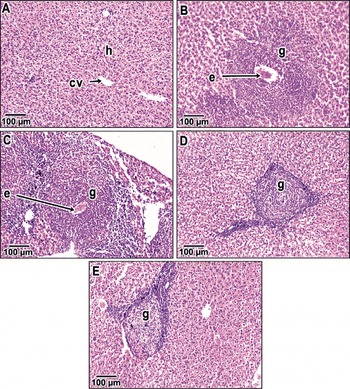
The infected untreated group showed large granulomatous lesions, consisting of collagenous fibrous tissue surrounding an intact egg and a peripheral zone of inflammatory cells. Treatment with silymarin decreased the granuloma size, with minimal degenerative changes in the liver tissue (fig. 1). Moreover, the most curative effect was observed in groups treated with mefloquine and silymarin, which led to a reduction in granuloma size, inflammatory cells and condensed connective tissue (table 1, fig. 1).
The histopathology of the mouse liver: (A) uninfected controls with central vein (cv) and hepatocytes (h); (B) a fibrocellular granuloma (g) around an egg (e) in group I; (C) a granuloma (g) surrounds a degenerated egg (e) in group I+ SI; (D) a granuloma (g) of reduced sized with a degenerated egg in group I+ ME; (E) a small-sized granuloma (g) with a disintegrated egg and normal hepatocytes in group I+ SI + ME. Mice groups: I, infected; SI, treated with silymarin; ME, treated with mefloquine.

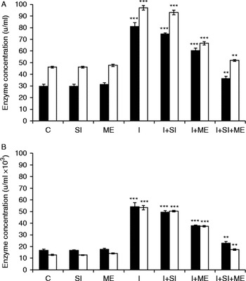
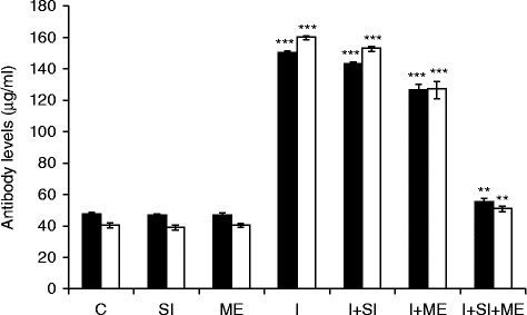
Insignificant changes (P > 0.05) in serum levels of ALT, AST, ALP, γ-GGT and total immunoglobulins IgG and IgM were observed in groups treated with silymarin or mefloquine alone compared with the healthy control group (figs 2 and 3). On the other hand, infection with S. mansoni led to a significant increase (P < 0.001) in all parameters compared with the healthy control group, indicating the establishment of liver fibrosis. The mean values of all the above-mentioned parameters were reduced in groups treated with either silymarin or mefloquine alone compared with infected untreated group. However, the highest improvement in the mean values of these parameters were observed in the group treated with both mefloquine and silymarin compared with the infected untreated group (figs 2 and 3).
Mean values ( ± SE) of liver enzymes: (A) alanine (black bars) and aspartate (white bars) aminotransferases; (B) alkaline phosphatase (black bars) and γ-glutamyl transferase (white bars) in experimentally infected mice (I) and those treated with mefloquine (ME), silymarin (SI) and both ME + SI. Mouse groups: C, healthy control; I, infected; SI, treated with silymarin; ME, treated with mefloquine. The labelling of the x-axis in (B) is in line that in (A). Level of significance: *P < 0.05, **P < 0.01, ***P < 0.001 compared with the healthy control.

Mean values ( ± SE) of serum IgM (black bars) and IgG (white bars) in experimentally infected mice (I) and treated with mefloquine (ME), silymarin (SI) and both ME+SI. Mouse groups: C, healthy control; I, infected; SI, treated with silymarin; ME, treated with mefloquine. Level of significance: *P < 0.05, **P < 0.01, ***P < 0.001 compared with the healthy control.

Discussion
Although chemotherapy eliminates mature worms successfully and prevents the accumulation of schistosome eggs, less effective drugs are directed to reversing the existing hepatic fibrosis, especially at the chronic and advanced stages of schistosomiasis. Therefore, treatment targeting hepatic fibrosis of schistosomiasis remains a challenging proposition (Andrade, Reference Andrade2008).
The present results showed that the administration of silymarin led to insignificant changes (P > 0.05) in worm burden, egg load and percentages of eggs at different developmental stages compared with the infected untreated group, indicating that silymarin did not influence oviposition or the arrival of eggs in the liver. However, treatment of S. mansoni-infected mice with mefloquine alone, or in addition to silymarin, showed a significant decrease (P < 0.001) in worm load, with complete disappearance of the egg load in the liver and intestine compared with the infected untreated group. More healing of hepatic granulomatous lesions and diminished granuloma size were also observed. These results were in parallel with those reported by Fakahany et al. (Reference Fakahany, Younis, El Hamshary, Fouad, Hassan and Ali2014), who stated that administration of mefloquine to mice infected with adult S. mansoni induced extensive and severe damage to the worm tegument, musculature, parenchymal tissues, digestive and reproductive systems.
In the present study, the infection of mice with 60 ± 10 cercariae of S. mansoni led to a significant increase (P< 0.001) in the liver function enzymes ALT, AST, ALP and γ-GGT in the sera of mice, compared with the healthy control group. These results are in line with those of Kadry et al. (Reference Kadry, Mohamed, Farrag and Fayed2013), who confirmed that the increment of such enzymes in the serum may be due to the destruction of hepatocytes by the action of toxins of the parasite ova, leading to their release into the circulation. Treatment with silymarin at the acute phase of infection was capable of reducing ALT, AST, ALP and γ-GGT levels in serum. These results were in accordance with those reported by Mata-Santos et al. (Reference Mata-Santos, Lino, Rocha, Paiva, Castelo Branco and Pyrrho2010, Reference Mata-Santos, Dutra, Rocha, Lino, Xavier, Chinalia, Hossy, Castelo-Branco, Teodoro, Paiva and Pyrrho2014), who indicated that silymarin interferes with the overall hepatic disease, reducing hepatic injury, fibrosis and ameliorating morbidity. This amelioration in liver enzyme activities could be attributed to the reduction in hepatic granuloma size and fibrosis, as well as the absence of necrotic liver tissue in infected treated mice, as observed in the histopathological examination.
Treatment with mefloquine alone caused a remarkable decrease in the activities of serum AST, ALT, ALP and γ-GGT when compared with the infected untreated group, indicating the maintenance of the functional integrity of hepatic cell membranes. The anti-schistosomal drug, mefloquine, causes worm tegument damage that consequently enhances the immune response of patients and generates a reversion of the level of fibrosis (Fahmy et al., Reference Fahmy, Rabia and Mansour2014). Regarding the immunological results, the mean values of anti-SEA immunoglobulins, total IgG and IgM, were increased in infected mice compared to the healthy control group. However, treatment with either silymarin and/or mefloquine caused a decrease in the mean values of anti-SEA immunoglobulins, total IgG and IgM. Doenhoff et al. (Reference Doenhoff, Wheeler, Tricker, Hamilton, Sturrock, Butterworth, Ouma, Mbugua, Kariuki and Koech2003) revealed a correlation between antibody levels and egg excretion, which could be explained by the fact that mefloquine induces changes in the antigenicity of the parasite, causing the release of concealed parasite antigen with binding of IgG and IgM to the surface of the parasite and finally lysis of schistosomal tissues by phagocytic cells. The interaction of host antibody with the exposed antigens leads to death of the parasite.
In addition, the improvement of liver function enzymes, histopathology and immunity in mice treated with both mefloquine and silymarin may be due to the blocking of liver fibrosis through the killing of parasites and complete eradication of eggs and their toxins by mefloquine, to alleviate liver inflammation, as well as the anti-oxidative properties, the anti-inflammatory and the anti-fibrotic actions of silymarin. Although the hepatoprotective properties of silymarin have been reported from both in vitro and in vivo studies, its mechanism of action still has not been established (Song et al., Reference Song, Deaciuc, Song, Lee, Liu, Ji and McClain2006).
In conclusion, treatment of murine schistosomiasis with silymarin combined with mefloquine was able to reduce granulomatous reactions and hepatic fibrosis. Since mefloquine is known to be effective in destroying the parasites in schistosomiasis, and silymarin proved to be a convenient and promising anti-fibrotic agent, their combination would be an efficient tool in the treatment of schistosomal liver fibrosis. Further studies on the mechanisms of action of both silymarin and mefloquine during schistosomal liver fibrosis or other chronic liver diseases may focus on developing therapeutic procedures in clinical practice.
Acknowledgements
The author would like to thank Dr Elham Ahmed Hassan, Department of Zoology, Faculty of Women for Arts, Science and Education, Ain Shams University and Dr Hoda Abu Taleb, Biostatistics and Demography, Medical Statistician, Department of Environment Research, Theodor Bilharz Research Institute, for assisting with the statistical analysis.
Conflict of interest
None.
Ethical standards
The author asserts that all procedures contributing to this work comply with the ethical standards of the Animal House of Schistosome Biological Supply Program Unit of Theodor Bilharz Research Institute and have been approved by the institutional committee of Theodor Bilharz Research Institute, Giza, Egypt.