Implications
Numerous studies have demonstrated that isoflavones have beneficial effects on lowering serum lipids and decreasing body fat deposition (Bhathena and Velasquez, Reference Bhathena and Velasquez2002). Cederroth et al. (Reference Cederroth, Vinciguerra, Kühne, Madani, Doerge, Visser, Foti, Rohner-Jeanrenaud, Vassalli and Nef2007) reported that male CD-1 mice fed from conception to adulthood a high soy-containing diet significantly increased energy expenditure and decreased adiposity (Cederroth et al., Reference Cederroth, Vinciguerra, Kühne, Madani, Doerge, Visser, Foti, Rohner-Jeanrenaud, Vassalli and Nef2007). Daidzein is a major isoflavonic phytoestrogen and has a variety of biological functions (Cassidy, Reference Cassidy2003; Hwang et al., Reference Hwang, Kwak, Lim, Lee, Kang, Choe, Hur and Han2006). As a metabolite of daidzein, equol (Eq) possesses more biological activities than its precursor (Matthies et al., Reference Matthies, Clavel, Gütschow, Engst, Haller, Blaut and Braune2008). Eq exists in hens’ blood and accumulates into eggs, preferentially into egg yolk (Saitoh et al., Reference Saitoh, Sato, Harada and Takita2001). However, whether Eq existing in eggs would affect lipid metabolism of the hatchlings is unclear. Therefore, the aim of this study was to investigate the effects of Eq directly injected in ovo on lipid metabolism and the related gene transcription in the liver of broilers.
Introduction
Phytoestrogens are a large group of plant-derived compounds, and they have attracted considerable interest in both medical and agricultural scientific research for decades because of their wide array of bioactive functions. Phytoestrogens include isoflavonoids, lignans, coumestanes, stilbenes and flavonoids (Price and Fenwick, Reference Price and Fenwick1985). Numerous studies in humans and rodents have suggested that, like estrogens, dietary phytoestrogens and their metabolites play a beneficial role in reducing obesity and body mass index (Bhathena and Velasquez, Reference Bhathena and Velasquez2002). In contrast, however, other studies have reported no beneficial effects of these chemicals on fat mass and serum lipid profiles (Yeung and Yu, Reference Yeung and Yu2003; Anderson et al., Reference Anderson, Fuller, Patterson, Blair and Tabor2007). These discrepancies are probably related to differences among the experimental designs and protocols (e.g. route of administration, composition, dose and duration), as well as the capacity of individuals to produce equol (Eq; Setchell, Reference Setchell2001).
Biotransformation is an important factor in regulating the biological activity of dietary isoflavones. Daidzein is known to be converted into Eq or O-desmethylangolen sin (O-DMA) by bacterial enzymes in the intestine (Matthies et al., Reference Matthies, Clavel, Gütschow, Engst, Haller, Blaut and Braune2008). Eq is absorbed more efficiently and has higher estrogenic activities than its precursor daidzein. Eq has been found in the blood of hens and accumulates predominantly in eggs (Saitoh et al., Reference Saitoh, Sato, Harada and Takita2001 and Reference Saitoh, Sato, Harada and Matsuda2004). Nevertheless, the effect of Eq in eggs on the growth and lipid metabolism of post-hatch chickens has not yet been elucidated. The fertilized egg provides a good model because it is enclosed by an eggshell and is relatively unaffected by external factors.
The liver is known to be the primary site of fatty acid synthesis in birds (Xu et al., Reference Xu, Wang, Mao, Zhan and Hu2003), and most of the body's endogenous lipids are hepatic in origin (O'Hea and Leveille, Reference O'Hea and Leveille1969). As in mammals, enzymes in the avian liver control sterol and lipid metabolism, and the levels of mRNA expression of lipogenic genes have been reported to parallel changes in enzyme activity, such as fatty acid synthase (FAS; Back et al., Reference Back, Goldman, Fisch, Ochs and Goodridge1986). In contrast to the large number of studies of hepatic lipid metabolism and the effects of isoflavones on lipogenic gene expression in rodents and humans, little is known about the effects of isoflavones on hepatic lipid metabolism in poultry, and especially about the effects of Eq on lipid liver metabolism in broiler chickens. Therefore, the aim of the present study was to investigate the effects of in ovo Eq administration on growth and lipid metabolism in post-hatch broilers. The study focused on changes in gene expression in the liver, with particular emphasis on the genes involved in fatty acid synthesis and cholesterol metabolism, including sterol regulatory element binding protein-1c (SREBP-1c), acetyl-CoA carboxylase (ACC-α), FAS, carnitine palmitoyl transferase I (CPTI), malic enzyme (ME), 3-hydroxy-3-methylglutaryl coenzyme A reductase (HMGR), low-density lipoprotein receptor (LDLR) and cholesterol-7alpha-hydroxylase (CYP7A1).
Material and methods
Animals
Animals and Experimental Protocol have been well described previously (Wei et al., Reference Wei, Ni, Lu, Grossmann and Zhao2011). In brief, on the 7th day of embryonic age, fertile eggs (Chinese local yellow broiler) were randomly allotted to three groups (146 eggs/group): 0 μg (control, Con), 20 μg (low dose, L) and 100 μg (high dose, H) Eq dissolved in 100 μl mixture of white mineral oil and ethanol were injected into the albumen of the fertile eggs, respectively. After hatching, broilers were raised in three pens (one pen/group) and split into 12 pens (four pens/group, 30 chickens/pen) at 21 days of age. Chickens were raised under the same standard conditions with free access to water and fed until 49 days of age.
At 49 days of age, birds were deprived of feed for 12 h and weighed just before slaughter; 20 broilers were taken randomly from each group and slaughtered in the morning by cervical dislocation for carcass analysis, blood and tissue sampling. Sera were stored at −20°C until triacyglycerol (TG) analysis. The liver was immediately removed from the body and weighed, and the same portion was sampled and snap-frozen in liquid nitrogen and stored at −70°C before analysis. Subcutaneous fat and abdominal fat were collected and weighed.
Animal care and use as described above was approved by the Institutional Animal Care and Use Committee of Nanjing Agricultural University.
Materials
Eq (purity ⩾99%, Catalog No. E-5880) was bought from Shanghai EQUL company (Shanghai, China) and synthesized from LC laboratories in the United States of America (Woburn, MA, USA).
Measurement of lipid parameters
Serum total cholesterol (TC) and TG concentrations were determined using enzymatic kits based on the ‘CHOD-PAP’ and ‘GPO-PAP’ reactions, respectively (Watson et al., Reference Watson, Butterwick, McConnell and Markwell1995). High-density lipoprotein cholesterol (HDLC) values were measured after magnesium chloride–heparin sulfate precipitation of apo-B-associated lipoproteins (low-density lipoprotein – LDL and very-low-density lipoprotein – VLDL). The relevant reagents were purchased from the Nanjing Jiancheng Bioengineering Institute (NJBI, China). The optical density of the samples was measured using a spectrophotometer three times at a wavelength of 546 nm (Biotech, Foster, USA).
Measurement of gene expression
RNA extraction
Total RNA was extracted from 100 mg of liver (10 for each group) using TRIZOL reagent (Takara, Kyoto, Japan) according to the manufacturer's protocols. In brief, 100 mg liver tissue was disrupted and homogenized immediately using 0.5 ml of 100% isopropanol and 1 ml of TRIZOL reagent. After incubation for 10 min at room temperature, the lysate was centrifuged at 12 000 × g/min for 10 min at 4°C. Subsequently, the RNA was washed with 1 ml of 75% ethanol and was dissolved in RNase-free water. The RNA concentration was then quantified by measuring the absorbance at 260 nm in a photometer (Eppendorf Biophotometer, Hamburg, Germany). Ratios of absorption (260/280 nm) of all preparations were between 1.8 and 2.0. Aliquots of RNA samples were subjected to electrophoresis with 1.4% agarose–formaldehyde gels stained with ethidium bromide to verify their integrity.
Real-time quantitative reverse transcription (RT)-PCR
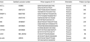
Reverse transcription was performed using the total RNA (2 μg) described above in a final volume of 25 μl containing 1 × RT-buffer, 100 U Moloney Murine Leukemia Virus reverse transcriptase (M-MLV; Promega, Madison, USA), 8 U RNase inhibitor (Promega, USA), 5.3 μM random hexamer primers and 0.8 mM dNTP (TaKaRa, Japan). After incubation at 37°C for 1 h, the reaction was terminated by heating at 95°C for 5 min and quickly cooling on ice. Real-time PCR was performed in Mx3000P (Stratagene, USA). Mock RT and No Template Controls were set to monitor the possible contamination of genomic DNA both at RT and PCR. The pooled sample obtained by mixing equal quantity of total RT products (cDNA) from all the samples was used for optimizing the PCR condition and tailoring the standard curves for each target gene, and melting curves were performed to insure a single specific PCR product for each gene. A volume of 2 μl of 16-fold dilution of RT product was used for PCR in a final volume of 25 μl containing 12.5 μl SYBR Green Real-time PCR Master Mix (TOYOBO Ltd, Japan) and 0.4 to 0.8 μM of each forward and reverse primers for SREBP-1c, ACC-α, FAS, CPTI, ME, HMGR, LDLR and CYP7A1 genes (the information of primers was shown in Table 1). Chicken β-actin mRNA was used as a reference gene for normalization purpose. The following PCR protocols were initial denaturation (1 min at 95°C), followed by a three-step amplification program (20 s at 95°C, 20 to 30 s at 60 to 64°C, 30 s at 72°C), which was repeated 45 times. The PCR products for each gene were sent to Haojia Biotech Ltd, China for sequencing to verify the specificity. The reported sequences exactly matched those published in GenBank. The method of 2−ΔΔCt was used to analyze the real-time RT-PCR data (Livak and Schmittgen, Reference Livak and Schmittgen2001). All the samples were included in the same run of RT-PCR and repeated in triplicate.
Summary of the oligonucleotide PCR primer sets

ACC-α = acetyl CoA carboxylase; FAS = fatty acid synthase; SREBP-1c = sterol regulatory element binding protein-1c; CPTI = carnitine palmitoyl transferase I; ME = malic enzyme; HMGR = 3-hydroxy-3-methylglutaryl coenzyme A reductase; CYP7A1 = cholesterol-7alpha-hydroxylase; LDLR = low-density lipoprotein receptor.
Statistical analysis
The results are expressed as mean ± s.e.m. Data were analyzed by ANOVA using the General Linear Models of SPSS 11.0 for Windows (StatSoft Inc., USA). Growth performance, tissues weight and serum lipid parameters were analyzed for differences due to gender, Eq and gender–Eq interactions. The multiple comparisons of the interactive means were performed using the Tukey correction. The effects of Eq administration on the hepatic metabolism gene expression data were analyzed by one-way ANOVA followed by least significant difference test post hoc analysis. Data were considered statistically significant when P < 0.05.
Results
Growth performance and weight of tissues
The results showed that males presented higher liver weight (24.59 ± 0.80 for males v. 17.29 ± 0.43 for females) and BW (966.40 ± 14.40 for males v. 642.18 ± 12.36 for females) and lower total fat (9.656 ± 0.80 for males v. 13.58 ± 1.17 for females) and relative liver weights (2.54 ± 0.03 for males v. 2.70 ± 0.02 for females) compared with females (P < 0.01; Table 2). Except a trend for an increase in the weight of total fat (P = 0.09), there was no significant effect of Eq treatment on BW, liver weight or the relative weight of liver to BW. In addition, there was no significant effect of Eq and Eq–gender interactions on growth performance or tissues weight (P > 0.05).
Effect of Eq injected in ovo on growth performance of male and female broilers at 49 days of age

Eq = equol; BW = body weight; LW = liver weight; FW = fat weight.
Values are means ± s.e.m. Con, L and H stand for control, low and high dosage of Eq injected into the albumen of the fertile eggs on the 7th day of embryonic age, respectively.
1LW/BW: the relative weight of liver to BW.
2FW containing subcutaneous and abdominal fat tissues.
Serum lipid metabolic parameters
The results of serum lipid metabolic parameters of broilers are presented in Table 3.
Effect of Eq injected in ovo on TG, TC, LDLC and HDLC in serum of male and female broilers at 49 days of age

Eq = equol; TG = triacyglycerol; TC = total cholesterol; LDLC = serum low-density lipoprotein cholesterol; HDLC = high-density lipoprotein cholesterol.
Values are means ± s.e.m. Con, L and H stand for control, low and high dosage of Eq injected into the albumen of the fertile eggs on the 7th day of embryonic age, respectively.
Male broilers presented significantly lower serum TG concentration than females (0.84 ± 0.30 for males v. 1.44 ± 0.10 for females; P < 0.05). Eq showed a significant effect on decreasing serum TG concentration of broilers at 49 days of age (0.97 ± 0.07 in H group and 1.19 ± 0.12 in L group v. 1.32 ± 0.12 in Con group; P < 0.05), whereas there were no interactions between gender and Eq (P > 0.05). Eq significantly increased serum TC (3.41 ± 0.07 in H group and 3.34 ± 0.05 in L group v. 3.09 ± 0.07 in Con group) and HDLC concentrations (2.43 ± 0.11 in H group and 2.22 ± 0.07 in L group v. 1.90 ± 0.11 in Con group; P < 0.05), whereas Eq did not affect serum low-density lipoprotein cholesterol (LDLC) concentration (P > 0.05). Except a significant gender difference of serum LDLC concentration (0.77 ± 0.04 for males v. 0.87 ± 0.11 for females), there was no gender difference in serum TC (3.38 ± 0.06 for males v. 3.17 ± 0.99 for females) or HDLC concentrations (2.19 ± 0.05 for males v. 2.14 ± 0.09 for females; P > 0.05). There were no gender–Eq interactions on the level of TC, HDLC or LDLC concentrations in the serum of broilers at 49 days of age (P > 0.05).
Hepatic lipid metabolism gene expression
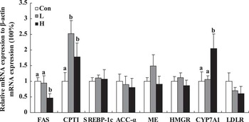
As shown in Figure 1, the gene expression of FAS in the liver of female broilers at 49 days of age was significantly downregulated by high-dose Eq (P < 0.05), whereas hepatic CPTI mRNA expression was significantly upregulated by both low- and high-dose Eq treatment (P < 0.05). High-dose Eq significantly increased hepatic CPY7A1 mRNA expression (P < 0.05). However, the expression levels of SREBP-1c, ACC-α, ME, LDLR and HMGR genes were not affected by Eq treatment (P > 0.05).
Effects of Eq on the mRNA expression of FAS, CPTI, CYP7A1, SREBP-1c, ACC-α, ME, HMGR and LDLR genes in liver of female broilers. Con, L and H stand for control, low and high dosage of Eq injected into the albumen of the fertile eggs on 7th day of embryonic age, respectively. Data are means ± s.e.m. from three different experiments in which each treatment was performed in triplicate within the experiment. Mean values without common superscript (a and b) differ significantly among the Con, L and H groups (P < 0.05, n = 10). FAS = fatty acid synthase; CPTI = carnitine palmitoyl transferase I; CYP7A1 = cholesterol-7alpha-hydroxylase; SREBP-1c = sterol regulatory element binding protein-1c; ACC-α = acetyl-CoA carboxylase; ME = malic enzyme; HMGR = 3-hydroxy-3-methylglutaryl coenzyme A reductase; LDLR = low-density lipoprotein receptor.

Discussion
Our results showed that except a trend increase in the weight of total fat, there was no significant effect of Eq treatment on BW, liver weight or the relative weight of liver to BW ratio, whereas there was a significant gender effect on BW and the tissues weight of broilers at 7 weeks of age. Eq showed significant effects on serum TG, TC and HDLC concentrations of broilers at 49 days of age, whereas there were no interactions between gender and Eq. On the basis of the results of hepatic genes expression measured in female chickens by real-time PCR, the changes in sterol and lipid metabolic parameters in the serum were accompanied by significant increases in CPTI and CYP7A1 1 gene expression and decreased FAS mRNA transcription in the liver.
Numerous studies have demonstrated that isoflavones have beneficial effects on lowering serum lipids and decreasing body fat deposition (Bhathena and Velasquez, Reference Bhathena and Velasquez2002). Supplementation of the soy protein diet with isoflavone in rats has been reported to dose-dependently decrease serum TG concentration (Takahashi et al., Reference Takahashi, Odbaya and Ide2009). Several studies have also reported that soy protein rich in isoflavones reduces fat-pad mass in rats, accompanied by decreased serum TG concentrations and cholesterol levels (Akahoshi et al., Reference Akahoshi, Koba, Ichinose, Kaneko, Shimoda, Nonaka, Yamasaki, Iwata, Yamauchi, Tsutsumi and Sugano2004; Torres et al., Reference Torres, Villalvazo and Tovar2006). It has been suggested that isoflavones are the principal physiologically active components responsible for the lipid-lowering effects of soy (Bhathena and Velasquez, Reference Bhathena and Velasquez2002). The results of the present study showed that serum TG concentrations in broilers decreased following in ovo Eq injection during the early embryonic period, without changes in BW or fat deposition. These results indicate that isoflavones and the metabolite Eq have lipid-lowering effects in mammals, as well as in chickens. This was in contrast to the results in chickens fed estradiol, which led to a marked elevation in plasma lipids, with the greatest increases in TG and LDLC concentrations (Hermier et al., Reference Hermier, Forgez, Williams and Chapman1989; Park and Cho, Reference Park and Cho1990). Estrogen use in mammals, however, has been reported to decrease TG and LDLC levels and to increase HDLC levels (Haffner and Valdez, Reference Haffner and Valdez1995). This discrepancy could be because of the different reproductive characteristics of birds and mammals. In addition, the different effects of estradiol and phytoestrogens in chickens suggest that phytoestrogens regulate lipid metabolism through anti-estrogenic bioactivity (Hwang et al., Reference Hwang, Kwak, Lim, Lee, Kang, Choe, Hur and Han2006), or through other non-estrogen receptor pathways, such as via adipogenic transcription factors, as has been suggested in rodents (Ørgaard and Jensen, Reference Ørgaard and Jensen2008). With respect to the cholesterol metabolism, dietary supplementation with soy isoflavones in quail significantly reduced the levels of cholesterol in the liver and muscles (Yilmaz et al., Reference Yilmaz, Guvenc, Cetintas, Tuzcu, Dayangac and Sahin2008). Blair et al. (Reference Blair, Appt, netau-Pelissero, Clarkson, Anthony, Lamothe and Potter2002) reported that soy or isoflavone extract fed to female hamsters resulted in a significant increase in plasma HDLC concentrations, but no changes in plasma LDLC and VLDLC concentrations, compared with those fed the control diet (Blair et al., Reference Blair, Appt, netau-Pelissero, Clarkson, Anthony, Lamothe and Potter2002). In addition, it has been reported that the intake of soy isoflavones appeared to improve serum cholesterol levels in female, but not in male mice, rats and hamsters (Kishida et al., Reference Kishida, Mizushige, Nagamoto, Ohtsu, Izumi, Obata and Ebihara2006; Mezei et al., Reference Mezei, Li, Mullen, Ross-Viola and Shay2006). In this study, TC and HDLC concentrations in broilers were significantly increased by Eq treatment; however, there was no significant interaction between sex and Eq treatment.
In the light of a significant decrease in serum TG concentration induced by Eq injection in ovo on the 7th day of incubation, we speculate that the changes in TG levels might generate the programming effects on reproduction, especially in female broilers, because egg production is an energy-intensive process, requiring a large increase in supplying of lipids (TG) to support the demands of new yolk formation. On the basis of this consideration, the mechanism behind the changes of TG concentration in serum of female broilers was elucidated in this study. The biological effects of soy phytochemicals on lipid metabolism are likely to be regulated through changes in gene expression (Torres et al., Reference Torres, Villalvazo and Tovar2006). Soy protein diets in rodents suppressed the expression of hepatic lipogenic enzymes and lowered the expression of SREBP-1, FAS and ME in the liver (Iritani et al., Reference Iritani, Hosomi, Fukuda, Tada and Ikeda1996; Ascencio et al., Reference Ascencio, Torres, Isoard-Acosta, Gómez-Pérez, Hernández-Pando and Tovar2004). The liver in birds is known to be the main site of fatty acid synthesis (Xu et al., Reference Xu, Wang, Mao, Zhan and Hu2003), and liver fatty acid metabolism has been recognized as the main source of variation in fat deposition and blood TG concentrations in poultry (O'Hea and Leveille, Reference O'Hea and Leveille1969). Previous studies have shown that SREBP-1c can directly stimulate the transcription of genes encoding ACC, the rate-limiting enzyme in fatty acid synthesis (Stoeckman and Towle, Reference Stoeckman and Towle2002; Yin et al., Reference Yin, Zhang and Hillgartner2002), making it a good candidate for a common regulator of the lipogenic genes expressed in avian liver. However, the results of the present study revealed no evidence that either SREBP-1c or ACC genes was directly responsible for the effect of Eq on lipid synthesis at the transcriptional level.
CPTI catalyzes the formation of long-chain acylcarnitine from activated fatty acids and free carnitine, thus controlling the rate of β-oxidation and regulating the deposition or oxidation of fatty acids (Zammit, Reference Zammit1999). FAS is a key enzyme in fatty acid synthesis that catalyzes the synthesis of long-chain fatty acids through the condensation of acetyl-CoA and malonyl-CoA (Back et al., Reference Back, Goldman, Fisch, Ochs and Goodridge1986). In rats, dietary soybean protein enriched with isoflavones reduced the concentrations of TG in plasma and liver, which is accompanied by decreases with lipogenic enzyme activities in the liver, including ME, FAS and ACC enzymes (Iritani et al., Reference Iritani, Nagashima, Fukuda, Katsurada and Tanaka1986). Morifuji et al. (Reference Morifuji, Sanbongi and Sugiura2006) reported that feeding soy protein to rats decreased hepatic TG levels and epididymal fat mass, and increased hepatic CPTI activity and mRNA levels in the liver (Morifuji et al., Reference Morifuji, Sanbongi and Sugiura2006). Similarly, when compared with casein, soy protein upregulated CPT activity to accelerate hepatic fatty acid oxidation in rats (Banz et al., Reference Banz, Davis, Peterson and Iqbal2004). Expressions of the hepatic SREBP-1 and its target gene FAS were also significantly reduced by soy protein feeding in obese Zucker fa/fa rats (Tovar et al., Reference Tovar, Torre-Villalvazo, Ochoa, Elias, Ortiz, Aguilar-Salinas and Torres2005). In the current study, the expression of CPTI gene in the liver of female broilers was significantly upregulated by in ovo Eq injection, which paralleled the significant reduction in hepatic FAS mRNA expression. Although mRNA levels are not always directly correlated with the activity of the encoded functional protein, there is evidence to suggest that the transcriptional levels of several hepatic lipogenic genes, including FAS, ME and CPT genes, reflect the corresponding enzyme activity levels (Banz et al., Reference Banz, Davis, Peterson and Iqbal2004; Torres et al., Reference Torres, Villalvazo and Tovar2006). Our results indicated that in ovo Eq injection reduced serum TG levels in female broilers by suppressing fatty acid synthesis and accelerating β-oxidation in the liver.
The liver is an important site of both cholesterol anabolism and catabolism, under the control of the HMGR and CYP7A1 enzymes, respectively. CYP7A1 regulates the removal of substantial amounts of cholesterol from the body by its excretion into bile. Moreover, the liver is a key element in the control of plasma cholesterol regulation by hepatic LDLRs (Spady et al., Reference Spady, Cuthbert, Willard and Meidell1998; Sato et al., Reference Sato, Ohuchi, Sook, Toyomizu and Akiba2003). Expressions of both HMGR and CYP7A1 genes in the liver in hens are increased during sexual maturation to support the enhanced cholesterol metabolism required for egg production (Sato et al., Reference Sato, Ohuchi, Sook, Toyomizu and Akiba2003). However, these two genes were not modulated by dietary estradiol supplementation, indicating that estradiol does not affect cholesterol metabolism at the transcriptional level during egg production (Sato et al., Reference Sato, Ohuchi, Sook, Toyomizu and Akiba2003). However, there is evidence to suggest that the gene expression of hepatic HMGR and CYP7A1 is regulated by cholesterol in the diet and by HDLC and LDLC in the plasma, indicative of feedback regulation (Goldstein and Brown, Reference Goldstein and Brown2009). HMGR mRNA expression in the liver in hens was significantly reduced by feeding a 3% cholesterol-supplemented diet, whereas CYP7A1 mRNA expression levels were markedly increased compared with those in hens fed the control feed (Sato et al., Reference Sato, Ohuchi, Sook, Toyomizu and Akiba2003), as seen in mammals. In the present study, in ovo Eq administration significantly increased serum TC and HDLC levels in female broilers and CYP7A1 mRNA expression in the liver, without affecting serum LDLC concentrations or hepatic LDLR and HMGR gene expression. The hepatic CYP7A1 gene was significantly upregulated by high dosage of Eq treatment, possibly as the result of feedback regulation by higher serum levels of TC in female broilers. However, the mechanism underlying the changes of TC in serum still needs further study.
In conclusion, the present study revealed that Eq decreased blood TG in broilers. For female broilers, the decrease in serum TG concentration induced by Eq injection in ovo might be caused by upregulating CPTI and downregulating FAS mRNA expression in the liver, and that high serum cholesterol levels stimulated the increase in hepatic CYP7A1 gene expression. The mechanism behind the long-term effects of Eq on lipid and cholesterol metabolism, as well as the relevant genes expression, may be associated with some kinds of programming process involved in chicken embryonic development; however, the exact mechanisms still need further study.
Acknowledgment
This work was supported by the National Nature Science Foundation of China (Project No. 30800809) and a project funded by the Priority Academic Program Development of Jiangsu Higher Education Institutions (PAPD).