

Introduction
Biopsies of the equine middle gluteus muscle have made it possible to determine the characteristics of this muscle, and subsequently the effects of trainingReference Essén-Gustavsson and Lindholm1–Reference Ronéus, Persson, Essén-Gustavsson and Arnason4 and influence of age and sexReference Essén, Lindholm and Thornton5, Reference Ronéus6. Muscle fibre types may be differentiated into type I, IIA and IIB fibres using myosin ATPase stainingsReference Brooke and Kaiser7, whereas immunohistochemical staining with myosin heavy chain (MHC) antibodies identifies the fibre types based on the different MHC isoforms – MHCI, MHCIIA and MHCIIXReference Rivero, Talmadge and Edgerton8. The immunohistochemical method has been the preferred method in recent studies as it is more sensitive in detecting the changes in the fibre type composition resulting from training or other stimuliReference Rivero, Talmadge and Edgerton8–Reference Dingboom, van Oudheusden, Eizema and Weijs10. With this method, hybrid fibres, i.e. fibres that express more than one MHC isoform simultaneously, can be identified. Type I and IIA fibres mainly correspond to MHCI and MHCIIA fibres, whereas type IIB fibres usually include both MHCIIX and MHCIIAX fibresReference Rivero, Talmadge and Edgerton8, Reference Karlström and Essén-Gustavsson9.
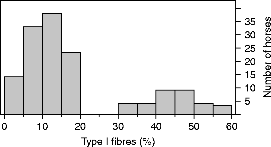
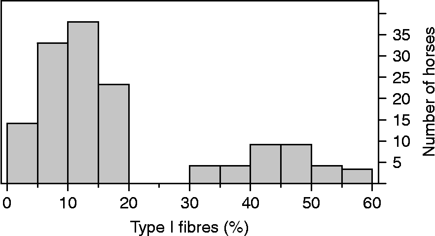
In a recent study, muscle biopsies from 141 Norwegian–Swedish coldblooded trotters (NSCT), aged 17–24 months, were investigated for their muscle characteristicsReference Revold, Ihler, Karlström, Larsen and Essén-Gustavsson11. The distribution of type I fibre percentages in the middle gluteus muscle of these horses is illustrated in Fig. 1. This distribution is split into two distinct portions, where the majority (108 horses) of the horses had a range from 2 to 20% of type I fibres. However, 33 horses had a range from 32 to 58% of type I fibres. The apparent subpopulation of the muscle with a high percentage of type I fibres was a notable finding, and not in line with previous findings at a comparable sampling depth of the muscle, in this breed or othersReference Karlström, Lindholm, Collinder and Essén-Gustavsson12–Reference Snow and Guy14. In one study of NSCT, the range was found to be between 8 and 28% in 2-year-old horses and between 9 and 18% in 4–8-year-old racing horsesReference Karlström, Lindholm, Collinder and Essén-Gustavsson12. The suspected subpopulation of the muscle in the study of NSCT of Revold et al. Reference Revold, Ihler, Karlström, Larsen and Essén-Gustavsson11 was best identified by the high percentages of type I fibres, and therefore the authors have focused on this fibre type in the present study.
Distribution of percentage of type I fibres from biopsy samples of gluteus medius muscle in 141 young Norwegian–Swedish coldblooded trottersReference Revold, Ihler, Karlström, Larsen and Essén-Gustavsson11
One objective was to investigate whether the described method of analysing the fibre type composition in a muscle sample was reproducible. If the type I fibre type percentages could be reproduced by resectioning and re-staining the muscle sample, the described distribution of type I fibre percentages would be the representative for the sample. In that case, the reason for the high percentage of type I fibres might rely on the positioning of the biopsy needle or a breed-specific topographic heterogeneity in this region of the muscle. The observed high proportion of type I fibres might also be a finding restricted to young horses in this particular breed. Therefore, it was of interest to study whether the fibre type composition in a new biopsy from the gluteus medius muscle showed a similar pattern for type I fibres in the same horses at the age of 3 years. Another explanation for the high type I fibre proportion might be a muscular abnormality present in these individuals. To evaluate this hypothesis, the present study also includes a comparison of the performance in the 3-year season between the horses that had a high percentage of type I fibres (group H) and those that had a normal percentage of type I fibres (group N).
Material and methods
Horses and biopsy technique
Biopsy samples from the gluteus medius muscle of nine NSCT was obtained at the age of 17–24 months. The horses were given mild sedation (detomidin 5 μg kg− 1 and butorphanol 0.05 mg kg− 1, both intravenously) prior to the biopsy procedure. A percutaneous sample was taken according to the method described by Lindholm and PiehlReference Lindholm and Piehl15. The biopsy sample was collected from the midpoint of an imaginary line from the tuber coxae to the root of the tail. The biopsy needle was directed perpendicular to the skin and inserted to a fixed depth of 4 cm, which had previously been marked on the needle. All samplings were performed by the same person. The samples were rolled in talcum powder and frozen in liquid nitrogen within 3–5 min following sampling and stored at − 80°C until analysed.
Histochemical and immunohistochemical analysis
The muscle biopsies were cut transversely in 10 μm sections in a cryostat (Reichert-Jung, Cambridge Instruments GmBH, Nussloch, Germany) at − 20°C. Serial sections were collected for immunohistochemical and histochemical analysis. The sections were stained for myosin ATPase at pH 4.6 for detection of fibre types I, IIA and IIBReference Brooke and Kaiser7. Type IIB fibres correspond to type IIX fibres in the gluteus medius of the horseReference Karlström, Lindholm, Collinder and Essén-Gustavsson12. Identification of myosin isoforms was performed with the commercially available monoclonal antibody, N2-261 (Alexis Biochemicals, Lausen, Switzerland) for detection of MHC I and MHC IIA. A4-74 (Alexis Biochemicals) was used for detection of MHC IIAReference Karlström, Lindholm, Collinder and Essén-Gustavsson12, Reference Karlström and Essen-Gustavsson16.
Since no commercial antibody against MHC IIX was available, fibres were classified as I, IIA, IIAX or IIX based on a combination of immunohistochemical and myosin ATPase (mATPase) staining. Fibres demonstrated as being type I by this method consistently showed staining with N2-261 and not with A4-74, and were classified as type I. Fibres stained as IIX with mATPase staining were classified as hybrid IIAX fibres if stained with MHC IIA antibodies, and as pure IIX if not stained with MHC IIA antibodies.
Each of the nine biopsy samples was cut and stained at two different occasions (T1 and T2), and the fibre composition was determined according to the described method after being stored at − 80°C. Approximately, 200 adjacent cross-sectioned fibres from each biopsy sample were typed and counted in representative areas of the sections without artefacts at both occasions. The same procedure was used in biopsies taken at the age of 3 years.
Measures and performance records
Measures of chest circumference (girth) and height at withers of the 141 horses were recorded. Height at withers was measured by tape measure from the highest point of the withers to the ground following the chest contour, which corresponds to a measure that is approximately 10 cm longer than the conventional height at withers. The height was adjusted by − 1 cm in horses wearing shoes. Performance information regarding attendance at regular race, total earnings and best racing times in the 3-year season was obtained for the same horses from The Norwegian Trotting Association. Best racing times were recorded for all horses that raced in the 3-year season (36 from group N and 11 from group H).
Statistical methods
The repeatability of the fibre type I classification was evaluated since this fibre type was best suited for description of the apparent subpopulation. The repeatability was evaluated by using agreement analysisReference Altman and Bland17, Reference Bland and Altman18. The mean difference between type I fibres at occasions T1 and T2 (DiffT1–T2) was calculated and tested against zero (paired Student's t-test). The standardized agreement index (AI) is defined as: 1 − (2 SDdiff/MeanT1–T2), where SDdiff is the standard deviation of the differences in percentage type I fibres at T1 and T2 and MeanT1–T2 is the mean of the percentage type I fibres at T1 and T2Reference Aarås, Veierød, Larsen, Ortengren and Ro19. A positive AI supports agreement and a value >0.5 indicates good agreementReference Kampen, Tollersrud, Larsen, Roth, Frank and Lund20. The agreement limits, DiffT1–T2 ± SDdiff, are estimated and DiffT1–T2 outside these limits are considered as outliers. A correlation analysis was performed to evaluate a possible correlation between the difference of type I fibres percentages at T1 and T2 and the level of type I fibre counts.
Data on chest circumference and height and the performance variables, total earnings and best racing time for group H and group N were compared. These comparisons were performed two-tailed, and the differences were considered significant for P-values ≤ 0.05. Comparisons within and between groups were performed using ANOVA. Whether the categorical variable starts in regular racing was compared between the two groups using Fisher's exact test.
Results
The type I fibre percentages of the nine samples at T1 and T2 are given in Table 1. The high type I fibre percentages from T1 were confirmed at T2 in horses 3, 6, 8 and 9. Likewise, the type I fibre percentages at T1 were reproducible at T2 in five horses with assumed normal findings (horses 1, 2, 4, 5 and 7). The agreement analysis showed an AI of 0.70 and all observations of DiffT1–T2 were within the agreement limits, i.e. no outliers were observed. The mean percentage of type I fibres at T1 and T2 and the absolute difference between type I percentages at T1 and T2 were not correlated (correlation coefficient − 0.47, P = 0.21), all of which are shown in Table 2. This means that absolute differences are not related to the level of type I fibres.
Percentages of muscle fibre type I in nine gluteus medius muscle biopsy samples from nine horses, stained and examined at two different occasions (T1 and T2)

Agreement analysis of the method of determination of fibre type composition from the same biopsy sample at two different occasions (T1 and T2) based on results given in Table 1

No horse showed muscle fibre type I proportion >30% (mean 15.2%, range 3–26) from the muscle biopsies of the eight horses at 3 years of age. The data on total earnings, best racing time and size variables for group H and group N are presented in Table 3. No statistically significant differences were detected between the groups for these variables, and there was also no difference regarding attendance to regular start or not between the two groups. However, there was a tendency for horses in group H to have greater height measures than those in group N (P = 0.06).

Discussion
There is no evidence of methodical problems in handling biopsy samples that would explain the notably high proportion of type I fibres in young NSCT. The agreement analysis showed that the repeatability of the described method is good and revealed no outliers. Further, the differences between the proportions of type I fibres at T1 and T2 are the same at any level of type I fibres.
The high AI of the method, in addition to the distribution, shown in Fig. 1, confirms that some muscle samples contained high proportions of type I fibres. As shown in Fig. 1, the horses in the study of Revold et al. Reference Revold, Ihler, Karlström, Larsen and Essén-Gustavsson11 had no type I fibre proportions between 20 and 30%, and horses having >30% type I fibres were normally distributed. These observations strengthen the hypothesis of a subpopulation of muscle fibres in this region of the gluteus medius muscle and thus a heterogenic muscle. A topographic heterogeneity of the equine middle gluteal muscle has been well documentedReference Serrano, Petrie, Lopez-Rivero and Hermanson21–Reference Lopez-Rivero, Serrano, Diz and Galisteo23, but it was surprising to detect such heterogeneity at a standardized sampling depth of 4 cm. The young horses having the high proportions of type I fibres showed no difference in performance in the 3-year season concerning the performance indicators attendance in regular race, best racing time and total earnings. It is therefore unlikely that the high type I proportions are related to muscular abnormalities in the gluteus muscle of these horses.
The high proportions of type I fibres in some muscle samples might be associated with the biopsy technique/sampling method. There might be special anatomical conditions in the NSCT that may bring the biopsy needle to other parts of the muscle when using the described method of placing the biopsy needle. The sedation of the horses may also have influenced the position of the biopsy needle. Even if the sedation was mild, some horses might relinquish the normal standing position and the muscular tension might be altered. From previous studies, higher type I proportions are reported from deeper parts of the gluteus medius muscleReference Serrano, Petrie, Lopez-Rivero and Hermanson21–Reference Lopez-Rivero, Serrano, Diz and Galisteo23. The constant depth of 4 cm was used in the present study as well as in the study of Revold et al. Reference Revold, Ihler, Karlström, Larsen and Essén-Gustavsson11. In a large muscle belly, a depth of 4 cm will be relatively superficial. One might therefore expect that a constant depth of sampling might influence the relative depth in the muscle when not taking the size of the horse into account. Subcutaneous fat in this region might also have influenced the sampling depth in the muscular tissue.
If the high type I proportions were caused by a relatively deep sampling site in some horses, we would expect this phenomenon to be more common in the smallest horses. On the contrary, there was a tendency for horses with high fibre type I proportions (group H) to have a greater height than the others (group N), while no difference was seen in girth measures.
Nevertheless, at the age of 3 years, when the muscle belly is larger due to both growth and training, all eight sampled horses had type I fibre percentages within the normal range.
In conclusion, the gluteus medius muscle in young NSCT is heterogeneous, as also described in other breeds. Whether the large heterogeneity identified in this breed is age dependent is not known, and further studies with sampling from different places and depths in the gluteus medius muscle at different ages must be performed before drawing any exact conclusions. Muscular disorders are not likely as an explanation for heterogeneity, because horses with a high type I proportion did not differ from others in later performance.