Introduction
Breast cancer is the commonest malignancy among women internationally. With its increased prevalence, more surgical options are propagated for its treatment. Breast conserving surgery (BCS) has become the standard surgical treatment option in approximately 60–75% of patients with early breast cancer in countries where the radiotherapy facilities are readily available.Reference Katipamula, Degnim and Hoskin1
Multidisciplinary preoperative patient selection and planning optimise the surgical resection techniques while maintaining cosmesis.Reference Bertozzi, Pesce, Santi and Raposio2
Seroma is the fluid that fills cavities post tissue excisions. It is considered as a non-serious condition that may lead to substantial morbidity in the form of wound failure, sepsis and subsequent delays in adjuvant therapy.Reference Woodworth, McBoyle, Helmer and Beamer3–Reference Aitkin and Minton5
In the breast, seroma may occur following either BCS or mastectomy. Depending on the size of the cavity, its reported occurrence varies from 2·5 to 57% of cases.Reference Bryant and Baum6
The introduction of the one-step intraoperative radiotherapy (IORT) aims to shorten the radiation treatment time, eliminates the geographical misses and potentiates the radio-biological effects leading to tumour cell apoptosis.Reference Belletti, Vaidya and D’Andrea7,Reference Sindelar and Kinsella8
Its emergence as the treatment for early breast cancer has documented few uncommon local complications ranging from seroma to fibrosis of the index site.Reference Ruano-Ravina, Cantero-Munoz and Eraso Urien9
Fluid collection may appear in the immediate postoperative period as a hemato-seroma followed by resolution as the healing process continues, occasionally it may persist as an encysted seroma for many years.Reference Epstein, Silverstein and Lin10
In this study, we attempt to review the occurrence, the course and the challenges encountered in the post operative IORT seroma.
Materials and Methods
Informed consent was obtained from all patients undergoing IORT. The ethical approval for the study was obtained from the Institutional Review Board at Imam Abdulrahman Bin Faisal University (number IRB-2013-01-035). Based on strict selection criteria, all eligible patients who received IORT\whole breast irradiation (WBIR) treatment between 2012 and 2019 in the university setting were included. The selection criteria for exclusive IORT were patient’s age > 45 years, tumour size less than 3 cm, invasive ductal carcinoma (IDC) tumour grade I–II, negative margins on the initial excision, negative lymphovascular invasion and less than three lymph nodes involvement. IORT as a boost was considered in patients whose age is less than 45 years, tumour size 4 cm, received neoadjuvant chemotherapy, invasive lobular carcinoma (ILC), tumour grade III, positive margins on the first excision, presence of lymphovascular invasion and more than three lymph nodes involved.
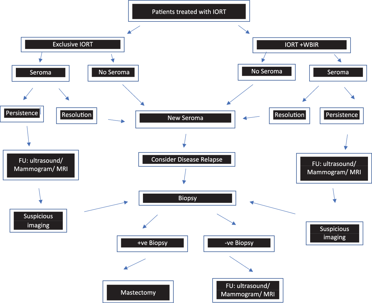
The mobile device Intrateam (Carl Zeiss Surgical, Oberkochen, Germany), a miniature X-ray source with 50 kV, was used to treat the selected patients after approval of the multidisciplinary team and the informed consent obtained. A standard management protocol was strictly applied for all patients undergoing IORT\WBIR treatment (Figure 1).
Summary of the patients’ management protocol for IORT + WBIR.

A prophylactic single dose of antibiotics using 1·5 g cefuroxime was administered after sensitivity testing. Prolonged treatment with antibiotics was considered in selected patients with comorbidities or those who received neoadjuvant chemotherapy.
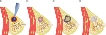
The observation of the changes that occurred at the postoperative index site based on the natural course of healing following BCS and IORT\WBIR is demonstrated in Figures 2a–2d.
(a) IORT treatment, (b) immediate postoperative seroma formation, (c) intermediate to late changes with the persistence of seroma with partial organisation and architectural distortion and (d) seroma resolution with tissue remodelling and post-operative scarring.

Postoperative inclusion criteria were strictly directed towards those who continued to show persistent index site seroma 12 weeks after the surgical excision.
All data were collected in an Excel™ (Microsoft Corporation, Redmond, Seattle, USA) datasheet. Collected data were analysed using statistical package for social science software (SPSS) version 19.
Results
The total number of patients who received IORT was 86. Patients treated with exclusive IORT were 39 (45%) while those treated with IORT\WBIR were 47 (55%). Age was between 31 and 75 years with a median age of 51 years. Forty-seven (55%) showed complete resolution of seroma on the first routine 12 weeks follow-up; thus, they were excluded.
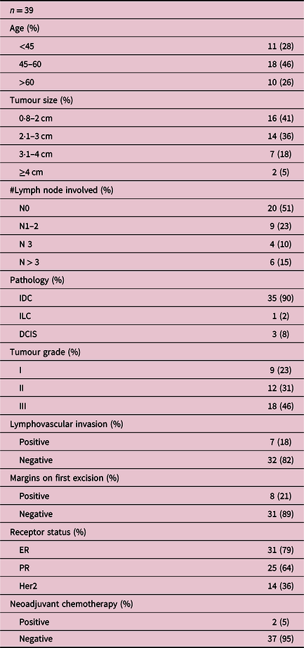
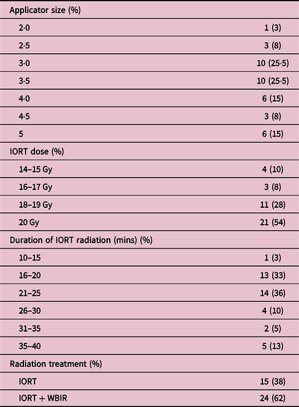
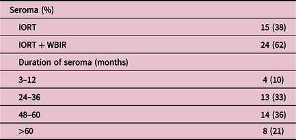
Seroma was observed in 39 (45%) patients, 15 (38%) were exclusively treated with IORT while 24 (62%) received additional WBIR. Tumour size ranged between 0·8 and 4 cm. Histological diagnosis included IDC in 35 (90%), ILC in 1 (2%) and ductal carcinoma in situ (DCIS) in 3 (8%). Tumour grade analysis showed grade I in 9 (23%), grade II in 12 (31%) and grade III in 18 (46%). Twenty (51%) were reported as node-negative and 19 (49%) as node-positive. Lymphovascular invasion was observed in 7 (18%) while the remaining 32 (82%) were reported as negative. Positive immunohistochemistry was reported as estrogen (ER) in 31 (79%), progesterone (PR) in 25 (64%) and Her2 in 14 (36%). Positive margins on the initial excision were documented in 8 (21%) patients, while 31 (79%) were reported as negative. All patients with positive margins underwent re-excision. Tumour size of ≥ 4 cm in 2 (5%) patients was successfully downsized by neoadjuvant chemotherapy (Table 1). The planned treatment for exclusive IORT dose was 14·0–20·1 Gray (Gy), while IORT as a boost was followed by WBIR 50 Gy in 25 fractions. Applicator sizes selection was based on the designed cavity and ranged from 2·0 to 5 cm with the duration of treatment based on the applicator size (Table 2). Minimal postoperative complications were observed ranging from mild self-limiting erythema/mastitis to partially gaping wound lasting for 2 weeks postoperatively. The development of the persistent seroma was observed in routine follow-up documenting the duration that ranged from 12 weeks to 6 years. Patients treated by IORT and the IORT\WBIR have both shown an increased risk of developing postoperative seroma. However, a higher risk of one- and half-time increase was observed in those treated with IORT\WBIR (Table 3).
Clinical characteristics of patients who developed seroma

IORT treatment details of the 39 patients

Seroma occurrence and duration

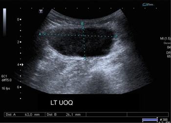
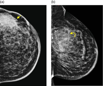
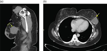
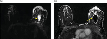
All seromas were both clinically and radiologically assessed, and the progress was documented during routine follow-up. Ultrasound at 3 months interval was utilised to assess the seroma progression/regression course and also aided in documenting the seroma wall thickness which ranged from 2 to 6 mm (Figure 3). Sequential annual mammogram documented parenchymal scarring, architectural distortion associated with thickening of the overlying skin possibly related to WBIR (Figure 4). Contrast-enhanced computed tomography of the chest mainly utilised for metastatic workup has inevitably captured the well-circumscribed round-shaped hypodense collection with irregular enhancing outer wall in a background of post-surgical changes (Figure 5). Magnetic resonance imaging (MRI) of the breast further enhanced the accuracy of diagnosis in cases where a clinical suspicion of recurrence was suspected (Figure 6). Repeated aspiration was performed for 2 (5%) patients who presented with symptomatic seromas. Routine follow-up course continued for all patients from the immediate postoperative period to 6 years. Three (8%) patients had developed index site relapse with positive fluid cytology at 1 and 2 years, respectively. All three patients were treated with mastectomy and adjuvant therapy.
Ultrasound showing an oval-shaped seroma with thickened wall and turbid echogenicity.

(a) Mammogram demonstrated a partially obscured oval-shaped density surgical bed associated with architectural distortion and overlying skin thickening related to EBRT. (b) Resolving seroma with tissue remodelling with surgical clips noted at the tumour bed.

(a, b) Contrast-enhanced CT-scan CHEST soft tissue window shows a well-circumscribed round-shaped hypodense collection with thickened and irregular enhancing outer wall in a background of post-surgical changes.

(a, b) Breast MRI (T2 FS and post gadolinium T1) revealed breast oedema with a well-circumscribed oval-shaped breast lesion which shows post contrast rim enhancement.

Discussion
The adoption of BCS is based on the basic principle of performing adequate excision while maintaining satisfactory cosmesis in early breast cancer. Factors such as tumour location, tumour-breast ratio and the extent of excision may be predictive factors that contribute to the cosmetic outcome.Reference Epstein, Silverstein and Lin11 BCS is accompanied by variable degrees of deformities such as asymmetry, skin and nipple retractions, and delayed radiotherapy effects in up to 30% of cases.Reference Matory, Wertheimer and Fitzgerald12–Reference D’Aniello, Grimaldi, Barbato, Bosi and Carli14 The standard treatment of BCS followed by WBIR has contributed to the reduction of local recurrences of 10% as compared to 30% without irradiation sparing many patients from mutilating mastectomies.Reference Urban, Lima, Schunemann, Spautz and Rabinovich15,Reference Vernonesi, Marubini and Mariani16
Seroma is a common complication presenting as a fluid collection that occupies cavities post tissue excisions. It is considered a minor complication in many instances; however, it may lead to substantial morbidity such as wound failure, sepsis, prolonged wound healing and delays in subsequent adjuvant therapy.Reference Fisher, Anderson and Bryant17,Reference Budd, Cochran, Sturtz and Fouty18
The introduction of accelerated partial breast irradiation (APBI) has gained popularity over recent years, its potential to enhance the accuracy of delivering a focused irradiation beam to the tumour bed had aided in abolishing the geographical miss which is reported in 20–90%.Reference Aitkin and Minton19
Currently, the various used modalities for APBI are brachytherapy, low voltage X-ray delivering photons (Intrabeam-IORT) and linear accelerators delivering electrons at different energies (Novac7, Liac).Reference Blank, Kraus-Tiefenbacher and Welzel20 This targeted radiotherapy to the tumour bed of 10–20 Gy further endorses the reduction of local recurrences.Reference Veronesi, Orecchia and Luini21
The acute toxicity reported with APBI complications is uncommon and non-life-threatening. Early evaluation within 4 weeks after surgery has reported erythema, mastitis, dry desquamation, hematomas, wound infections, lipo-necrosis, and delayed wound healing.Reference Blank, Kraus-Tiefenbacher and Welzel22–Reference Ruano-Ravina, Cantero-Munoz and Eraso Urien24
The development of postoperative seroma is not uncommon. The majority of diagnosed cases are treated conservatively as spontaneous fluid resorption and tissue remodelling take place.Reference Tuschy, Berlit and Romero25
Occasionally, a quarter of surgically treated patients may require repeated aspiration to relieve the local discomfort and expedite the healing process.Reference Kraus-Tiefenbacher, Scheda and Steil26
Many conflicting reports regarding the incidence of seroma have been documented in many publications. Some studies have documented no significant difference in the incidence of seroma between the use of IORT as compared to conventional WBIR,Reference Tuschy, Berlit and Romero27 while others have shown that seroma was observed more frequently in the exclusive IORT patients as compared to WBIR (22% versus 4%).Reference Kraus-Tiefenbacher, Welzel and Brade28
On the contrary, this current study has documented a higher incidence of seroma in the IORT\WBIR 24 (62%) as compared to the exclusive IORT treatment 15 (38%).
The timing of the administration of WBIR following IORT is critical. Early treatment of less than 6 weeks may carry a higher risk of up to 51% of long-term toxicity as compared to the delayed interval of more than 12 weeks.Reference Kraus-Tiefenbacher, Welzel and Brade28,Reference Wenz, Welzel and Keller30
One-third of patients undergoing early WBIR may develop complications requiring treatment intervention.Reference Wenz, Welzel and Blank31,Reference Senthi, Link and Chua32
In this study, minimal postoperative complications were observed ranging from mild self-limiting erythema/mastitis to partially gaping wound. Seroma was observed in 39 (45%) while 47 (55%) showed complete resolution in both IORT and IORT\WBIR.
All patients treated with IORT and IORT\WBIR have shown an increased risk of developing postoperative seroma. However, in our series, we observed a one and half fold increase in IORT\WBIR-treated patients as compared to the exclusive IORT treatment. Patients developed seroma independent of the age, disease characteristics and technical applications.
The late effects of the low-energy photons may result in fibrosis surrounding the tumour bed. This may be dose-related as the suggested median dose for fibrosis to occur is estimated at the depth of 3–6 mm depending on the applicator diameter.Reference Falco, Masojć and Rolla33,Reference Bartelink, Horiot and Poortmans34 Not many studies have addressed the local effects on the cavity and its relation to dose or the applicator size. These can be considered as contributing factors in the aetiology of local complications.Reference Ruano-Ravina, Cantero-Munoz and Eraso Urien24,Reference Collette, Collette and Budiharto35
Many authors affirm that the use of a prophylactic antibiotic in patients undergoing IORT contributed to lowering the incidence of mastitis and wound infections from 25 to 11% in some series.Reference Tuschy36
It is known that BCS with irradiation results in structural changes, tissue scarring, local oedema, fat necrosis and dystrophic calcification at the index site. Mammography coupled with ultrasound is the recommended standard imaging modalities in postoperative follow-up.Reference Joseph, Bydder and Jackson37 Mammographic diffuse changes are more apparent following WBIR as compared to IORT. However, local parenchymal thickening, seroma formation and architectural distortion resulting from IORT may be challenging in follow-up imaging.Reference Wenz, Sedlmayer and Herskind38 Sequential mammograms demonstrate the reduction of the lesion, yet the irregularity and the increased density as the seroma retracts and is replaced by fibrous tissue result in architectural distortion, which may pose diagnostic challenges.
Caution should be practiced, when an increase in the size of a regressing seroma is observed, it raises the suspicion of local recurrence. Detection of seroma details by ultrasound has proven its accuracy in outlining the early development of a mixed echogenic collection with variable fluid (anechoic) and haemorrhagic (echogenic) contents.Reference Elsberger, Romsauerova and Vinnicombe39
Ultrasound has proven an effective noninvasive modality that can be liberally used for serial follow-up in BCS–IORT cases which allows documentation of the natural course of local changes. It is also a valuable tool in accurately demonstrating the seroma wall thickness.Reference Dershaw40
The increased tissue thickness surrounding the tumour bed impairs the local interstitial serous fluid circulation, thus contributes to the seroma formation and its persistence. Tissue scarring and structural distortion alerts to the possibility of local recurrence.Reference Goble, Drukteinis, Lee, Khakpour, Kiluk and Laronga41,Reference Rotaru, Punga, Crivceanschii and Codreanu42
This warrants the use of other diagnostic modalities that can enhance the radiological evaluation such as magnification mammography, spot compression mammography and MRI. Persistent cavities may occasionally be outlined by a rim contrast enhancement.Reference Herskind, Steil, Kraus-Tiefenbacher and Wenz43 It provides surgical bed details regarding the cavity internal component, permeability mapping and vascularity assessment hence the differentiation between local recurrence and postoperative changes. MRI is not recommended in the early postoperative or post-irradiation periods as false-positive reports may be encountered.Reference Wasser, Schoeber and Kraus-Tiefenbacher44
The recognition of the post IORT parenchymal changes and the precise radiological interpretation aid in the successful follow-up.Reference Engel, Schnitzer and Brade45 A high incidence of oil cysts following the complete resolution of hematoma/seroma is currently more recognised in large partially organised wound cavities.Reference Wasser, Ruch and Brade46
Multiple scattered round calcification following BCS and IORT treated patients may correspond to tungsten deposits that should be reported as benign and do not require biopsies.Reference Wasser, Ruch and Brade47
The positive impact imposed by the recognition and the understanding of the local effects of IORT on tissues in the form of the postoperative seroma and the parenchymal distortion will certainly spare many patients have been subjected to excessive imaging and unnecessary invasive procedures.
The limitations encountered in this study are the small sample size, and the difficulty encountered in defining standard guidelines in assessing post IORT complications in the existing studies, as different investigators adopt different approaches of evaluating the toxicities.
Conclusion
Seroma is a common non-life-threatening complication that may result in local morbidity. Clinical and radiological modalities should be utilised liberally to ensure a thorough interpretation of post IORT structural changes. IORT has proven a safe and efficient technique when used as an adjuvant treatment to BCS.
Acknowledgements
Special appreciation is extended to Dr. Dina Lutfi, Department of Graphic Design and Multimedia, College of Design, for hand illustrations and assistance with the infographics.