Introduction
The aim of this study is to correlate EEG beta band power in an eyes-closed resting state and an eyes-closed condition during simple cognitive demands with the outcome of a common memory test. Beta activity has repeatedly been shown (Claus et al., Reference Claus1998; Babiloni et al., Reference Babiloni2004) to be positively correlated with cognitive ability, but to our knowledge no memory specific correlation analysis has been published so far.
Cognitive decline is becoming more prevalent as life expectancy expands (The Lancet, 2015). In particular, Alzheimer´s disease (AD) and vascular dementia are on the increase. Thus, fast and accurate screening methods will be needed in the future. While typical symptoms of dementia are loss of memory as well as aphasia, apraxia, agnosia, and impaired executive functions (American Psychiatric Association, 2000), mild cognitive impairment (MCI) is considered a prodrome of dementia characterized by significant cognitive impairments, but in contrast to dementia, activities of daily living are preserved (Petersen et al., Reference Petersen, Smith, Waring, Ivnik, Tangalos and Kokmen1999; Petersen et al., Reference Petersen, Caracciolo, Brayne, Gauthier, Jelic and Fratiglioni2014). MCI with memory impairment has a higher risk for conversion to dementia than MCI without memory impairment (Petersen et al., Reference Petersen2010). Compared to other cognitive tests, memory impairments have been reported to have the highest sensitivity and specificity in distinguishing normal controls (NC) from MCI participants (Chandler et al., Reference Chandler2005). In the field of geriatric research there is a growing body of evidence for psycho-physiological changes in the brain (van der Hiele et al., Reference van der Hiele2007) indicated, for example, by the decrease of relative theta power and alpha reactivity in AD. Biomarkers for different kinds of dementia are of highest importance for adequate treatment selection, thus the EEG can be considered a helpful diagnostic tool (Vecchio et al., Reference Vecchio2013; Al-Qazzaz et al., Reference Al-Qazzaz, Ali, Ahmad, Chellappan, Islam and Escudero2014). Alpha and theta power are correlated with memory decline (Klimesch, Reference Klimesch1999; Hanslmayr et al., Reference Hanslmayr, Staudigl and Fellner2012) as well as with cognitive decline (van der Hiele et al., Reference van der Hiele2007). EEG beta power is positively correlated with several cognitive abilities, as assessed, for example, by use of the Montreal Cognitive Assessment (MoCA) (Wang et al., Reference Wang, Zhang, Huang, Zhu, Guan and Liu2013). Thus, beta activity may be considered important in cognitive processes (Guntekin et al., Reference Guntekin, Emek-Savas, Kurt, Yener and Basar2013). Beta oscillations and event-related target responses are more pronounced in NC as compared to MCI patients (Guntekin et al., Reference Guntekin, Emek-Savas, Kurt, Yener and Basar2013). In addition, beta was also found to be correlated with alertness (Gola et al., Reference Gola, Kaminski, Brzezicka and Wrobel2012). An interesting finding here is that beta power is decreased after cerebral hemorrhage in the thalamus or basal ganglia (Liu et al., Reference Liu, Lai, Mu and Zhou2013). With respect to this study, it is important to outline that EEG beta was also reported to have a predictive value for the conversion from MCI to AD (Poil et al., Reference Poil, de Haan, van der Flier, Mansvelder, Scheltens and Linkenkaer-Hansen2013; Gu et al., Reference Gu, Chen, Lu and Pan2014), which was found for dementia in Parkinson's disease as well (Olde Dubbelink et al., Reference Olde Dubbelink2014).
Furthermore, a decrease in beta synchronization was reported to be correlated with mild AD during resting condition as well as under cognitive effort (Pijnenburg et al., Reference Pijnenburg, v d Made, van Cappellen van Walsum, Knol, Scheltens and Stam2004). These results emphasize the importance of beta band synchronization as a predictor of cognitive decline. The subdivision of EEG beta band into beta 1 and beta 2 could shed light on differences between these poorly defined bands. Although guidelines suggest the subdivision of EEG beta (Jobert et al., Reference Jobert, Wilson, Ruigt, Brunovsky, Prichep and Drinkenburg2012), many studies use only one beta band.
Since memory impairments are dominant in most types of dementia, we were interested in possibilities of how to predict the outcome of a memory test through EEG beta power measures.
Of interest could be the use of a backward counting condition instead of resting state EEG power measure, since backward counting seems to be a potential EEG protocol for differentiating MCI and AD from NC (McBride et al., Reference McBride2014). Based on previous findings of higher dementia conversion rates among amnestic MCI and the respective correlations with EEG beta power, we conclude that under cognitive demands EEG beta power should be correlated with memory performance and that there is a difference between resting state and counting condition. Most of the presented studies have used eyes-closed resting state EEG data, which do not include specific behavioral instructions while recording the EEG. Thus, we are interested in data of an eyes-closed condition with an accompanying cognitive task. This issue was already found to be of high importance in a recent study that reported highest discriminative potential of EEG measures when a simple counting task was used (McBride et al., Reference McBride2014). The authors reported an overall accuracy of a three-way classification between MCI, AD, and NC of 83.3% in resting state and even better with 85.4% in eyes-closed counting condition. Especially for geriatric patients who suffer from aphasia or who are not resilient, such an EEG diagnostic measure could be an option for assessing cognitive abilities and planning further therapies. EEG parameters would presumably be less affected by motivational factors, which is often the case for classical neuropsychological testing. The use of specific EEG surrogate markers could possibly help to predict treatment response in Alzheimer´s and other dementias (Katz, Reference Katz2004).
Concluding, we hypothesized that EEG beta power under cognitive effort and in eyes-closed resting state is correlated with memory test outcomes. A comparison of resting state condition and cognitive effort condition was conducted to analyze differences and possible advantages. We focused on correlations between the memory test of the Consortium to Establish a Registry for AD (CERAD) (Morris et al., Reference Morris, Mohs, Rogers, Fillenbaum and Heymann1988) with frontal and parietal beta 1 and 2 power.
Materials and methods
Subjects
A six channel EEG of 28 female right-handed geriatric in-patients with subjective cognitive impairment (SCI) or MCI was recorded. Patient characteristics are given in Table 1. Inclusion criteria were right-handedness and female gender as well as MCI or SCI, which was diagnosed clinically by a neurologist and a clinical psychologist on the basis of neuropsychological tests and the common criteria (American Psychiatric Association, 2000). Age range was 67–90 years. MCI was defined as a prevalent objective cognitive impairment and z scores <−1.3 standard deviations (SD) below mean (Sollberger and Monsch, Reference Sollberger and Monsch2007) but preserved activities of daily living (Dilling et al., Reference Dilling, Mombour and Schmidt1991; American Psychiatric Association, 2000). All paricipants involved were geriatric patients of the Department of Geriatric Medicine Salzburg. Exclusion criteria were chronic diseases like dementia as well as acute stroke or acute confusional state or increased pain levels. All participants came to the hospital for medical reasons, such as vertigo, falls, neuralgia, internistic diseases, and orthopaedic diseases.
A sound attenuated room was used for placing the patient in a comfortable armchair for recording of the EEG, which took place in one session. To prevent falling asleep during recording, we have chosen a short recording time. Vigilance was visually controlled during the recording. Sleep spindles in the EEG during recording were considered sign of sleepiness and would have led to interruption of the recording, but were not prevalent in the sample.
Written informed consent was obtained from all participants. In Table 1, subject characteristics are presented. Mean age of participants was 80.6 (SD = 6.2) years. Years of education were 10.3 (SD = 1.8), with a range from 7 to 16 years.
Patient characteristics

MMSE (Mini-Mental State Examination); GDS (Geriatric Depression Scale).
Neuropsychological test
The CERAD battery (Morris et al., Reference Morris, Mohs, Rogers, Fillenbaum and Heymann1988), including the Mini-Mental Status Examination (Folstein et al., Reference Folstein, Folstein and McHugh1975) was the basis for cognitive assessment. The Geriatric Depression Scale GDS (Sheikh and Yesavage, Reference Sheikh, Yesavage and Brink1986) and Hospital Anxiety and Depression Scale HADS (Zigmond and Snaith, Reference Zigmond and Snaith1983) were used to measure depressive and anxiety symptoms.
The CERAD is used to measure semantic word fluency (animals), confrontational naming (Boston Naming Test), 10-item word list learning, visuoconstructive figure copying (circle, rhomb, rectangles, and cube), word list recall, word list recognition, figure recall and recopy, phonemic word fluency (s-words), and the Trail Making Tests A (cognitive speed) and Trail Making B (divided attention). In detail, in the word fluency tasks patients have to name as many animals as possible within one minute. The Boston Naming Test is used to assess the ability to name simple objects. The verbal memory test consists of ten words, which have to be learned in three trials. After every trial the list has to be recalled immediately. The third trial of the verbal memory test is followed by a figure copying test, which asks the patients to draw simple figures. When the figure copying is finished, the patient is asked to recall the ten words previously learned. Then the figures have to be recalled and recopied. For assessing phonemic word fluency, the patient should name as many words as possible with an initial “s”. Trail Making Test “A” is a simple number linking test, while in Trail Making Test “B” the patient has to switch between numbers and letters (1 – A – 2 – B. . .).
Commonly, CERAD measures are transformed into z-scores based on a validation sample. For simplification of the CERAD outcome, we calculated the Chandler total score (Chandler et al., Reference Chandler2005), which allows transforming the complex CERAD profile into an easily interpretable score. Since we hypothesized that memory is the strongest predictor of cognitive decline and evidence for correlations between EEG beta power, we focused on the memory tests of the CERAD battery for statistical analysis. The MMSE score was used for screening of dementia and for measure of global cognitive performance. In detail, we correlated immediate word list recall, which in our case was the third free replication of the learning list. Furthermore, we correlated free recall of the wordlist after a few minutes of distraction and the score of the delayed figure recall.
EEG recording
A ProComp Infinity System with 6Ag–AgCl electrodes was used, which were placed on F3, Fz, F4, P3, Pz, and P4 according to the international 10/20 system. Sample frequency was 256 Hz. Reference and ground electrodes were placed on the right and left mastoid. A notch filter at 50 Hz was enabled. EEG was recorded during four minute counting backwards in thought condition with eyes closed, three minute resting state EEG condition with eyes closed, and one minute eyes opened condition.
During the eyes-closed counting condition, participants were instructed to count backwards in thought from 100 to 0. In case of interruptions the participant was instructed again. Only artifact-free data was used for further analysis.
EEG analysis
For EEG analysis, we used Brain Vision Analyzer 2.0. Blinking artifacts were visually detected and deleted. All artifacts greater than 100 μV were removed. At least two minutes of artifact-free EEG data from resting state conditions and counting conditions were analyzed.
A Fast Fourier Transformation (FFT) with a Hanning Window (10%) and 1Hz resolution was calculated for analysis of EEG power spectra. As outlined in the introduction, we were interested in the beta frequencies defined as beta 1 (12.9–19.2 Hz) and beta 2 (19.2–32.4 Hz) as used by Moretti et al. (Reference Moretti2011, Reference Moretti, Paternicò, Binetti, Zanetti and Frisoni2012) on mild cognitive impaired patients. For each of these two EEG bands, we calculated the mean power, which was used for correlation analysis.
Medication
To rule out pharmacological effects, we analyzed correlations between taken medication, EEG power, and memory test outcome. Therefore, benzodiazepine, antidepressant, and antidementia drug-use was coded and analyzed. Since we had no information about the duration of taking and possible addiction, we categorized into different classes of medication. This information is commonly recorded in the patient documentation and could easily be transferred to our data base.
Statistical analysis
We used Microsoft Excel 2007 and SPSS 21 for statistical analysis. Effects of age and education were controlled by correlation analysis. We used only non-parametric statistics, since the memory test outcomes and the EEG data are poorly normally distributed and pharmacological data were binary-coded. Point to point correlations were calculated for beta 1 and beta 2 power at F3, F4, Fz, P3, P4, and Pz with immediate wordlist recall, delayed wordlist recall, recognition of learned words, and delayed figure recall (Table 3). Additionally, we checked for correlations between EEG and the rest of the CERAD tests (semantic word fluency, phonemic word fluency, Boston Naming Test, figure copy, TMT A, and TMT B).
A binary-logistic regression model with memory impairment as dependent variable and EEG beta 2 as independent variable was calculated for classification of memory impaired and intact patients. Memory impairment was defined as z < −1.3 in delayed wordlist recall or immediate wordlist recall. We additionally calculated a partial correlation for our main findings and age as control variable.
Results
Mean MMSE score was 27.6 (SD = 1.7) in our sample; mean Chandler total score was 88.2 (SD = 10.8). GDS mean was 2.9 (SD = 2.1); HADS-depression mean 56.2 (SD = 9.7), and HADS-anxiety 46.1 (SD = 9.4). The CERAD profile was as follows: semantic word fluency (z = −0.2; SD = 0.9); Boston Naming Test (z = 0.1; SD = 1.4); immediate word list recall (z = −1.2; SD = 1.0), delayed word list recall (z = −0.6; SD = 1.1), word recognition (z = −0.5; SD = 1.6), figure copying (z = 0.4; SD = 1.3), delayed figure reproduction (z = 0.0; SD = 1.2), phonemic word fluency (z = −0.3; SD = 0.9), Trail Making Test A (z = −0.4; SD = 1.2), and Trail Making Test B (z = −0.5; SD = 1.0).
There were no correlations between EEG power and education or age in any of our recorded conditions. Furthermore, there were no correlations between age and education and any of our memory test outcomes. There were no significant correlations between EEG data and semantic word fluency, phonemic word fluency, figure copy, TMT A, and TMT B. We found a significant positive correlation between Boston Naming Test (r (26) = 0.41, p < 0.03) and beta 2 power in the counting condition at P4.
Effects of medication
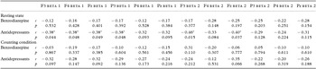
There were no relevant correlations (Spearman rho) between benzodiazepine or antidepressant use and immediate wordlist recall or delayed word list recall. Only small negative correlations between benzodiazepine use and figure recall (r (26) = −0.39, p = 0.04) and antidepressant use and word recognition (r (26) = −0.40, p = 0.04) can be reported. There was no significant correlation between EEG beta 1 power in counting condition and benzodiazepine or antidepressant use, the same holds true for beta 2 power. In contrast to counting condition, in resting state a negative correlation between antidepressant use and EEG Beta 1 at F3, F4, Fz, and P3 as well as in Beta 2 at F3 and Fz was found (Table 2). Antidementia drugs were taken by only one participant (Rivastigmin).
Point to point correlations between benzodiazepine or antidepressant use and EEG in resting state and counting condition

Note. Significant negative correlations can be reported between beta 1 power and antidepressant use at F3, F4, Fz, and P3 is resting state. There are no correlations between benzodiazepine or antidepressant use and EEG in counting condition. Abbreviations. r: Point to point correlation. *p value if < 0.05; **p value if < 0.01.
Beta 1
We found no significant correlations between beta 1 power and immediate word list recall, delayed word list recall, word recognition, and figure recall.
Beta 2
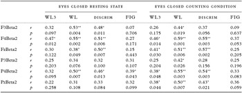
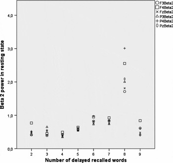
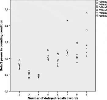
In contrast to beta 1, we found significant positive correlations between frontal and parietal EEG beta 2 power and memory test outcome (Table 3), indicating that higher beta amplitudes correlate with better memory test outcome. Immediate wordlist recall (third trial of the ten word learning phase) was significantly positively correlated with F4 (r (26) = 0.47, p < 0.012) in resting state. The EEG power of the counting condition was also significantly positively correlated with immediate word list recall at F4 (r (26) = 0.46, p < 0.014), Fz (r (26) = 0.41, p < 0.030), P4 (r (26) = 0.38, p < 0.048), and Pz (r (26) = 0.38, p < 0.044). Very strong positive correlations were found between resting state and delayed wordlist recall and F3 (r (26) = 0.53, p < 0.004), F4 (r (26) = 0.55, p < 0.002), and P4 (r (26) = 0.50, p < 0.007), as well as between counting condition and delayed wordlist recall and F4 (r (26) = 0.59, p < 0.001), Fz (r (26) = 0.51, p < 0.006), P4 (r (26) = 0.55, p < 0.003), and Pz (r (26) = 0.50, p < 0.007) (Figures 1 and 2). Recognition of learned words was also strongly correlated with F4 (r (26) = 0.55, p < 0.003), Fz (r (26) = 0.57, p < 0.002), and P4 (r (26) = 0.54, p < 0.003) in counting condition and in resting state F4 (r (26) = 0.51, p < 0.006), Fz (r (26) = 0.5, p < 0.007), and P4 (r (26) = 0.46, p < 0.013).
Spearman correlations of frontal and parietal EEG beta 2 power in resting condition and while counting backwards with memory scores

Note. Significant positive correlations can be reported between frontal and parietal beta 2 power and immediate and delayed verbal-memory recall. Discrimination of previously learned verbal information is also correlated with frontal and parietal beta 2 power in both conditions.
Abbreviations; r: Spearman Rho; *p value if < 0.05; **p value if < 0.01; WL3 Third trial of immediate wordlist recall. WL Delayed wordlist recall. FIG delayed Figure recall.
There was no correlation between free figure recall and beta 2 power in counting condition, but small correlations in resting state at P4 (r (26) = 0.385, p < 0.43). Taking into account multiple hypothesis testing with 14 variables, only the correlation between F4 Beta 2 and delayed wordlist recall would be significant after Bonferroni correction (p < 0.05). Therefore we additionally calculated a partial correlation for our main finding. Controlling for age revealed a significant positive correlation between F4 Beta 2 in counting condition and delayed free wordlist recall (F4 (r (25) = 0.53, p < 0.005).
Binary regression
To evaluate possibilities for surrogate measures of memory through EEG, we used EEG beta 2 power at F4 under cognitive effort. Beta 1 was not used for the regression model because it was not correlated with memory test outcome, neither was beta 2 in resting state because it was confounded with pharmacological treatment. In detail 13 of 28 participants were characterized as memory impaired. Six participants had impaired immediate wordlist recall, three only impaired delayed wordlist recall, and four a deficit in immediate and delayed wordlist recall. Mean years of education in the non-impaired group were 10.2 years compared to 10.38 years in the impaired group. The mean age was 80.3 years in the intact and 80.9 years in the memory impaired group.
[Verbal memory impairment = 1.704 + F4Beta2 Power * −1.795]
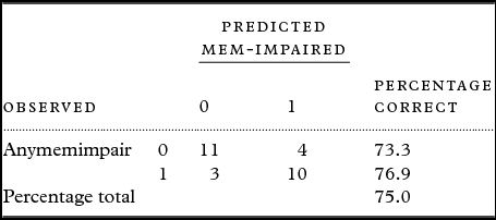
The resulting model, which includes a constant (1.704) and Beta 2 power at F4 multiplied with −1.795 allows for the classificatory separation of verbal-memory impaired and verbal-memory unimpaired patients from each other. Depending on the rounded off outcome (0 or 1), we can simply categorize our patients into impaired and unimpaired. According to the guideline of the validated European Version of the CERAD (Sollberger and Monsch, Reference Sollberger and Monsch2007) we used a cut-off of z < −1.3. The sensitivity of the model was 76.9 in detecting memory impairments, while specificity was 73.3 (Table 4). The calculation of a sum score for all recorded positions did not reveal stronger correlations with memory test outcomes.
Beta 2 mean power (F3, F4, Fz, P3, P4, Pz) in resting state and total number of delayed recalled words.

Beta 2 mean power (F3, F4, Fz, P3, P4, Pz) in counting condition and total number of delayed recalled words.

Binary-logistic regression model with verbal-memory impairment as dependent variable and frontal beta 2 power at F4 under cognitive effort as independent variable

Anymemimpair = immediate or delayed word list z < −1.3 SD.
Discussion
In this pilot study, we analyzed correlations between EEG beta power and memory impairments, which are dominant in MCI patients and ongoing dementia (Petersen et al., Reference Petersen2010). Especially EEG beta power is positively correlated with cognitive ability, but has not been specifically analyzed in respect of memory deficits, which we focused on. While our findings point out that EEG beta 2 power is positively correlated with cognitive abilities (Wang et al., Reference Wang, Zhang, Huang, Zhu, Guan and Liu2013) and that it is important in cognitive processes (Guntekin et al., Reference Guntekin, Emek-Savas, Kurt, Yener and Basar2013), we could also further specify these findings. The predictive value of EEG beta band in the conversion from MCI to AD (Poil et al., Reference Poil, de Haan, van der Flier, Mansvelder, Scheltens and Linkenkaer-Hansen2013; Gu et al., Reference Gu, Chen, Lu and Pan2016) seems to be supported by the strong correlation with memory function only, a core feature of dementia. The results are in line with McBride, who found discriminative power of EEG, when a simple counting task was used to discriminate between NC, MCI, and AD (McBride et al., Reference McBride2014). In comparison, we found slightly higher correlations between counting condition and memory than those in resting state. The subdivision of EEG beta into two different bands as recommended (Jobert et al., Reference Jobert, Wilson, Ruigt, Brunovsky, Prichep and Drinkenburg2012), yielded specific results for Beta 2 and not for Beta 1. Furthermore, pharmacological treatment seems to have effects on EEG resting state, but not under cognitive effort.
While there was no correlation between EEG Beta 2 and age nor EEG Beta 2 and education, we were also able to further specify that only verbal-memory is correlated with counting task EEG, but not with visual recall. We found a significant positive correlation between beta 2 power and delayed verbal-memory recall, which is in line with findings of several studies on MCI patients (Poil et al., Reference Poil, de Haan, van der Flier, Mansvelder, Scheltens and Linkenkaer-Hansen2013; Wang et al., Reference Wang, Zhang, Huang, Zhu, Guan and Liu2013; Gu et al., Reference Gu, Chen, Lu and Pan2016).
Conclusion
In conclusion, we investigated recent findings on EEG as a biomarker for memory impairments and found that EEG beta 2 is correlated with memory test outcome, while beta 1 is not. We were able to shed light on pharmacological effect, which seem to affect only resting state EEG and not EEG under cognitive effort. The use of a counting task yielded stronger correlations than resting state. While previous studies focused on global cognition, we focused on memory only and detected very strong and specific correlations. The use of specific surrogate markers could help to predict treatment response in future studies (Katz, Reference Katz2004). The presented specific surrogate marker could be useful in dementia. Since memory is a core feature of most dementias, which is mostly progredient, this surrogate marker could also be a useful predictor of therapeutic treatment response. For patients who are speech impaired or resilient this surrogate marker could be a fast and accurate alternative for diagnosis and planning of therapeutic treatment.
Further studies are needed to replicate the findings of this pilot study and make them feasible for clinical use. A larger sample size, including men and younger ages will be necessary for replication of the results. Furthermore, detailed information about duration of medication and possible addiction should be considered.
Conflict of interest
None.
Ethical requirements
Written informed consent was obtained from all participants. The study was approved by the local ethics committee.
Description of authors´ roles
Mr A.K. Kaiser is responsible for conception and performance of the study. He is also responsible for most of the written text. Mr. M. Doppelmayr was responsible for the general concept of the paper and methodological issues. Mr B. Iglseder is responsible for the general study design and final version of the manuscript.
Acknowledgments
I want to thank my wife and my children for supporting my scientific work.