The term “inflammatory bowel disease” (IBD) refers primarily to two major disorders, ulcerative colitis (UC) and Crohn′s disease (CD). Both entities are chronic conditions characterized by recurrent inflammatory lesions involving the gut mucosa. Although UC and CD show a considerable degree of etiological and epidemiological similarities, their pathology is entirely different. Ulcerative colitis is essentially a disease involving only the mucosa and the inflammatory disorder is limited to the large intestine. Crohn′s disease is a transmural inflammatory process involving the intestinal wall, from mucosa to serosa, and may affect any point along the gastrointestinal tract from the mouth to the rectum (Steidler, Reference Steidler2001).
Although the pathophysiology of IBD remains the subject of much debate, recent experimental and clinical studies suggest that the initiation and pathogenesis of these diseases are a confluence of multifactorial events involving interactions among genetic susceptibility, altered mucosal immunity and environmental challenges, mostly related to the acquisition and establishment of the enteric microflora (Fiocchi, Reference Fiocchi1998; Shanahan, Reference Shanahan2001).
The concept that the cause of IBD is immunological arose from the observation that IBD is characterized by massive cellular infiltrates and is associated with abnormalities of the immune system that include inappropriate production of antibodies and T-cell dysfunctions. An imbalance in T-helper cells (Th1 and Th2) differentiates CD from UC on an immunological basis. In UC an over expression of Th2 type cytokines (IL-4, IL-5) has been demonstrated, whereas in CD, Th1 type cytokines such as IFN-γ predominate as well as IL-12 produced by macrophages (Blumberg & Strober, Reference Blumberg and Strober2001; Steidler, Reference Steidler2001).
Animal models of IBD have been useful in potential therapeutic strategies because IL-12 drives Th1 responses. Several animal models with 2, 4, 6-trinitrobenzene sulphonic acid (TNBS)-induced colitis and treated with anti IL-12 showed that treatment was effective to ameliorate the disease (Fuss et al. Reference Fuss, Marth, Neurath, Pearlstein, Jain and Strober1999). This model of inflammation is associated with a Th1 response.
Intestinal T-lymphocytes are hyperactive in IBD. Pirzer et al. (Reference Pirzer, Schänhaar, Fleischer, Hermann and Meyer zum Büschenfelde1991) showed that local tolerance mechanisms are abrogated in Crohn's disease. Patients with UD or UC show an impaired tolerance towards commensal bacteria of the resident microbiota. These findings suggest that commensal bacteria present in the intestinal lumen might induce immune-inflammatory responses that could damage the intestinal mucosa. The intestinal inflammation is accompanied by an imbalance in the microflora and a strong inflammatory response might be mounted toward the gut microbiota, leading to perpetuation of the inflammation and gut barrier dysfunction. These pathologies are treated with anti-inflammatory drugs or surgery.
Current research is addressed to the identification of probiotics strains for bacterial antagonism therapies. Probiotics are live microbial food supplements or components of bacteria which have been shown to have beneficial effects on human health (Guarner & Schaafsma, Reference Guarner and Schaafsma1998; Salminen et al. Reference Salminen, Bouley, Boutron-Ruault, Cummings, Franck, Gibson, Isolauri, Moreau, Roberfroid and Rowland1998). A recent experimental study has shown that colonization with a Lactobacillus reuteri strain can prevent the development of colitis in genetically susceptible mice (Madsen et al. Reference Madsen, Doyle, Jewell, Tavernini and Fedorak1999).
Some probiotics may naturally exhibit anti-inflammatory properties when interacting with the human gut mucosa (Salminen et al. Reference Salminen, Bouley, Boutron-Ruault, Cummings, Franck, Gibson, Isolauri, Moreau, Roberfroid and Rowland1998). In our laboratory, we demonstrated that conventional yoghurt may exert antitumour activity by a decrease in the inflammatory immune response (Perdigón et al. Reference Perdigón, de Moreno, Valdez and Rachid2002) and that the dietary supplementation of yoghurt may also play a role in modulating cell proliferation, inducing apoptosis during the development of colorectal carcinoma (Rachid et al. Reference Rachid, Gobbato, Valdéz, Vitalone and Perdigón2002). In normal hosts yoghurt was able to stimulate the immune cells associated with the gut (Perdigón et al. Reference Perdigón, Rachid, De Budeguer and Valdez1994).
Many papers have reported the possible mechanisms underlying the beneficial effects of probiotics in IBD although the observations in some of them are controversial (Guarner & Schaafma, Reference Guarner and Schaafsma1998). The main role of probiotic bacteria would be to counteract the inflammatory process by stabilizing the gut microbial environment and the intestine permeability barrier, with an improvement in the gut immunological barrier, particularly the IgA responses (Isolauri et al. Reference Isolauri, Sütas, Kankaanpää, Arvilommi and Salminen2001). The probiotic effect may also be mediated via control of the balance between pro-and anti-inflammatory cytokines (Isolauri, Reference Isolauri2001); thus, probiotics may offer a valuable tool for the prevention and control of inflammatory bowel diseases. As there are no reports concerning the prevention or alleviation of IBD using conventional yoghurt, the present work was carried out to study whether or not yoghurt administration has an effect on the amelioration of colitis induced by TNBS in mice.
Materials and methods
Animals
Six- to eight-week-old random-bred BALB/c mice, each weighing 28–30 g, were obtained from the random-bred closed colony kept of our institution (Instituto de Microbiología, Facultad de Bioquímica, Química y Farmacia, Universidad Nacional de Tucumán). Each experimental group consisted of 20 mice (5 for each experimental period). Five mice were also used as normal controls in each assay. All animals received standard pelleted chow and tap water ad libitum. They were used under the current laws for animal studies in our Institute of Microbiology.
Yoghurt preparation
A simulated commercial yoghurt was prepared from a stock culture of lactic acid bacteria containing Lactobacillus delbrueckii subsp. bulgaricus and Streptococcus thermophilus. These strains, mixed at a 1:1 ratio, were incubated in 10% non fat milk at 37°C for 4 h and then at 4°C for 24 h. At the end of this process the total bacterial count was 2×10⁸ cell/ml. Yoghurt was prepared every 24 h to insure that the numbers of bacteria were constant during the time of experimentation.
Colitis induction
Colitis was induced in mice as described by Chin et al. 1994 with TNBS. Briefly, after overnight fasting, mice were anesthetized by intraperitoneal injection of Pentothal Sodico (Tiopental Sodico) (Abbott Laboratories, Abbott Park, Illinois) (USA) (70 mg/Kg). A 100 μl enema of TNBS solution (Sigma Chemical Co., St. Louis, MO) at a dose of 20 mg/kg in 40% ethanol was instilled for 1 min into the colonic lumen by the anal route. 1 ml syringe fitted with a polyethylene cannula. After instillation the mouse was held by the tail in a vertical position for 30 seconds.
Feeding procedure and TNBS administration
Four groups of animals were used in the study.
Group 1:
Normal controls (C): animals given no treatment and fed ad libitum with a balanced diet. Animals were sacrificed on the 4th week, coinciding with the end of the experiment for the rest of the group.
Group 2:
Colitis control (TNBS): the animals were instilled in the colonic lumen with TNBS solution once a week for 3 consecutive weeks and fed ad libitum with a balanced diet. The samples were taken once a week for 4 weeks after the last TNBS instillation. Samples were taken on weeks 1, 2, 3 and 4.
Group 3:
Control yoghurt (Y): the mice were given 3 ml yoghurt as a dietary supplement for 10 consecutive days. This period of yoghurt administration was determined in our laboratory as the optimal dose to stimulate gut immunity (Perdigón et al. Reference Perdigón, Rachid, De Budeguer and Valdez1994). This group did not receive TNBS instillation. Yoghurt was not supplemented during the 3 weeks corresponding to TNBS administration. Then yoghurt was administered again for 10 consecutive days. The samples for this group were taken on week 4.
Group 4:
Test group (Y-TNBS-Y): before colitis induction, mice were given a daily supplement of 3 ml yoghurt for 10 consecutive days. At the end of the feeding period, the mice received 3 inoculations of 20 mg TNBS solution/kg body weight once a week for 3 consecutive weeks. After TNBS instillation, this group again received a daily supplement of 3 ml yoghurt for 10 consecutive days. The samples were taken on weeks 1, 2, 3 and 4.
Determination of peripheral blood mononuclear cells (PBMCs)
These determinations were performed once a week during 4 weeks only for the TNBS and Y-TNBS-Y groups. In group 1 and 3 the samples were taken at the end of the experiment on week 4. Blood was collected from 10 mice by heart puncture. The number of erythrocytes and leucocytes was determined by the haemacytometric method and expressed as mm3. Smears stained with Giemsa solution were examined to differentiate between granulocyte, lymphocyte and monocyte populations. They were expressed as percentages.
Histological studies
Five mice from groups 2 and 4 were killed by cervical dislocation on the 1st, 2nd, 3rd and 4th week. The large intestine was carefully removed and prepared for histological and immunological evaluation, fixed with formalin 10%, dehydrated and embedded in paraffin using standard methods (Sainte Marie, Reference Sainte-Marie1962). Serial paraffin sections of 4 μm were cut from each tissue and stained with haematoxylin-eosin for microscopic examination.
Determination of the number of Ig A+, CD4+ and CD8+ cells population
The number of IgA-secreting cells, CD4+ and CD8+ T population was determined from histological samples of large intestine using direct immunofluoresence assay.
The number of IgA secreting cells was measured using α-chain monospecific antibodies conjugated with fluorescent isothiocyanate isomer (Sigma). Monoclonal antibodies (MAbs) specific for CD4 or CD8 T-lymphocytes-FICT conjugate (Gibco BRD Life Technologies, Buenos Aires, Argentina) were used to determine these populations. Samples were incubated with 0·2 ml of the antibody diluted 1:100 for IgA and 1:50 for T cells for 30 min at room temperature and then washed three times (10 min each time) with 0·01 m-phosphate-buffered saline, pH 7·2. The results were expressed as the number of positive cells in ten colonic crypts of the large intestine.
Apoptosis determination
Apoptosis was evaluated on histological slices of the large intestine from each group of animals. The presence of DNA breaks was detected in situ by TUNEL test using fluorescein in a Promega apoptosis detection system kit (Madison, WI) (Rachid et al. Reference Rachid, Gobbato, Valdéz, Vitalone and Perdigón2002). It is based on the specific binding of terminal deoxinucleotidyl transferase to the 30-OH ends of DNA, ensuring the synthesis of a polydeoxynucleotide polymer. The fluorescein-12-dUTP nick end-labelled DNA can be visualized directly by fluorescence microscopy. The cells were defined as apoptotic if the whole nuclear area of the cell labelled positively fluorescent. The number of apoptotic cells was determined by counting 30 fields at 40× magnification using a fluorescence microscope.
Statistical Analysis
Results are expressed as means±sd. Student's test and ANOVA test were used to assess the statistical significance among groups.
Results
Determination of peripheral blood mononuclear cells (PBMCs)
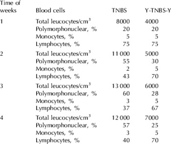
We observed a marked increase in polymorphonuclear cells and in the number of total leucocytes. from the 2nd week after inoculation of TNBS, in the TNBS group, relative to the Y-TNBS-Y group. The values obtained with the Y-TNBS-Y group were similar to the control animals during the period assayed. These results are shown in Table 1.
Study of haematological modifications in mice from groups TNBS and Y-TNBS-Y. For normal control the values were: total leucocytes/cm3 6000, comprising polymorphonuclear cells 20%, monocytes 4%, lymphocytes 76%. Normal controls are animals given no treatment and eating a convencional balanced diet throughout. For group Y the values were: total leucocytes/cm3 7000, comprising polymorphonuclear cells 25%, monocytes 5%, lymphocytes 70%

Histological studies
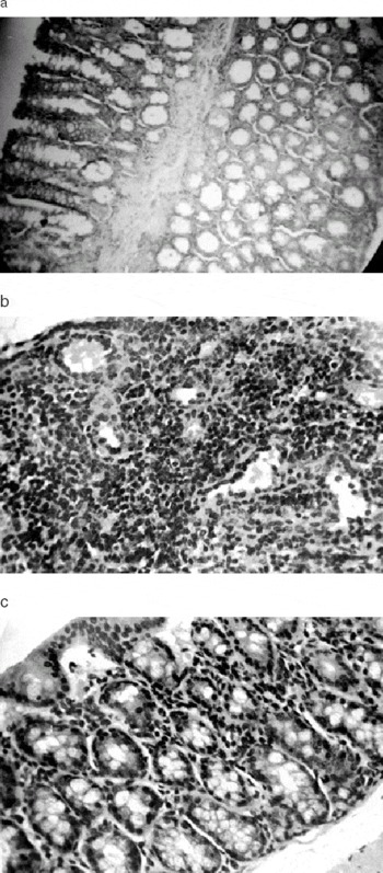
In TNBS group the large intestine showed changes as early as assay (week 1). These changes continued increasing until week 4 with a distortion of crypts, loss of the histological structure, great infiltration of mononuclear cells in the colonic interstitium, and focal ulcers with an acute inflammatory cell exudates and/or the presence of mucosal edema. (Fig. 1b).
Light micrograph of haematoxilin-eosin stained section from the large intestine of mice. (a) Large intestine of normal control mice without treatment, (b) large intestine of mice treated with TNBS (TNBS group) on week 4, (c) large intestine of Y-TNBS-Y group on week 4. Magnification 40×.

In the control group Y the large intestine did not show histological modifications throughout of feeding period. The test group Y-TNBS-Y showed an improved histological structure of the large intestine compared with TNBS group with a moderate infiltration of mononuclear cells. This result is expressed in Fig. 1c.
Determination of the number of IgA-producing cells, CD4+ and CD8+ T population
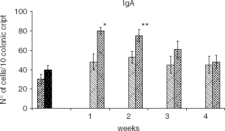
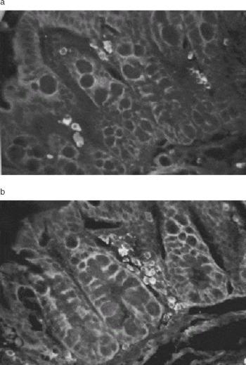
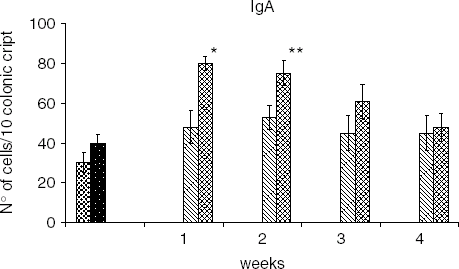
Figure 2 and Fig. 3a & b show the IgA producing cells in the large intestine. There was an increase in the number of IgA producing cells in the group Y-TNBS-Y in relation to the group TNBS during weeks 1 and 2, non-significant differences were found on week 3 and 4 for both groups. Values for the group Y did not show significant differences from the untreated control.
Determination of the number of IgA producing cells in the large intestine by direct inmunofluorescence assay. Values are the means of n=5±sd. *P<0·001 **P<0·01. , Control;
 , Y;
 , TNBS;
 , Y-TNBS-Y.
(a) Microphotograph of IgA+ cells in colonic crypts of the large intestine of mice treated with TNBS, on the week 2 (b) Microphotograph of IgA+ cells in colonic crypts of the large intestine in the Y-TNBS-Y group on the week 2. Magnification 40×.

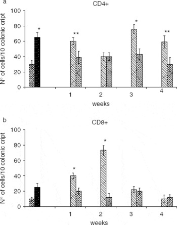
CD4+ T cells were increased in group Y in relation to the untreated control. For the Y-TNBS-Y group values for CD4+ were lower than the TNBS group in the 1st, 3rd and 4th weeks of treatment. In the second week the results were similar to the TNBS group, Fig. 4a.
(a) Determination of the number CD4+ T cells in the large intestine by direct inmunofluorescence assay. (b) Determination of the number CD8+ T cells in the large intestine. Values are the means of n=5±sd. *P<0·001 **P<0·01., Control;, Y;, TNBS;, Y-TNBS-Y.

The values for CD8+ T-cells were significantly lower in Y-TNBS-Y group during 1st and 2nd weeks compared with TNBS group. No significant differences were found in the 3rd and 4th week, values being similar for both groups (Fig. 4b).
Values for CD4+ and CD8+ cells in group Y showed significant differences from the control group.
Apoptosis Determination
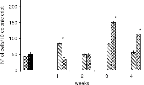
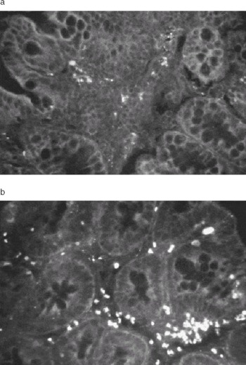
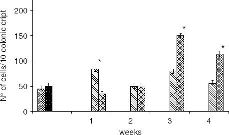
The percentage of apoptotic cells in large intestine in Y group and in the untreated controls groups were similar, while the number of apoptotic cells was decreased in the Y-TNBS-Y group compared with the TNBS group only for the 1st week. In the 2nd week the values were similar to those obtained for TNBS group. For the 3rd and 4th week values for Y-TNBS-Y group were significantly increased compared with the TNBS group. These results are showed in Fig. 5 and Fig. 6a & b.
Determination of the number of apoptotic cells in the colonic crypts in the large intestine. Values are the means of n=3±sd. *P<0·001. , Control; , Y; , TNBS; , Y-TNBS-Y.
Apototic cells determined in histological slices of the large intestine on week 4 (a) Apoptotic cells in mice from the TNBS group (b) Apoptotic cells in mice from the Y-TNBS-Y group. Magnification 40×.

Discussion
The oral introduction of probiotics has been shown to reinforce the various lines of gut defence: immune exclusion, immune elimination, and immune regulation (Perdigón et al. Reference Perdigón, Alvarez, Rachid, Agüero and Gobbato1995; Maldonado Galdeano et al. Reference Maldonado Galdeano, de Moreno, Vinderola, Bonet and Perdigón2007). Probiotics also stimulate non-specific host resistance to microbial pathogens (Gobbato et al. Reference Gobbato, Maldonado Galdeano and Perdigon2008).
Normal responses in the gut are based on the production of IgA antibodies that perform the immunoexclusion of microbes and antigens on the surface of the epithelium. These responses do not induce inflammation or tissue damage. In a previous work we demonstrated gut immunostimulation induced by yoghurt feeding (Perdigón et al. Reference Perdigón, Rachid, De Budeguer and Valdez1994).
IgA secretion is known to promote the gut immunological barrier and has been shown to be of great importance in the establishment of tolerance towards the indigenous microflora in a T-cell independent manner (Macpherson et al. Reference Macpherson, Gatto, Sainsbury, Harriman, Hengartner and Zinkernagel2000). In our laboratory we observed that dietary supplementation with yoghurt increased the number of IgA-secreting cells in the large intestine during the development of a colorectal carcinoma (Perdigón et al. Reference Perdigón, de Moreno, Valdez and Rachid2002). The increase in local IgA levels resulting after probiotic ingestion may also contribute to the protective effect of gut mucosal against an IBD process (Malin et al. Reference Malin, Suomalainen, Saxelin and Isolauri1996).
In this paper we demonstrated through histological studies in a TNBS induced colitis model, that feeding with yoghurt decreased the number of infiltrative immune cells in the large intestine. We did not observed transmural injury, crypt cell hyperplasia or ulcerations (Fig. 1c) as found in the TNBS group (Fig. 1b) in relation to the control (Fig. 1a). We did not find hematological modifications in the group Y-TNBS-Y even after TNBS instillation in relation to the group TNBS (Table 1). The low values found for the Y-TNBS-Y group would show a great diminution of the inflammatory response induced by TNBS.
When we analysed the number of IgA secreting cells we observed a significant increase for the 2nd and 3rd week in the group Y-TNBS-Y compared with the TNBS and control groups and it was less significant in the group Y (Fig. 2, Fig. 3a & b). In a previous report (Perdigón et al. Reference Perdigón, Rachid, De Budeguer and Valdez1994) the influence of yoghurt feeding on the number of IgA cells was demonstrated. The increase in the IgA+ cells in the group Y-TNBS-Y could have contributed to the anti-inflammatory effect determined by histological studies due to the anti-inflammatory property attributed to the IgA (Iaacson, Reference Isaacson1982). In a previous work we observed a similar effect of yoghurt with an enhancement in the IgA+ cells an experimental model colon carcinoma (Perdigón et al. Reference Perdigón, Valdez and Rachid1998 & Reference Perdigón, de Moreno, Valdez and Rachid2002). We believe that the strong stimulation induced by TNBS instillation induces a great inflammatory response with a high production of proinflammatory cytokines. This inflammatory response is not stimulated by yoghurt feeding which induced a clonal expansion change by proliferation of the IgA+ cell with an important increase in the number of these cells in the Y-TNBS-Y group, with a consequent diminution of such response.
During acute inflammatory episodes, the mucosal lining of the intestine displays a characteristic inflammatory infiltrate of mast cells, lymphocytes, macrophages, and activated neutrophils (Singh et al. Reference Singh, Read, Asseman, Malmström, Mottet, Stephens, Stepankova, Tlaskalova and Powrie2001). Damage to the epithelial gut mucosa in IBD has been linked to elevated levels of effecter immune cells such as activated CD4+ and CD8+ cytotoxic T cells, cytolytic intraepithelial lymphocytes (IELs), and perforin and granzyme-containing T cells (Muller et al. Reference Müller, Lory, Corazza, Griffiths, Z'graggen, Mazzucchelli, Kappeler and Mueller1998).
There is considerable evidence that IBD patients have inappropriate T-cell responses to antigenic components of their own intestinal microbiota. Thus, IBD patients not only suffer from hyper-reactiveness towards their normal intestinal microbiota but the reactivity of T cell populations driving inappropriate auto-immune reactions, this fact impairs the bacterial clearance. We determined that yoghurt feeding was effective by modulating the immune deregulation induced in IBD process. We found that TNBS administration produced an increase in CD4+ on weeks 1, 3 and 4 and in CD8+ T cells on weeks 1 and 2 in the large intestine, while in the Y-TNBS-Y group induced a diminution in the number of both T populations throughout the period assayed (Fig. 4a & b). We believe that TNBS instillation induces a strong inflammatory response with damage to the epithelial barrier, as a consequence, the immune cells as CD4+ and CD8+ increase and are activated to release proinflammatory cytokines increasing the bacterial translocation originate by disruption of the epithelium. This speculation allow us to explain the increase in CD4+ and CD8+ cells in the group TNBS where the great inflammatory response would be magnified by cytotoxic or cytolic activities of both T cells populations. When yoghurt was administrated we observed a diminution in the cellular infiltration of both T cells population, this fact would avoid the proinflammatory cytokines produced by CD4+ cells, or the cytotoxic effect mediated by CD8+ T cells. In this model yoghurt feeding induced normalization in the number of T cell, reaching values lower than the group Y, but similar to the values for the untreated control group. In group Y the number of CD4+ and CD8+ cells were enhanced in relation to the control values, we think that the effect of yoghurt administration is different in a normal host, perhaps due to the immune cells associated with the gut, are not receiving an inflammatory stimuli as occurs in IBD. The diminution in the CD8+ T population was also observed in a cancer colon model (Perdigón et al. Reference Perdigón, Valdez and Rachid1998).
Intestinal tissue homeostasis is maintained by a balance between cell proliferation and cell death, and a loss of this equilibrium, is involved in pathogenic disease (Saikumar et al. Reference Saikumar, Dong, Mikhailov, Denton, Weinberg and Venkatachalam1999). In physiology condition, T cells become more susceptible to programmed cell death (apoptosis) once they are activated. This fact leads to the cell elimination, a necessary feature, to allow re-establishment of the non-inflamed state. There is evidence that T cells of the lamina propria from CD and UC patients are more resistant to apoptosis (Boirivant et al. Reference Boirivant, Marini, Di Felice, Pronio, Montesani, Tersigni and Strober1999). In this experimental model of IBD we observed that feeding with yoghurt increased the number of apoptotic cells in large intestine (Fig. 5, 6); this increase was more significant toward the end of the period of yoghurt administration. These results agree with a previous work, where we showed that the dietary supplementation of yoghurt may play a role in modulating cell proliferation or apoptosis during the development of colorectal carcinoma (Rachid et al. Reference Rachid, Gobbato, Valdéz, Vitalone and Perdigón2002).
In the present paper we demonstrated that yoghurt consumption would be useful to alleviate IBD. The way by which yoghurt exerts such effect would be through the immune mechanisms, with an increased in IgA+ cells, capable of modulating the inflammatory response with a diminution in CD4+ and CD8+ population and an increase in the apoptosis of the infiltrative immune cells.
This work was financially supported by grants from the Consejo de Investigaciones Universidad Nacional de Tucumán, Argentina (CIUNT) D/351 and from the Consejo de Investigaciones Científicas y Técnicas, Argentina (CONICET) PIP 5445.