


INTRODUCTION
Ironstones are sedimentary rocks with >15% iron (Kimberley, Reference Kimberley1978), whose constituent iron oxides and hydroxides may be inorganic or bioinduced in origin (Almaraz et al., Reference Almaraz, Whitaker, Andrews and Duckworth2017). In the Amazon region, iron-rich deposits occur in upland terrains as Cenozoic lateritic soil profiles and economically exploited Precambrian banded iron formations (Horbe and Costa, Reference Horbe and Costa2005; Klein, Reference Klein2005; Vasconcelos et al., Reference Vasconcelos, Heim, Farley, Monteiro and Waltenberg2013). In addition, iron-rich incrustations, clearly distinct from laterite and banded iron formation, occur on fractured bedrock along the Xingu River, a major clearwater river draining eastern Amazonia (Sawakuchi et al., Reference Sawakuchi, Hartmann, Sawakuchi, Pupim, Bertassoli, Parra and Antinao2015), but have never been examined in detail.
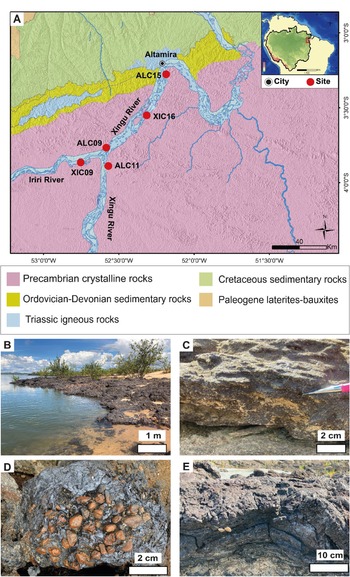
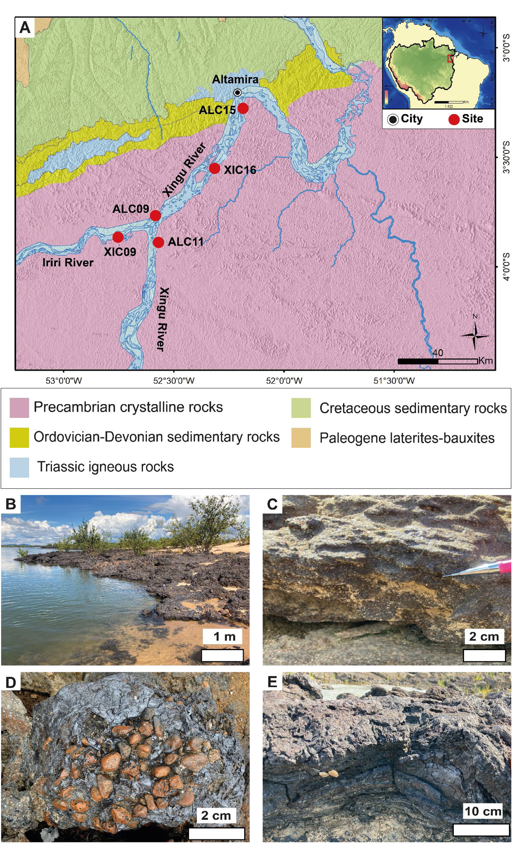
The Xingu ironstones developed predominantly on Precambrian crystalline rocks of the Brazilian Shield and, to a lesser extent, on Paleozoic sandstones of the Amazonas Basin (Fig. 1A). These ironstones are morphologically complex, marked by an array of cavities that define aquatic niches providing abundant shelter and highly diverse habitats for aquatic species in the Xingu River, including a unique fish fauna adapted to crystalline rocky substrates and fast-moving water (Sabaj Perez, Reference Sabaj Perez2015; Chamon and Sousa, Reference Chamon and Sousa2016; Winemiller et al., Reference Winemiller, McIntyre, Castello, Fluet-Chouinard, Giarrizzo, Nam, Baird and Darwall2016; Fitzgerald et al., Reference Fitzgerald, Winemiller, Sabaj Pérez and Sousa2017, Reference Fitzgerald, Sabaj Perez, Sousa, Gonçalves, Rapp Py-Daniel, Lujan, Zuanon, Winemiller and Lundberg2018; Sousa et al., Reference Sousa, Chaves, Akama, Zuanon and Sabaj2018; Reference Sousa, Lucanus, Arroyo-Mora and Kalacska2021). Thus, because they harbor endemic fish species currently threatened by extinction due to habitat loss, the Xingu iron crusts are of great ecological relevance to Amazonian ecosystems (Winemiller et al., Reference Winemiller, McIntyre, Castello, Fluet-Chouinard, Giarrizzo, Nam, Baird and Darwall2016).
Geological map and different types of Xingu ironstone (site ALC09). (A) Geology of the study area with sampling sites (red dots) in the northern Amazon Basin (inset map). Bedrock geology after Bahia et al. (Reference Bahia, Faraco, Monteiro, Oliveira, Schobbenhaus, Gonçalves, Santos, Abram, Leão Neto, Matos, Vidotti, Ramos and Jesus2004). (B) Ironstones exposure during the dry season. (C) Ferruginous sandstone. (D) Ferruginous conglomerate. (E) Ferruginous crust.
The distinct difference between these ferruginous deposits and other Amazonian ironstones motivated investigation as to their origin and age. For this, we combined field observations and analytical methods to characterize the morphology and mineralogy as well as the age and genesis of ironstones along the Xingu and Iriri rivers near Altamira (Pará, northern Brazil), focusing mostly upon laminated crusts (Table 1). Our results shed light on the roles of biotic and abiotic sedimentary processes in the genesis of the Xingu ironstones.
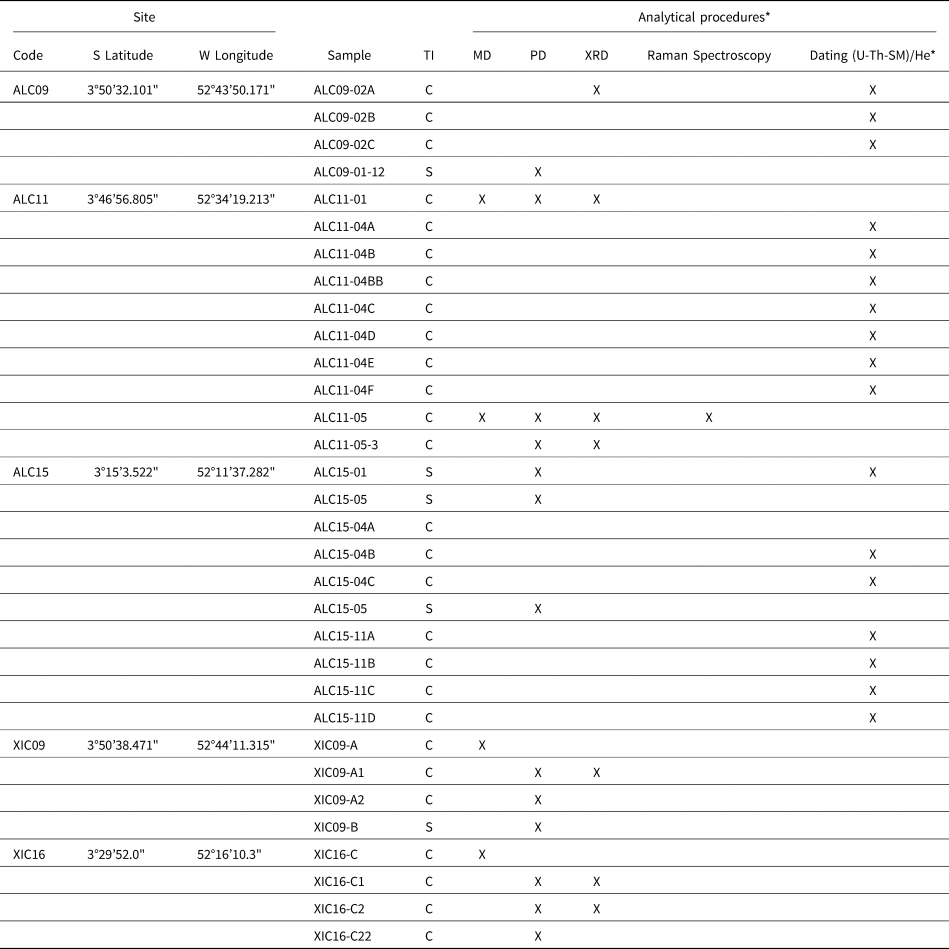
Information for location, sample numbers, ironstone type, and analytical procedures applied to the material studied here. Latitude/longitude according to World Geodetic System (WGS84).

* TI = Type of Ironstone: C = Crust; S = sandstone. MD = Macroscopic description. PD = Petrographic description. XRD = X-Ray Diffraction.
THE XINGU RIVER
The Xingu River is the second-largest clearwater tributary of the Amazon River (Sioli, Reference Sioli1984; Goulding et al., Reference Goulding, Barthem and Ferreira2003), flowing 2000 km through central and northern Brazil (Fig. 1A). River waters range from 6.0–7.0 in pH and are well oxygenated (Silva et al., Reference Silva, Ríos-Villamizar, Miranda, Ferreira, Bringel, Gomes, Silva, Pascoaloto, Santana and Cunha2019). Rainfall in the Xingu River basin is controlled by the strength of the South American Monsoon System (SAMS) and north-south migration of the Intertropical Convergence Zone (ITCZ) (Marengo, Reference Marengo2004; Garreaud et al., Reference Garreaud, Vuille, Compagnucci and Marengo2009). These factors promote marked seasonality, not only on Xingu River discharge (ANA, 2020) but also on major element composition of suspended sediment in the Xingu River. Relative Fe concentration increases during the wet season while P and K increase during the dry season (Bertassoli et al., Reference Bertassoli, Sawakuchi, Sawakuchi, Pupim, Hartmann and McGlue2017).
Downstream from the Iriri River, the Xingu's largest tributary, the Xingu River flows NE and then abruptly deflects, in sequence, SE, NE, and finally NW, over 130 km to form the “Volta Grande do Xingu,” (aka, the Great Bend of the Xingu). Here, the riverbed is steeper, with large rapids, waterfalls, and a complex array of anastomosing channels and bars (Sawakuchi et al., Reference Sawakuchi, Hartmann, Sawakuchi, Pupim, Bertassoli, Parra and Antinao2015). Downstream from the Volta Grande do Xingu, the river cuts through increasingly younger (Ordovician to Cretaceous) strata of the intracratonic Amazonas Basin. Paleocene lateritic-bauxite (Fig. 1A) and Quaternary alluvial sediments overlie these rocks. In this sector, the Xingu River exhibits a “ria” morphology characterized by a single broad funnel-like channel (Archer, Reference Archer, Blum, Marriott and Leclair2005; Fricke et al., Reference Fricke, Nittrouer, Ogston, Nowacki, Asp, Souza Filho, da Silva and Jalowska2017).
MATERIAL AND METHODS
Sampling
Sampling sites were selected with a preference for thicker ironstone deposits up river from Altamira along the middle Xingu and lower Iriri rivers (Fig. 1A). Sampling was conducted during the dry season of 2014 when the ironstones were well exposed. The Xingu ironstones consist of ferruginous crusts, sandstones, and conglomerates. The ferruginous crusts dominate the samples because they offer the greatest potential for studying ironstone origin owing to their greater concentration of iron oxides/hydroxides. Accompanying the 26 samples of ferruginous crusts that we analyzed (Table 1) are five sandstone samples collected for comparative petrographic analysis of the mineralogy and texture of the iron oxides and hydroxides that cement the sand grains and form the crusts.
Mineralogy and texture
Iron oxide and hydroxide minerals and biogenic features were characterized incorporating the following analytical techniques: Polarized Light Microscopy (PLM), scanning electron microscopy (SEM) coupled to X-ray energy dispersive spectroscopy (EDS), and X-ray diffraction (XRD) analyses, all carried out at the Institute of Geosciences of the University of São Paulo (IGc-USP), Raman spectroscopy at the Institute of Chemistry of USP, and SEM with mineral liberation analyzer (MLA) in the Technological Characterization Laboratory of the Polytechnic School of USP (LCT-USP).
The mineralogical composition was determined by XRD in seven samples (Table 1). Whole-rock fragments (1 cm3 in volume, weighing ~4 g, and crushed to a powder) were analyzed in a Brucker D8 Advance diffractometer with a 40 kW and 40 mA copper K-α radiation tube. Quantitative XRD analysis employing the Rietveld method was carried out on three samples using TOPA 5.0 software, with quality adjustment via evaluation of the GOF = 1.10 and Dw = 1.70 parameters (Table 1).
Texture and mineralogy of ferruginous crusts and sandstones were described in petrographic thin sections 30 μm thick by PLM using a Carl Zeiss Axioplan 2 transmitted light microscope with LAS-Leica Application Suite software for imaging (Table 1).
Morphological features and chemical composition of mineral phases were characterized by SEM/EDS analyses, including secondary electron (SE) and backscattered electron (BSE) modes. These analyses were carried out on fresh surfaces of uncontaminated portions of fragments, ~1 cm3 in volume, of sample ALC11-05 (Table 1) at LCT-USP, using the Oxford FEI Quanta 250 detector model with INCA 300 software. Semi-quantitative ESD was collected using Xact detector with INCA software and a 40 mm window, by constant acceleration voltage 20 kV at 300Kcp of free counting in quartz and probe calibration in cobalt. Quantitative mineralogical domains in a thin section were determined by MLA image analysis software—Mineral Liberation Analyzer with remote microscope control and EDS system, using a Thermo/FEI Quanta 650 FEG. Complementary analyses were performed at IGc-USP using an Oxford LEO440i Electron Microscopy detector and LEO v03 software.
Organic matter was investigated using a micro-Raman Renishaw inVia spectrometer, together with a confocal microscope, in a fragment ~1 cm3 in volume and in a thin section of sample ALC11-05 (Table 1). A diffraction net of 1200 lines and excitation wavelengths of 532 nm, 633 nm, and 785 nm were used with a 20 LW lens. Software WiRE™ 4.1 and Origin®8 were used for the interpretation of data.
(U-Th)/He goethite dating
Goethite in 17 layers of iron crust samples from sites ALC09, ALC11, and ALC15 (Tables 1, 2) were dated by the (U-Th)/He method, which is based on the accumulation of 4He within the goethite structure due to alpha decay of 238U, 235U, 232Th, and 147Sm (Ault et al., Reference Ault, Gautheron and King2019). Samples were broken and fragments 200–500 μm in size were selected under a binocular microscope. These fragments were then disaggregated with a jaw crusher and a disc mill, and the 63–500 μm fraction isolated by sieving. In general, for each rock specimen, three to seven aliquots of goethite were encapsulated in previously weighed 99% pure niobium tubes 600 μm in diameter and 1 mm in length, and then reweighed. Individual goethite fragments selected for dating varied from 5–228 mg in weight.
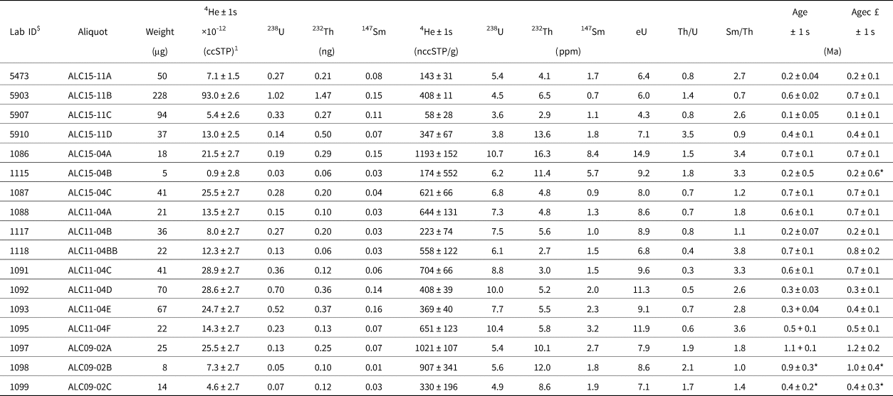
Results of age determinations on goethite using the (U-Th-Sm)/He method. Observations: $ = Laboratory ID number; £ = (U-Th)/He corrected age; ccSTP = cubic centimeters at standard temperature and pressure; * = (U-Th)/He ages not considered because of large uncertainties associated with very small U-Th-Sm and He content measured in the smallest aliquots.

Helium content was measured at Geoscience Paris Saclay (GEOPS—Université Paris Saclay, Orsay, France), following the protocol described by Gautheron et al. (Reference Gautheron, Pinna-Jamme, Derycke, Ahadi, Sanchez, Haurine and Monvoisin2021). Each encapsulated fragment was degassed using a diode laser under vacuum for 30 minutes at <1000°C, mixed with a known amount of 3He, and, after gas purification, the gas was analyzed in a Pfeiffer Prisma Quadrupole mass spectrometer. After degassing, the samples were dissolved in Teflon-capped vials using different acid solutions. First, 50 μL of 5M HNO3 containing a known amount of 235U, 230Th, and 149Sm was introduced into each vial, followed by 200 μL of 30% HCl, and a few drops of 38% HF. The vials were closed and heated at 70°C overnight. The solutions were evaporated at 180°C to concentrate the sample. Second, 1 mL of 5M HNO3 was added to the final solution and heated for 1h at 100°C. U, Th, and Sm were measured on an ELEMENT XR ICPMS. More analytical details can be found in Gautheron et al. (Reference Gautheron, Pinna-Jamme, Derycke, Ahadi, Sanchez, Haurine and Monvoisin2021).
The raw (U-Th)/He age is calculated using the He, U, Th, and Sm content and the radioactive decay constants of 238U, 235U, 232Th, and 147Sm. No correction of alpha ejection was made here because the mean alpha stopping distance of ejected He in goethite, ~15 μm (Ketcham et al., Reference Ketcham, Gautheron and Tassan-Got2011), is small compared to the sample size (> 1 mm). On the other hand, between 2–10% of He is estimated to have been lost by diffusion in goethite because of the polycrystalline nature of the millimetric samples (e.g., Shuster et al., Reference Shuster, Vasconcelos, Heim and Farley2005; Vasconcelos et al., Reference Vasconcelos, Heim, Farley, Monteiro and Waltenberg2013; Hofmann et al., Reference Hofmann, Reichen and Farley2017). In this study, to account for possible He loss by diffusion and U-Th disequilibrium, ages have been corrected by adding 10% to the original value (Table 2).
RESULTS
Mineralogy and texture of the ironstones
We studied laminated ferruginous crusts as well as goethite-cemented sandstones and conglomerates from permanently submerged zones and marginal areas of seasonally inundated riverbeds (Fig. 1B). The ironstones are black to brown in outcrop and dark reddish or yellowish on freshly broken surfaces. They occur in discontinuous mound-like, lenticular or tabular (sheets or plates) morphologies from 10–100 m in extent and up to 4 m thick, and are referred to as mocororô by the local population.
These ferruginous sedimentary rocks comprise intercalated deposits of laminated crusts, sandstone, conglomeratic sandstone, and conglomerate (Fig. 1C–E), but the dominant lithotype varies locally. Clastic ironstones may be massive or incipiently cross-bedded or horizontally bedded, and, except for their ferruginous cement, they resemble present-day channel bar deposits.
Platy, poorly to moderately sorted ferruginous conglomerates (Fig. 1D) overlying sandy riverbed can break up, allowing underlying sand to be washed away, thereby producing caves and stacked plates of a ferruginous conglomerate. When deposited on solid bedrock, the ferruginous conglomerate fills gaps between boulders of crystalline rocks.
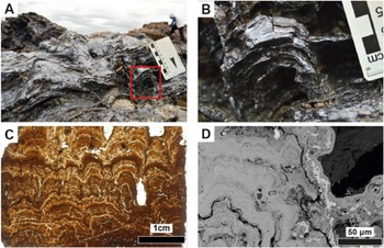
As for the ferruginous crust, the most notable structure observed at all scales, from macroscopic to microscopic, is a wavy millimetric to micrometric lamination, typically characterized by two or more orders of irregularly undulating to crenulated surfaces (Fig. 2). PLM shows laminae to be convex-upward with alternating color patterns of brown, red, and yellow, varied thickness, and stacking modes from laterally continuous to domal, pseudocolumnar, and columnar (Fig. 2C). Similar patterns are also evident in the tones of grays in images of laminae obtained by SEM (Fig. 2D).
Laminated ferruginous crusts at different scales. (A) Ferruginous crust with irregularly wavy lamination. (B) Detail of dome in red rectangle in (A). (C) Photomicrograph (PLM) of lamination of ferruginous crust in thin section showing alternating color pattern of iron hydroxides, variation in laminar thickness, convex-upward laminae with several orders of irregular undulation or crenulation, and laterally continuous to domal, pseudo-columnar and columnar laminar stacking morphologies; ALC11-01. (D) SEM/SE image showing similar patterns of laminar morphology and tonal variation (of gray) as observed in (C) at the sub-millimetric scale.

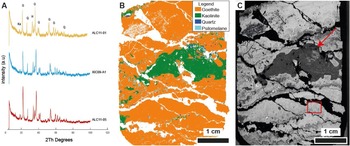
XRD analyses revealed that the crusts are composed mainly of goethite, with some quartz, kaolinite, and subordinate hematite (Fig. 3A). Mineralogical mapping confirms that goethite is the main component and that it hosts kaolinite and quartz (Fig. 3B). Psilomelane is present in fractures (Fig. 3B). Note that the goethite mapping (Fig. 3C) also presents the irregularly wavy lamination pattern evident in PLM (Fig. 2).
Mineralogical and morphological analyses of ferruginous crust. (A) Qualitative X-ray diffraction analyses showing mineralogical composition; samples ALC11-01, XIC09-A1, and ALC11-05. Goethite (G) is dominant, followed by quartz (Q), hematite (H), and kaolinite (Ka). (B) Mineralogical map (MLA) showing goethite domain (orange), an irregular band of kaolinite (green), quartz (dark blue), and psilomelane (light blue); ALC 11-05. (C) SEM/SE image corresponding to (B) showing textural, structural, and morphological variation in the goethite and the contact with the kaolinite band (red arrow); red rectangle is the site of Raman analysis (see Fig. 7E); ALC11-05.

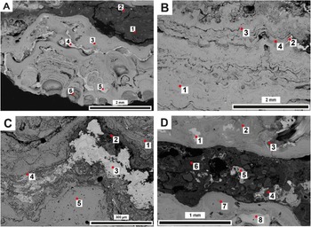
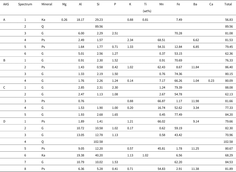
The composition of the goethite is heterogeneous because Fe shows substitution not only by Al but also by P, Si, Mn, Ba, and Ca, as detected by semi-quantitative EDS analysis (Table 3). The darker and lighter tones of gray in SEM images reflect the amount of substitution of Fe (Fig. 4), ranging from 34 to 57% in dark bands and generally less in light bands, from 21 to 41%. Where Fe replacement is greatest, Mn and Ba exhibit their greatest concentration (Fig. 4, Table 3).
(A–D) Semi-quantitative EDS sites (see results in Table 3) and SEM/SE of laminated ferruginous crust. Variation in isomorphic Fe substitution by other cations in the goethite is greater in the darker bands, as indicated by spectrum 6 in (A), 4 in (B), 2 and 4 in (C), and 3 in (D), and less in lighter bands, as indicated by spectrum 3 in (A), 1 and 3 in (B), 1 and 5 in (C), and 2 and 7 in (D). Mn, Ba, and Ca are greater in the dark bands than in the light bands of the goethite (see Table 3). Psilomelane is white in SEM/SE images; semi-quantitative analysis (see Table 3) indicates variations in its composition, with replacement of Mn by Al, K, Ba, Si, and Fe in almost all occurrences, and less so by P: (A) spectrum 5, (B) spectrum 2, (D) spectrum 8. Kaolinite is discontinuous and lenticular in SEM/SE here: (A) spectrum 1, (D) spectrum 6; semi-quantitative EDS analysis (see Table 3) shows intergrowth with iron oxides (~6–7%); it also indicates isomorphic substitution by Mg, K, and Ti (~1%). Quartz appears mixed with kaolinite in the ferruginous crust: (A) spectrum 2, (D) spectrum 4.

Semi-quantitative EDS data showing the doses of chemical elements present in minerals making up the laminated ferruginous crust (see Table 1). AAS = four areas analyzed in sample ALC11-05, as shown in Figure 4. Ka = kaolinite, Q = quartz, G = goethite, Ps = psilomelane. The values do not add up to 100% because EDS does not dose light elements (e.g., OH).

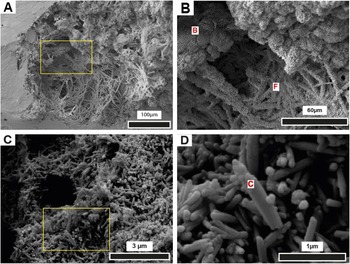
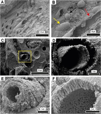
The smallest features observable in the goethite under high magnification in SEM are nanometric rounded equidimensional to rod-like bodies, rarely larger than 0.1 μm, here referred to as “nanorods” (Fig. 5A, B). In open spaces, they may form thin coatings on other structures (minerals and microbial remains) and may be aggregated into larger clumps, which themselves may form larger bunches (Fig. 5B). They also comprise the massive, compact groundmass or matrix of the ironstone (Figs. 6, 7A–D) that commonly hosts uncompacted vestiges of microbial remains, which are mostly clusters of tubular microbial filaments from <3 μm (Fig. 5C, D) to ~20 μm in diameter (Figs. 6, 7A–D). The tubes represent smooth hollow external molds and commonly contain cylindrical internal casts or molds. A very thin lining evident in some of the filament molds represents initial incrustation of the outer wall of sheaths prior to massive entombment by iron hydroxides (Figs. 6A, B, 7A). All filaments are simple, with no evidence of branching, and may occur gregariously in a meshwork (Fig. 5A), closely packed and parallel (Fig. 6A, B), or jumbled (Fig. 7A). Coccoid, barrel-shaped to rounded rod-like cells 5–20 μm long and <10 μm in diameter are less common, and most appear to occur within filaments. They have been preserved by delicate surficial incrustation or impregnation (permineralization) (Figs. 5A, 6A, B, 7A, B).
SEM/SE micrographs of ironstone crust showing filamentous (F), bunched (B), and cylindrical (C) features, ALC11-05. (A) Meshwork of filaments encrusted by individual to aggregated nanorods, originally HFO, now goethite; detailed (yellow rectangle) in (B). (B) Filaments (F) are continuous, straight to curved cylinders of similar diameter, lightly to massively incrusted by nanorods. Permineralized short, barrel-shaped cells appear in the filament just left of the bar scale at the bottom of the image. Bunches (B) of variously sized, rounded aggregates of nanorods in the upper half of the image thickly encrust the filaments and partially fill the open space. (C, D) Different forms of filament incrustation (circular void in C) by straight cylindrical goethite crystallites in various stages of growth (yellow rectangle), detailed in (D), from elsewhere in sample ALC11-05.

SEM/SE micrographs showing microbial structures preserved in the ferruginous crust, ALC11-05. (A, B) External filament/sheath molds (hollows), very thin initial external incrustation of filaments (red arrow), coccoid (black arrow) to bacillar (yellow arrow) cells amid and apparently within filaments, and thin cylindrical internal molds of filaments/sheaths (in the largest tube and broken tube in the upper left of B); (B) shows magnified section from central part of (A). (C, D) Additional external filament molds (hollows) and a second example (yellow rectangle) of a permineralized incipiently degraded wrinkled wall or sheath, detailed in (D). (E) Spiral structure in mineralized tube interpreted as permineralized incipiently degraded sheath or wall of the filament. (F) Internal filament mold formed by a 2 μm-thick centripetal incrustation of prismatic goethite. Compare this to the permineralized tube in (D).

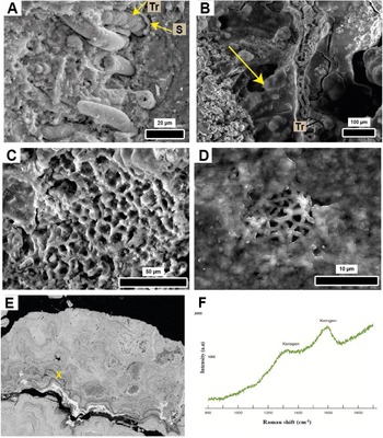
Trichome and biofilm morphologies (SEM/SE), and Raman evidence for kerogen in the ferruginous crust; ALC11-05. (A) Abundant solid to tubular internal casts of microbial filaments, as well as several permineralized cells of a trichome (Tr) surrounded by a fine, incrusted sheath (S). Cylindrical depressions in the matrix are external molds of filaments. (B) Permineralized amorphous mucilaginous biofilms (vertical at the center of the image, yellow arrow), originally EPS (extracellular polymeric substances), associated with tubular structures (filaments?) and a few permineralized cells of a trichome (Tr). (C, D) Enigmatic meshwork, possibly representing mucilaginous biofilm (EPS) once encasing coccoidal microbes (not preserved) in (C), and a network of very narrow filaments or cylindrical cells covered by a permineralized amorphous biofilm in (D). Neither structure is assuredly biogenic. (E) SEM/SE image showing point (X) analyzed by Raman. (F) Raman spectrum with peaks indicative of kerogen (organic matter) at 1350 and 1600 cm−1.

Once entombed, yet prior to decay, original organic walls and/or sheaths served as templates for internal incrustation that produced tubular to solid internal casts of empty sheaths. Kerogen has been identified by Raman spectroscopy within ironstone containing such features (Fig. 7E, F), although no original organic matter remains within the molds and casts. Fig. 6C–E shows mineralized concentric tubes that exhibit external wrinkles (Fig. 6C, D) and spiral structures (Fig. 6E), which suggest that the actual sheath or wall may have been impregnated (permineralized), rather than incrusted. This appears to be the case as well for smooth sheet-like structures interpreted as biofilms of amorphous extracellular polymeric substances (EPS), secreted by many kinds of microbes (Fig. 7B–D).
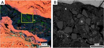
Detrital kaolinite and quartz are also found within the ferruginous crust. Nanometric kaolinite occurs in lenses (Figs. 3B, C, 4A, D, 8) containing up to ~6–7% iron hydroxides, as shown by quantitative EDS (Table 3, A-spectrum 1 and D-spectrum 6). Interestingly, kaolinite itself also presents isomorphic substitution by Mg, K, and Ti (~1%; Fig. 4A, D, Table 3). Angular to subangular quartz grains, 60–600 μm in size (Fig. 8B) occur scattered throughout the iron hydroxides or in lenticular bands with kaolinite/iron hydroxides (Reference Armstrong and BrasierFigs. 4A, D, 8).
SEM/SE/EDS showing ironstone crust mineralogy and mineral distribution and morphology; ALC11-05. (A) Mineralogical map (MLA): Fe hydroxide (pink) dominates; quartz (green) is scattered throughout the lens of kaolinite (dark gray) in the center; psilomelane (light blue) occupies pores in the goethite, detailed in (B) (yellow rectangle). (B) Anhedral and angular to subangular quartz grains (Q) within nanometric kaolinite (Ka).

(U-Th)/He geochronology
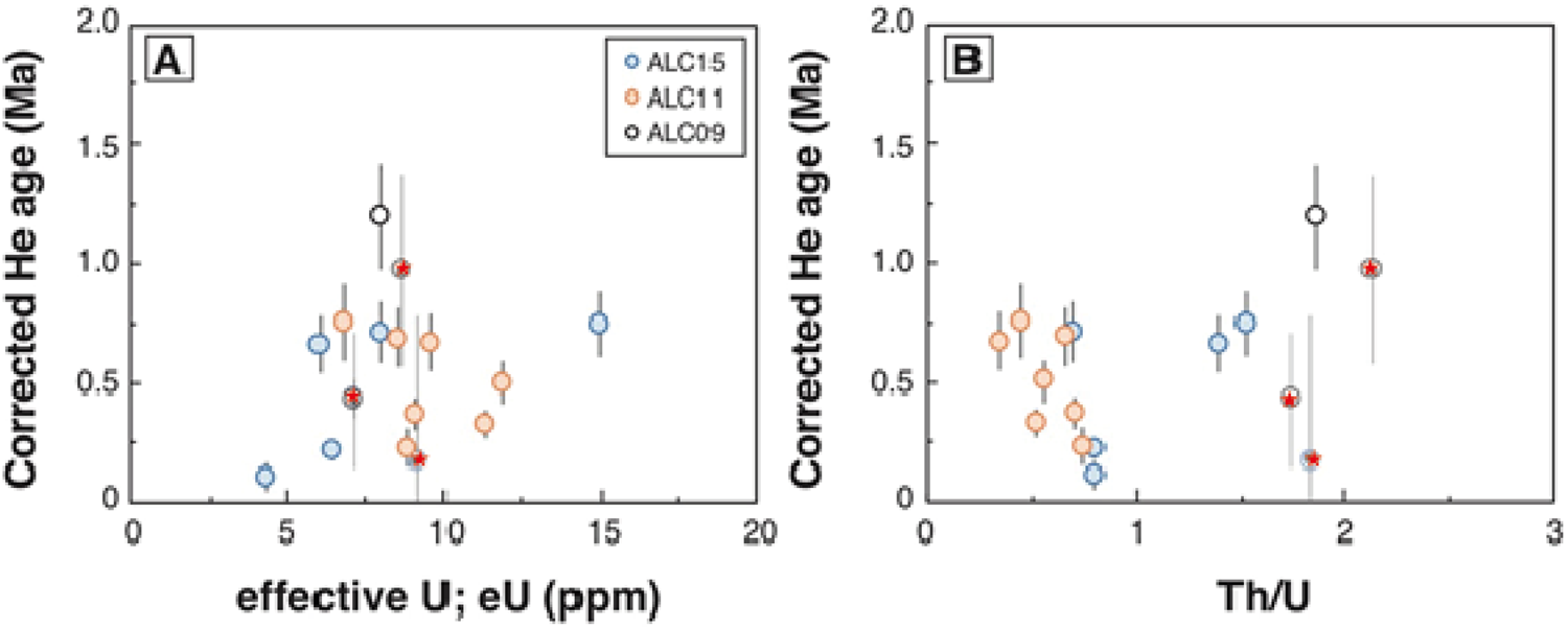
Table 2 presents the results of all (U-Th)/He age determinations. For some samples with very low He concentration, close to that of the blank, large errors are reported. The error for U-Th-Sm content is 1.5% for each concentration at 1σ. Corrected (U-Th)/He ages range between 0.1 ± 0.06 and 1.2 ± 0.2 Ma, with an effective U content (eU = U + 0.235Th) ranging from 4–15 ppm and Th/U ratio of 0.3–3.5 (Fig. 9). Three aliquots show large errors that reflect their very low He and low U-Th-Sm contents, and thus will not be considered in the following discussion. Sample ALC11, with an age of 0.8 ± 0.2 to 0.2 ± 0.1 Ma, presents a more homogeneous eU value and a smaller Th/U ratio than the other two samples (ALC09 and ALC15).
(U-Th)/He age evolution as a function of the effective uranium concentration (eU = U + 0.0234 × Th + 0.0046 × Sm) in (A) and Th/U ratio in (B). The points marked with stars have not been considered here because of large uncertainties associated with the very low U-Th-Sm and He content measured in the smallest aliquots.

The (U-Th)/He dating method, in the current analytical form, is not adapted to calculate precise age, because, for ages younger than 1.1 Ma, the U and Th series are not in secular equilibrium, leading to underestimation of the (U-Th)/He age (Farley, Reference Farley2002). To account for the possible error, we added an error of 20%. Thus, exact age values should be considered with caution, but they do indicate an age of <1 Ma.
DISCUSSION
Goethite is the principal component of both the ferruginous crusts and the very early-diagenetic cement that transformed fluvial sandstones and conglomerates into clastic ironstones. Goethite in the ferruginous crusts, however, occurs not as a cement, but rather as a rock-forming mineral, either a primary precipitate or an eodiagenetic alteration product of a precursor hydrous ferrous oxide (HFO) (Emerson and Weiss, Reference Emerson and Weiss2004). The extensive and varied isomorphic substitution of Fe by Al, Si, P, Mn, Ba, and Ca (Fig. 4, Table3) favors the latter hypothesis. All points analyzed exhibited substitution by Al, Si, and P, and, in ~80% of the points, by Mn as well. Although substitution of goethite by Al, Si, and Mn is common in supergene systems (Fysh and Clark, Reference Fysh and Clark1982; Schwertmann and Carlson, Reference Schwertmann and Carlson1994; Carlson, Reference Carlson1995; Deer et al., Reference Deer, Howie and Zussman2013), replacement in goethite by the other elements mentioned above is unusual (Schwertmann and Carlson, Reference Schwertmann and Carlson1994). Moreover, the elements most frequently substituting for Fe in the ironstone were Al, Si, P, and Mn, which, except for Mn, have also been observed in biogenic iron (oxyhydr)oxides (BIOs) (Whitaker et al., Reference Whitaker, Austin, Holden, Jones, Michel, Peak, Thompson and Duckworth2021, and references cited therein). Other BIOs associated with chemolithoautotrophic Fe-oxidizing bacteria (FeOB) are known from wetland environments at the surface and around roots in water-logged soils, as well as in some marine settings (Emerson et al., Reference Emerson, Fleming and McBeth2010; Johnson et al., Reference Johnson, Carmichael, McDonald, Rose, Pitchford, Windelspecht, Karatan and Brauer2012; Schmidt et al., Reference Schmidt, Sánchez, Fretschner, Kreps, Ferrero, Sineriz and Szewzyk2014).
Another important characteristic of the goethite in the Xingu ironstone is that it occurs as nanorods (Fig. 5A, B) about the size (~100 nm) of initial granules of amorphous ferric hydroxide (ferrihydrite) observed encrusting decaying bacteria in microbial mats from hydrothermal waters in Iceland (Konhauser and Ferris, Reference Konhauser and Ferris1996). Indeed, HFOs, including ferrihydrite, are very common, but unstable in surface environments, occurring exclusively in the form of nanocrystals (Cornell and Schwertmann, Reference Cornell and Schwertmann2003) that transform easily into more stable iron oxides and hydroxides, such as goethite and hematite (Konhauser, Reference Konhauser1998; Cornell and Schwertmann, Reference Cornell and Schwertmann2003; Chan et al., Reference Chan, Fakra, Edwards, Emerson and Banfield2009). We conclude, therefore, that the ironstone crusts are an early-diagenetic alteration product and probably pseudomorphic after ferrihydrite crystallites. Given its inherent instability (Emerson and Weiss, Reference Emerson and Weiss2004), it is not surprising that ferrihydrite was not identified in our analyses.
The most notable feature of the ferruginous crusts, as previously stated, is their wrinkled convex lamination, commonly exhibiting several orders of undulations (“undulations upon undulations”) and domed, columnar, and pseudo-columnar stacking morphologies (Fig. 2), which naturally invite comparisons with microbialites in the geological record, although similar morphologies also appear in abiogenic speleothems and tufas (Walter, Reference Walter1976; Salama et al., Reference Salama, El Aref and Gaupp2013; Grey and Awramik, Reference Grey and Awramik2020). The crusts show several other features common to microbialites, as well: (1) smooth mineralized amorphous films reminiscent of mucilaginous (EPS) biofilms secreted by microbial communities for protection and fixation to the substrate (Riding, Reference Riding2000; Fig. 7 B–D); (2) detrital quartz and kaolinite immersed in the ferruginous crust (Fig. 8), as if bound or entrapped within originally sticky microbial mats (Noffke et al., Reference Noffke, Gerdes, Klenke and Krumbein2007; Fig. 8); (3) the presence of kerogen in the ironstone (Fig. 7E, F), which is consistent with the findings of Sowers et al. (Reference Sowers, Holden, Coward and Sparks2019) that FeOB have a high capacity to adsorb dissolved organic matter; and (4) dense concentrations of narrow (1 to <20 μm in diameter), unbranched, smooth filamentous microbes in meshwork (Fig. 5A, B) and parallel (Fig. 6) or jumbled groupings (Fig. 7A), typical of most modern and ancient microbial mats and stromatolites (Knoll et al., Reference Knoll, Swett and Mark1991; Armstrong and Brasier, Reference Armstrong and Brasier2005; Gallardo et al., Reference Gallardo, Espinoza, Fonseca and Musleh2014; Williams et al., Reference Williams, Alpers, Sumner and Campbell2017).
Practically all filaments are preserved as uncompressed cylindrical outer molds (hollow tubes in the goethite), many of which contain narrower tubular to solid internal filament casts or molds (Figs. 6A, B, F, 7A), and a few exhibiting a very thin external incrustation (Fig. 6A, B). Outer molds show no evidence of decay or degradation prior to entombment. Cells apparently occupy some filaments (Fig. 6A, B), and internal casts (or petrifactions) of cells can be seen within trichomes (Fig. 7A, B).
Mucilaginous biofilms (Fig. 7B–D) and degraded and wrinkled sheath or wall remains (Fig. 6C–E) appear to have been preserved three dimensionally by impregnation (permineralization) (Schopf, Reference Schopf1975) rather than by surficial incrustation (compare with Fig. 6F). Clearly, most, if not all, microorganisms were quickly encrusted and entombed in the HFO prior to compaction or significant physical degradation, quite possibly while still alive. Early decay of sheaths and cells offered abundant sites for further rapid permineralization. On the other hand, the very fine external coating in some filaments (Fig. 6A, B) quite possibly was bioinduced by living filaments themselves, which is exactly what many FeOB do (Zajic, Reference Zajic1969; Emerson and Weiss, Reference Emerson and Weiss2004). Once formed, these crusts likewise may have acted as templates for additional precipitation. Following entombment, the inner sides of the organic walls/sheaths were lightly to heavily encrusted by HFO, presumably ferrihydrite, to produce molds and casts prior to final organic decomposition that left the microscopic hollows and open spaces that now demarcate the filaments in the ferruginous crust. A similar pattern of initial very thin incrustation of outer cell envelopes followed by later internal permineralization by centripetal mineral growth has been described by Levett et al. (Reference Levett, Gagen, Rintoul, Guagliardo, Diao, Vasconcelos and Southam2020) in a vein of goethite cutting saprolite in weathered banded iron formation in the Quadrilátero Ferrífero of Southeast Brazil. The internal coating (Fig. 6F), however, evidently has a different, probably later origin, because it consists of uniform prismatic goethite crystals, which are very different from the nanorod pseudomorphs after ferrihydrite.
Microbial mats occupied the surface of the bedrock and were rapidly entombed by HFO precipitation, potentially promoted by the microorganisms themselves, probably filamentous iron-bacteria, such as Leptothrix, Gallionella, Sphaerotilus, and others (Emerson et al., Reference Emerson, Fleming and McBeth2010; Schmidt et al., Reference Schmidt, Sánchez, Fretschner, Kreps, Ferrero, Sineriz and Szewzyk2014). These FeOB consist of smooth, generally straight simple cylindrical filaments with single rows (trichomes) of cells having rounded ends and very thin sheaths that may become densely impregnated with HFO during autotrophic or heterotrophic growth. Cells in Leptothrix easily can slip out of the ferric-hydrate-incrusted sheath and initiate new growth and sheath production. Some filamentous iron bacteria can produce more than their weight in ferric hydrate and form centimeter-thick mats in a few weeks (Zajic, Reference Zajic1969). Emerson and Weiss (Reference Emerson and Weiss2004) describe flocculent HFOs associated with FeOB that filled a shallow patch, 20 cm deep, in the Contrary Creek wetland (USA), that grew back within a month after being washed away. It is significant therefore that, although reaching greater diameters, the Xingu filaments share many features with filamentous FeOB, specifically: (1) morphology of filaments and associated cells; (2) very thinly encrusted sheaths; (3) abundant undeformed abandoned filaments; and (4) faithful three-dimensional preservation of filaments and cells, possibly the consequence of incrustation in vivo. Hence, this suggests that the filamentous organisms in the Xingu ironstone crusts played an important role in HFO precipitation. Thus, it is reasonable to consider the Xingu crusts as ferruginous microbialites—after all, microbialites are known to occur in ironstones and lithologies other than carbonates (Walter, Reference Walter1976; Schieber and Glamoclija, Reference Schieber, Glamoclija, Schieber, Bose, Eriksson, Banerjee, Sarkar, Altermann and Catuneau2007; Preston et al., Reference Preston, Shuster, Fernandez-Remolar, Banerjee, Osinski and Southam2011; Salama et al., Reference Salama, El Aref and Gaupp2013).
Despite the low availability of dissolved Fe2+ in the water of Amazon rivers, including the Xingu (Silva et al., Reference Silva, Ríos-Villamizar, Miranda, Ferreira, Bringel, Gomes, Silva, Pascoaloto, Santana and Cunha2019), the circumneutral pH and oxygenated water of the middle Xingu fluvial system (Camargo et al., Reference Camargo, Giarrizzo and Isaac2004) would have been favorable for development of FeOB, even in the face of a challenging food supply (Emerson et al., Reference Emerson, Fleming and McBeth2010), in the seasonally isolated ponds that occur along the Volta Grande do Xingu. Goethite age data, based on the (U-Th)/He method, varies from 1.2 ± 0.2 to 0.10 ± 0.06 Ma (Fig. 9), which we interpret as very close to the time of precipitation of the primary HFOs and their very early alteration to goethite. This signifies that crust formation took place sometime since the Early Pleistocene.
Testing the hypothesis that the Xingu ironstone crusts are FeOB microbialites will require determining the relative amounts and relationships of microbial remains and the groundmass of the ironstones, as well the roles and significance of biological versus physical-chemical parameters in the origin of the oddly iterative lamination that characterizes the ironstone crust from the scale of a few microns to decimeters. If a relationship with climate can be shown, then the Xingu ironstone crusts may offer new insights into the Quaternary climate in the Amazon Basin.
CONCLUSIONS
Ferruginous crusts containing abundant microbial molds and casts along the Xingu River in the Amazon are here interpreted as ferruginous microbialites that developed in seasonally isolated ponds since the Early Pleistocene (<1.2 Ma). This is the first such occurrence documented in South America. The ferruginous crusts became lithified by very early transformation of bioinduced/bioinfluenced unstable HFO crystallites into goethite nanorods. Future study of the micrometer-to-decimeter lamination so striking in these microbialites may help decipher the Quaternary history of climate, sedimentation, and ecology of the Xingu River and Amazon Basin.
Acknowledgments
F. Haurine is thanked for help with goethite dissolution and ICP-MS analysis at GEOPS, France; C. Rodriguez, R. Damati, and T. Silva for illustrations; and E. Silva, F. Rodrigues, F. Machado, I. Sayeg, and R. Contessotto for laboratory support.
Financial Support
This research was supported by the São Paulo Research Foundation (FAPESP, grants 2016/02656-2, 2018/23899-2, 2018/15613-1, and 2018/15123-4) and the Brazilian Federal Agency for Support and Evaluation of Graduate Education (CAPES, grant AUXPE 2043/2014). MPF was awarded a Master's scholarship. AOS and GAH are supported by the National Council for Scientific and Technological Development (CNPq, grants 304727/2017-2 and 306527/2017-0). DJB acknowledges the support of FAPESP grants 2019/24977-0, 2018/15123-4, 2019/24349-9, and 2016/11141-2. (U-Th-Sm)/He analysis was funded by the Agence Nationale de la Recherche (France) Project 17-CE01-0012-01, “Reconstructing the influence of Climate change on Laterite formation.”