It is very difficult for both clinicians and morphologists to visualise and appreciate the anatomic features of Ebstein’s malformation in three dimensions. In the past, cross-sectional echocardiography has provided limited insights into the functional anatomy of the valvar leaflets, their tension apparatus, and the accompanying abnormalitiesReference Oechslin, Buchholz and Jenni1, Reference Seward2. According to conventional wisdom, the fundamental abnormality in Ebstein’s malformation is downward displacement of the hinge points of the leaflets of the tricuspid valveReference Becker and Anderson3, Reference Gasul, Arcilla and Lev4. More recent studies have shown this concept to be inaccurate. Because the leaflets of the tricuspid valve have failed to delaminate from the ventricular walls during ventricular developmentReference Kanani, Moorman and Cook5, their hinges, rather than being downwardly displaced, are rotated around the aortic rootReference Vettukattil, Bharucha and Anderson6. This feature, in turn, results in the effective valvar orifice being located at the junction of the inlet and apical trabecular components of the right ventricle, rather than being hinged at the atrioventricular junctionReference Schreiber, Cook, Ho, Augustin and Anderson7.
Tricuspid valvar dysplasia is often confused with Ebstein’s malformation, with the different prognosis of the two conditions likely contributing to the varying outcomes reported for patients reputedly having Ebstein’s malformation. Very often those with exclusively valvar dysplasia have larger right ventricular volumes, which are reduced in Ebstein’s malformation because of the atrialisation of the inlet ventricular component. Such problems of appreciation of the true morphology of Ebstein’s malformation, and its diverse functional characteristics, have now been resolved with the use of three-dimensional echocardiography. The purpose of our study was to investigate the ability of the multiplanar review modality of three-dimensional echocardiography to assess the dynamic morphology of malformed tricuspid valves, showing how this information serves to differentiate Ebstein’s malformation from exclusive tricuspid valvar dysplasia.
Methods
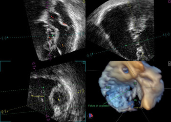
Approval was obtained from the local research ethics committee. We included in our study patients who had previously been diagnosed with Ebstein’s malformation by a paediatric cardiologist using cross-sectional echocardiography. All patients had concordant atrioventricular and ventriculo-arterial connections. Three-dimensional echocardiographic data was obtained with patients unsedated and spontaneously breathing. A commercial real-time three-dimensional imaging system (Philips Sonos 7500 or IE33, Philips Co, Netherlands), interfaced with a 3-5 MHZ matrix phased array Transducer, was used to acquire three-dimensional data sets. The three-dimensional data was then analyzed offline by two independent observers in the multiplanar review mode of commercially available analysis software Philips Q lab (version 5.0) and Tom Tec ultrasound data analysis (image arena version 3.0 Build 3.04.48, Tom Tec imaging systems, Germany). Analysis by multiplanar review allows the operator to cut the moving three-dimensional data sets in three orthogonal planes, each plane being moveable independently to allow examination of the entire data set. The three planes were positioned as demonstrated in Figure 1 to allow the operator to focus on the tricuspid valve, and each plane was then moved through the data set to allow examination of each valvar leaflet, its zones of coaptation with its neighbours, and the subcordal apparatus.
Multiplanar review of Ebstein’s malformation of the tricuspid valve. The left upper panel shows the heart in its long axis. The mitral and aortic valve are visualised in the same section in long axis, while the tricuspid valve is seen in short axis. The left lower panel shows the heart in short axis, with a small left ventricle and a large atrialised portion of right atrium. The right upper panel shows the heart in the long axis two chamber section. The septal leaflet of tricuspid valve is rotated along the axis of the aorta, with concomitant displacement of the mural leaflet. The effective valvar orifice is orientated towards the right ventricular outflow tract. The bottom right panel displays the reconstructed three-dimensional view, showing the failure of coaptation between the 3 leaflets of the tricuspid valve. Abbreviations: Ao: aorta; TV: tricuspid valve; LV: left ventricle; AML: anterior mitral leaflet; ASL: antero-superior leaflet, SL: septal leaflet, ML: mural leaflet.

Displacement of tricuspid valve
The valvar hinge points were carefully delineated by reference to all three planes, permitting measurement of the maximum distance from the crux of the heart to the hinge of the septal leaflet. The extent of valvar displacement was then calculated relative to the hinge of the mitral valve as previously described by Ports and colleaguesReference Ports, Silverman and Schiller8, as one minus the ratio of the tricuspid septal leaflet-to-apex length divided by the mitral valve leaflet-to-apex length.
Rotation of tricuspid valve
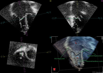
Rotation of the valvar hinge points was assessed as illustrated in Figure 2. In multiplanar review, the true long axis of the heart was identified by positioning the two longitudinal planes parallel to the atrial and ventricular septums. In this view, in a normal heart, the tricuspid and mitral valves are both seen in their long axes. Rotation of the hinges of the tricuspid valvar leaflets around the aortic root was accepted when the tricuspid valve was seen partially or completely in short axis in this view, or in the left ventricular outlet view, with the mitral valve remaining in long axis.
Multiplanar review of tricuspid valvar dysplasia. The left upper panel shows a four chamber view. There is a large right atrium and right ventricle. Both the mitral and tricuspid valves are seen in their long axis. There is apparent displacement of the septal leaflet due to enlargement of the right atrium. The left lower panel shows the short axis of both ventricles. The right ventricle is enlarged, and the tricuspid valve was seen in its short axis, confirming the absence of any rotational abnormality. The right upper panel shows a two chamber view, with the orifice of the tricuspid valve orientated towards the apex. The right lower panel is a 3 dimensional reconstruction view, illustrating the apparent displacement of septal leaflet along with the abnormal subcordal apparatus. Abbreviations: ASL: antero-superior leaflet; RA: right atrium; RV: right ventricle.

The direction of effective valvar orifice was determined by following this feature on the long axis plane when the short axis plane was moved anteriorly, bisecting the outflow tract of the right ventricle. Other morphological characteristics evaluated included abnormalities in delamination, subcordal apparatus, effective right ventricular volume, and presence or absence of pulmonary stenosis. The effective length of the right ventricle was measured from the point of separation of the leaflet to the ventricular apex, parallel to the ventricular septum, and compared to the measurement for the left ventricle.
Based on these features, patients were re-assigned to a diagnosis of either tricuspid valvar dysplasia or Ebstein’s malformation. Patients were considered to have Ebstein’s malformation if they had rotation of the hinge points around the aortic root in association with any of the other abnormalities, and considered to have exclusively tricuspid valvar dysplasia if they lacked rotation of the valvar hinge points.
Results
We examined 23 unselected patients, who presented sequentially to our service. The patients were aged from 1 day to 70 years, with a median age of 23 years 10 months. The quality of the images was sufficient to allow review by means of multiplanar analysis in all patients, although respiratory artefacts prevented further detailed subanalysis in some. Multiplanar review allowed detailed viewing of each tricuspid valvar leaflet, along with characterisation of the functional characteristics and the mechanism of failure of coaptation, if present, in every patient. Using the features of rotational abnormality, and the site of the effective valvar orifice, as criterions for diagnosis, we reassigned 12 patients (52%) to a diagnosis of tricuspid dysplasia.
To date, 10 patients have been submitted to surgery, 4 with tricuspid valvar dysplasia, and 6 with confirmed Ebstein’s malformation. The echocardiographic findings regarding valvar displacement, rotation, abnormalities of delamination, and abnormal morphology of the leaflets, were compared with immediate post-operative documentation of intra-operative findings by the surgeon. In all patients, the surgical findings correlated well with three-dimensional multiplanar review findings.
Inconsistent values were noted for tricuspid displacement when measured by 3 independent investigators, although all measurements were made using the standard method described at the crux of the heart.
Abnormalities in delamination were seen in 10 of 11 patients with tricuspid valvar dysplasia, and 11 of 12 with Ebstein’s malformation, while problems with the subcordal apparatus were identified in 6 of 11 with valvar dysplasia and 6 of 12 with Ebstein’s malformation. In one patient with Ebstein’s malformation, it proved difficult to visualize the effective valvar orifice, an off-axis anatomical plane being required to define the orientation of the tricuspid valve.
We showed that the volume of the right ventricle was greater in those with tricuspid valvar dysplasia, with mean ratio of ventricular lengths of 0.76, compared to a ratio of 0.63 for those with Ebstein’s malformation. Pulmonary stenosis was present in 5 patients, 3 (27%) with tricuspid valvar dysplasia, and 2 (17%) of those having Ebstein’s malformation.
Discussion
Our investigation shows that multiplanar review of three-dimensional data sets gives specific details regarding the functional anatomy of patients with Ebstein’s malformation when compared with findings obtained using cross-sectional echocardiography. Multiplanar review also has advantages over traditional “cropping” of three-dimensional data sets when displaying anatomy in three-dimensional views. During multiplanar review, the data set is viewed in three planes simultaneously, which allows continuous cross-referencing of the focused structure to the other planes. This allows precise and clear delineation of individual valvar leaflets and their cordal apparatus, rather than viewing them in solitary planes, when they may be hidden or overlain by other structures. The operator is able to position each plane so as to view the tricuspid valve in attitudinally appropriate manner, and with reference to the rest of the cardiac anatomy. In this way, the morphology of the individual leaflets, and their planes of coaptation, can be visualized without losing sight of their anatomical relationships to the remaining intracardiac structures.
Using the multiplanar review, we have shown that patients having exclusively tricuspid valvar dysplasia can readily be distinguished from those with Ebstein’s malformation, when the latter is defined on the basis of the rotational anomaly shown in recent morphological studiesReference Martinez, O’Leary and Anderson9. When using this criterion, we found that almost half of those diagnosed initially with cross-sectional echocardiography as having Ebstein’s malformation, in reality had exclusively valvar dysplasia. Our group of patients was unselected, and therefore likely to constitute a representative cross-section of patients with this diagnosis. This differentiation based on the rotational anomaly is a key factor not only for diagnosis, but also for surgical intervention. When downward displacement of the valvar leaflets had been used as the criterion for diagnosis, we found significant overlap between those having exclusive dysplasia and those with rotated valvar orifices, with the measurement of reputed downward displacement producing significant differences between observers. It is possible that this is the consequence of multiple adhesions secondary to abnormal delamination, adhesions being mistaken as insertions of the septal leaflet. Using the multiplanar review, such attachments were clearly delineated. Severe dilation of the right atrium, especially in the absence of an associated atrial septal defect, may cause stretching of the atrioventricular junction, also leading to the apparent displacement of the hinge points of the dysplastic tricuspid valve.
In tricuspid valvar dysplasia, the functional right ventricular cavity is larger, and the effective valvar orifice is directed towards the apex of the right ventricle, in contrast to a small right ventricular cavity in Ebstein’s malformation, with the opening of the valve directed towards the outflow tract.
Our study is limited by its retrospective nature, and the patients were selected in a nonrandomised fashion. We conclude, nonetheless, that multiplanar review provides increased detail of the anatomy and dynamic function of the abnormal tricuspid valve when compared to cross-sectional echocardiography, providing clear visualisation and understanding of all the features of Ebstein’s malformation.