I. INTRODUCTION
Based on a series of scientific studies and clinical practice in the past years, cardiovascular stent intervention has emerged as the most effective therapy for cardiovascular diseases.Reference Sheng, Liu, Min, Wang, Liu, Wang, Huang, Wu and Hou1 However, the present commercial stent materials, such as stainless steel, polylactic acid, and cobalt chromium alloy, may cause serious coagulation, inflammations, and hyperplasia after implantation and further lead to restenosis, which may require reoperation and endanger patients’lives.Reference Ramcharitar and Serruys2,Reference Serruys, Kutryk and Ong3 Thus, developing preferable biodegradable stents with better biocompatibility is an issue that needs to be addressed immediately.Reference Im, Jung and Kim4,Reference Zheng and Yang5 Magnesium, as an essential element in the human body, participates in normal physiological functions, metabolism, and maintaining cell viability, while its biodegradable and absorbable properties may play a crucial role in stent designing aiming at self-degradation after regeneration and repair of vascular intima.Reference Wacker and Vallee6,Reference Lu, Fan, Liu, Cao, Wu and Xu7 Yet mechanical properties of pure magnesium cannot meet the needs of the blocked blood vessels that require long-term support.Reference Galvin, Cummins, Yoshihara, Mac Donald and Lally8 Therefore, magnesium alloys have been developed with enhanced mechanical properties to develop new-generation biodegradable cardiovascular stents.Reference Li and Zheng9 Magnesium alloys with potential application in designing cardiovascular stents include Mg–Zn–Mn alloy,Reference Wang, Jang, Wan, Giridharan, Song, Xu, Koo, Qi, Sankar, Huang and Yun10 Mg–Zn–Y–Nd alloy,Reference Wu, Zhu, Wang, Liu, Yue, Wang and Guan11 Mg–Nd–Zn–Zr alloy,Reference Shi, Zhang, Chen, Zhang, Yuan, Shen, Chen, Pei, Li, Tan and Yuan12 Mg–Y–RE (Gd, Nd)–Zr alloy (WE43), and Mg–Al–Zn–Mn alloy (AZ31),Reference Lukyanova, Anisimova, Martynenko, Kiselevsky, Dobatkin and Estrin13,Reference Adekanmbi, Mosher, Lu, Riehle, Kubba and Tanner14 among which Mg–Zn–Y–Nd alloy has been reported widely for its superior performance, such as slower degradation ratio, better mechanical properties, and lower biological toxicity.Reference Feng, Liu, Cao, Yang, Xu and Guan15,Reference Hou, Zhang, Guan, Ren, Gao, Lu and Cui16 Mg–Zn–Y–Nd alloy also shows good blood compatibility and anti-inflammation function after fine surface processing.Reference Liu, Wang, Chu and Xi17 Nevertheless, many of its properties still need to be further improved to satisfy the requirements of cardiovascular stent designing, such as antihyperplasia functions, preferable degradation ratio, and pro-endothelialization.
Surface modification with functional coatings is recognized as an effective method to enhance the properties of stent materials, wherein immobilization of specific molecules is used to improve the materials’biocompatibility,Reference Strohbach and Busch18,Reference Li, Zhang and Huang19 which usually depends on the surface enrichment of conjugated groups, such as amine group and/or carboxyl group.Reference Li20 Polydopamine film (PDA) is well known for its mussel-inspired functions, i.e., its outstanding conjugation with metals and other molecules, which benefits from its complex reaction with metals and abundant amine groups.Reference Zhang, Bai, Wang, Li, Guan and Li21 Several studies also demonstrated that PDA possesses properties to promote vascular endothelial cells’ growth and that this property has obvious contributions to the surface endothelialization of cardiovascular stent materials.Reference Su, Xue, Ye, Wang, Zhao, Huang and Li22 The synthetic peptide sequence Arg-Glu-Asp-Val (REDV) is known for its specific recognition by integrin α4β1 which is abundant in endothelial cells and scarce in other cells.Reference Wei, Ji, Xiao, Lin and Ji23,Reference Zhang, Li, Wang, Liu, Wang, Chen, Huang and Guan24 Thus, immobilizing REDV molecules on the PDA-deposited Mg–Zn–Y–Nd substrate via covalent reaction between its carboxyl group and PDA’s amine group is a preferable strategy to improve surface endothelialization.
In this study, we deposited polydopamine film (PDA) on the finely processed Mg–Zn–Y–Nd surface and then immobilized REDV molecules via covalent reaction. The modified layers were labeled as PDA/REDV-1, PDA/REDV-2, and PDA/REDV-3, respectively (with the initial concentrations of REDV being 0.5 mg/mL, 1 mg/mL, and 2 mg/mL, respectively). Surface morphology and roughness of the PDA/REDV layers were studied using atomic force microscopy (AFM), and their wettability was examined with the water contact angle measurement. Dynamic blood test and static fibrinogen denaturation were performed to evaluate their blood compatibility. Endothelial cells, smooth muscle cells, and macrophages were cultured on the surfaces to investigate the cell compatibility of PDA/REDV layers.
II. MATERIALS AND METHOD
A. Conjugating REDV molecules on PDA-deposited Mg–Zn–Y–Nd
Mg–Zn–Y–Nd alloy smelted in our laboratory were cut into small disks with 10 mm diameter and 3 mm thickness and then subtly polished with metallographic abrasive papers of 100#, 200#, 400#, 600#, 800#, and 1000#, successively. Then, the disks were sonicated sequentially in acetone, ethanol, and deionized water (dH2O) and finally dried at room temperature. Clear Mg–Zn–Y–Nd disks were immersed in 2 mg/mL dopamine (PDA) solution (dissolved in pH 8.5 Tris buffer), deposited for 24 h at 25 °C, and then rinsed with dH2O for three times (5 min per time), and the samples were labeled as PDA.Reference Bai, Zhang, Xu, Liu, Guan, Liu, Chen and Li25 Next, PDA samples were immersed in activated Arg-Glu-Asp-Val (REDV, Shanghai Feather Flower Biological Science and Technology Co., Ltd., Shanghai, China) peptide solutions of various initial concentrations (0.5, 1.0, and 2.0 mg/mL in normal saline, pH 3.6, 2.7, and 2.5) at 25 °C for 6 h and then subsequently rinsed with dH2O for three times to remove the unconjugated REDV.Reference Li, Wu, Zhang, He, Zou, Luo, Fan, Yang, Zhao and Huang26 The samples were labeled as PDA/REDV-1, PDA/REDV-2, and PDA/REDV-3, respectively. The fabrication process of the PDA/REDV coating is shown in Fig. 1.
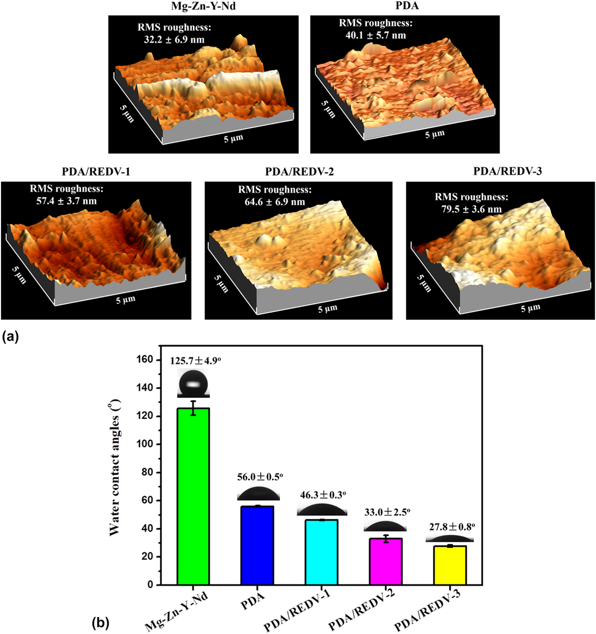
(a) AFM images and (b) water contact angles of the Mg–Zn–Y–Nd, PDA, PDA/REDV-1, PDA/REDV-2, and PDA/REDV-3 samples (mean ± SD, n = 6).

B. Surface characterization of PDA/REDV coating
Morphology and roughness of the PDA/REDV-1, PDA/REDV-2, PDA/REDV-3, PDA, and Mg–Zn–Y–Nd samples were observed by an atomic force microscope (AFM; Key Sight 7500, Santa Rosa, California) in tapping mode.Reference Zhang, Wang, Guan, Li and Li27 Their surface wettability was assessed by water contact angle measurement (DSA 100, Krüss GmbH, Hamburg, Germany) at room temperature followed by image processing of a sessile drop of 5 μL test ultrapure water with DSA 1.8 software; at least six contact angles in different areas were measured and averaged.Reference He, Li, Luo, Zou, Yang, Zhao and Huang28 The number of coatings of REDV conjugated on PDA/REDV was determined using acid orange II (AO II) colorimetric method.Reference Wu, Li, Zhang, He, Yang, Zou and Huang29 Electrochemical tests were carried out using a classical three-electrode cell, with a platinum rod as the auxiliary electrode, a saturated calomel electrode as the reference electrode, and the samples as the working electrode. Simulated body fluid (SBF) was prepared according to previous studies.Reference Chen, Li, Chang, Jin, Wu, Yan, Wang and Guan30 The electrolyte was buffered at pH 7.4 using Tris-hydroxymethylaminomethane ((HOCH2)3CNH2) and hydrochloric acid (HCl) and maintained at 37 °C. The sample area exposed to the solution was 1 cm2. Measurements were carried out at a scan rate of 0.5 mV/s using an electrochemical station, and the sample was kept in the solution for 1 h before polarization tests to establish the open circuit potential.Reference Chen, Li, Chang, Jin, Wu, Yan, Wang and Guan30
C. Blood compatibility test
To investigate the blood compatibility of PDA/REDV coatings, a dynamic blood test was performed for each sample by placing them in a 24-well culture plate and culturing on the shaking table in vitro at 37 °C for 1 h. Then, the samples were taken out, washed with normal saline (NS, pH = 7.4, 3 times, 5 min), fixed with 2.5% glutaraldehyde solution, dehydrated at increasing alcohol concentrations (50%, 75%, 90%, 100%;  ${{{V_{{\rm{alcohol}}}}} / {{V_{{\rm{d}}{{\rm{H}}_{\rm{2}}}{\rm{O}}}}}}$), and dealcoholized at increasing isoamylacetate concentrations (50%, 75%, 90%, 100%; V isoamyl acetate/V alcohol). All the samples were dried with critical point drying (CPD030, BALZERS) and coated with gold to undergo SEM in order to evaluate their morphology. The number of adherent platelets and activated platelets was examined using our previous method.Reference Li, Zhang, Xu, Chen, Yang, Zhao, Zhao and Huang31
Fibrinogen denaturation of the samples exposes their γ chain and makes them bind to the GPIIb/IIIa receptor on platelet membrane, further causing platelet aggregation.Reference Li, Yang, Qin, Maitz, Zhou and Huang32 Thus, the fibrinogen absorption and denaturation on the surface is generally accepted as a crucial evaluation for the blood compatibility of the material.Reference Li, Zhang, Wu, Zhang, Yang, Tu and Huang33 In this study, fresh whole blood provided by the Affiliated Hospital of Zhengzhou University was centrifuged at 3000 rpm to obtain platelet-poor plasma (PPP). Onto the surface of each sample, 50 μL PPP was added, followed by incubation for 1 h at 37 °C. Subsequently, 1% bovine serum albumin (BSA, Sigma, Ontario, Canada) was added onto the surface for 0.5 h at 37 °C after removing the remaining PPP and rinsing with NS for three times (5 min each time). After repeated rinsing, 50 μL HRP-conjugated goat anti-human fibrinogen absorption/denaturation antibody (diluted 1:2.5 in NS) was added onto each surface, followed by incubation at 37 °C for 1 h. The samples were washed three times again with NS. Further, 100 μL TMB solution was added onto the sample surface and allowed to react in the dark for 10 min, and then 50 μL 1 M H2SO4 (stop solution) was used to stop the peroxidase-catalyzed reaction. Finally, 140 μL supernatant was transferred to a 96-well plate, and the absorbance at 450 nm was measured on a microplate reader.Reference Li, Zhang, Wu, Zhang, Yang, Tu and Huang33
D. Culture of endothelial cells
Human umbilical vein endothelial cells (HUVEC, ECV304, Oligobio Co., Ltd., Beijing, China) of between third and fifth generations were cultured in a humidified incubator with 95% air and 5% CO2. The PDA/REDV-1, PDA/REDV-2, PDA/REDV-3, PDA, and Mg–Zn–Y–Nd samples were kept in a 24-well culture plate, and HUVEC were seeded onto the samples with a concentration of 5 × 10⁴ cells/ml and then cultured at 37 °C for 24 h. Then, a cell-permeable acridine orange (AO) in combination with a plasma-membrane-impermeable DNA-binding dye propidium iodide (PI) was used to detect apoptosis or necrosis of HUVEC. AO and PI excite green and red fluorescence, respectively, when they are intercalated into DNA. Only AO but not PI can cross the plasma membrane of a normal cell. Late apoptotic and necrotic cells take up the two dyes and show a predominant orange fluorescence. The cells were stained with a 1:1 mixture of AO (100 mg/mL) and PI (100 mg/mL) at 37 °C for 5 min and then immediately inspected in a fluorescence microscope (DMRX, Leica, Wetzlar, Germany). Criteria for identification are the following: (i) green intact nucleus, vital cells; (ii) dense green areas of chromatin condensation in the nucleus, early apoptosis; (iii) dense orange areas of chromatin condensation, late apoptosis; and (iv) orange intact nucleus, secondary necrosis.Reference Li, Zhang, Chen, Liu, Yang, Zhao and Huang34 The number of HUVEC on each sample was detected via CCK-8 method,Reference Xiang, Li, He, Wu, Yang and Huang35 and the functional factor, nitric oxide (NO) released from HUVEC on each sample, was measured by a typical ELISA.Reference Li, Li, Zhang, Liao, Yang, Maitz and Huang36
E. Culture of smooth muscle cells
Human umbilical arterial smooth muscle cells (HUASMC) were purchased from Haoyi Biotechnology Co., Ltd., of Chengdu in China and cultured in the standard condition elaborated above. HUASMC between second and fifth generations were seeded on the PDA/REDV-1, PDA/REDV-2, PDA/REDV-3, PDA, and Mg–Zn–Y–Nd samples with a concentration of 5 × 10⁵ cells/ml and cultured at 37 °C for 24 h. Then they were observed under fluorescence microscope after fixing step and stained with 4,6-diamino-2-phenyl indole (DAPI, Sigma).Reference Li, Qin, Zhang, Wu, Yang, He, Zhao and Huang37 The number of HUASMC was also examined by a typical CCK-8 assay.Reference Xiang, Li, He, Wu, Yang and Huang35
F. Attachment of macrophages
Peritoneal macrophages of SD rats (Haoyi Biotechnology Co., Ltd., Chengdu, China) were also cultured in the standard culture condition, were added into 24-well culture plates with a concentration of 5 × 10⁵ cells/ml, and cultured with DMEM medium (each sample immersed for 24 h) at 37 °C for 24 h. To study the cell adhesion and activation, fluorescence staining of the attached macrophages was performed using DAPI, and the number of cells on each sample was statistically counted from 15 random pictures.Reference Li, Xu, Zhou, Chen, Yang, Yang, Li and Huang38 Inflammatory factors (TNF-α and IL-6) released from the macrophages were also detected using typical ELISA methods.Reference Li, Zhang, Ma, Wu, Yang, He and Huang39
G. Statistical analysis
Mean values ± SD are displayed with their representative images. Statistical significance requires a p-value <0.05.
III. RESULTS AND DISCUSSION
A. Surface characterization
Figure 1(a) shows the surface morphology and roughness changes on the PDA/REDV-1, PDA/REDV-2, PDA/REDV-3, PDA, and Mg–Zn–Y–Nd samples. Mg–Zn–Y–Nd substrate showed an obvious smoother surface with lower roughness (32.2 ± 6.9 nm) because of the polishing process. Roughness increased slightly (40.1 ± 5.7 nm) after PDA preparation for 24 h. After REDV conjugation, the samples exhibited significantly rougher surfaces compared with both pure PDA surfaces and Mg–Zn–Y–Nd substrate, and roughness increased with the initial concentrations, indicating the following trend of roughness: Mg–Zn–Y–Nd < PDA < PDA/REDV-1 < PDA/REDV-2 < PDA/REDV-3. This phenomenon may be considered as an indirect evidence for REDV conjugation onto the PDA-deposited Mg–Zn–Y–Nd, and conjugation levels on PDA/REDV-1, PDA/REDV-2, and PDA/REDV-3 were different.
Surface wettability is a fatal property that affects the biological functions of the biomedical materials, and several studies indicated that surfaces with the water contact angles ranging from 10° to 90° may contribute better cell adhesion and spreading.Reference Yao, Li, Li, Wang, Li and Wang40 The present study [Fig. 1(b)] showed that Mg–Zn–Y–Nd presents a very high water contact angle (125.7 ± 4.9°), indicating a superhydrophobic surface, which is not prone to protein adsorption and endothelial cell growth, while PDA deposition significantly reduced the water contact angles and made it to a hydrophilic range (56.0 ± 0.5°), which may contribute to the adhesion and growth of endothelial cells. PDA/REDV coatings further increased the surface hydrophilicity, and the water contact angles decreased with the initial concentrations increasing: PDA/REDV-1 (46.3 ± 0.3°) > PDA/REDV-2 (33.0 ± 2.5°) > PDA/REDV-3 (27.8 ± 0.8°). Hydrophilicity of the PDA/REDV coatings showed positive correlation with their roughness, which is also an indirect proof for different REDV densities on each surface.
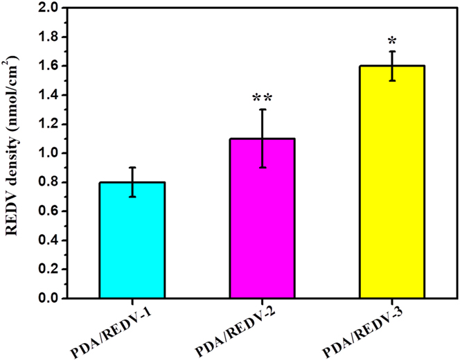
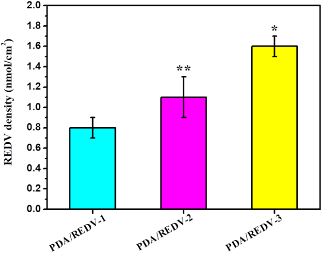
REDV density on the surface may control the specific adhesion and growth of the endothelial cells and may also influence the growth of other cells, even the blood compatibility.Reference Zhang, Li, Wang, Liu, Wang, Chen, Huang and Guan24 Figure 2 shows that the initial concentrations of the REDV solutions displayed positive correlation with the conjugated REDV density on the surface: PDA/REDV-1 < PDA/REDV-2 < PDA/REDV-3. In addition to Fig. 1, results indicated that a higher REDV density may lead to a rougher and more hydrophilic surface.
Conjugated REDV density on each sample (*p < 0.05 compared with PDA/REDV-1 and PDA/REDV-2, **p < 0.05 compared with PDA/REDV-1, mean ± SD, n = 3).
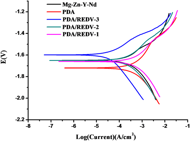
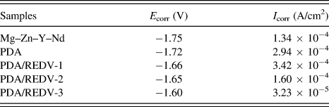
Corrosion resistance is a vital property for the modified coatings of the biomedical magnesium alloy, and the typical method to investigate the samples’ corrosion resistance is the electrochemical test.Reference Feng, Zhu, Wang, Chang, Yan, Song and Guan41 Figure 3 presents the polarization curves of the PDA/REDV-1, PDA/REDV-2, PDA/REDV-3, PDA, and Mg–Zn–Y–Nd samples in SBF. Corrosion potential (E corr) and corrosion current density (I corr) are summarized in Table I. A previous study indicated that the samples’ corrosion resistance was determined by their corrosion current density and resistance.Reference Chen, Li, Chang, Jin, Wu, Yan, Wang and Guan30 From Fig. 3 and Table I, PDA/REDV-3 coating obviously improved the corrosion resistance of the Mg–Zn–Y–Nd substrate, with the corrosion current density decreasing from 1.34 × 10⁻⁴ to 3.23 × 10⁻⁵ A/cm2.
Polarization curves of the PDA/REDV-1, PDA/REDV-2, PDA/REDV-3, PDA, and Mg–Zn–Y–Nd samples in SBF.

Values from the polarization curve of different samples in SBF.

B. Blood compatibility
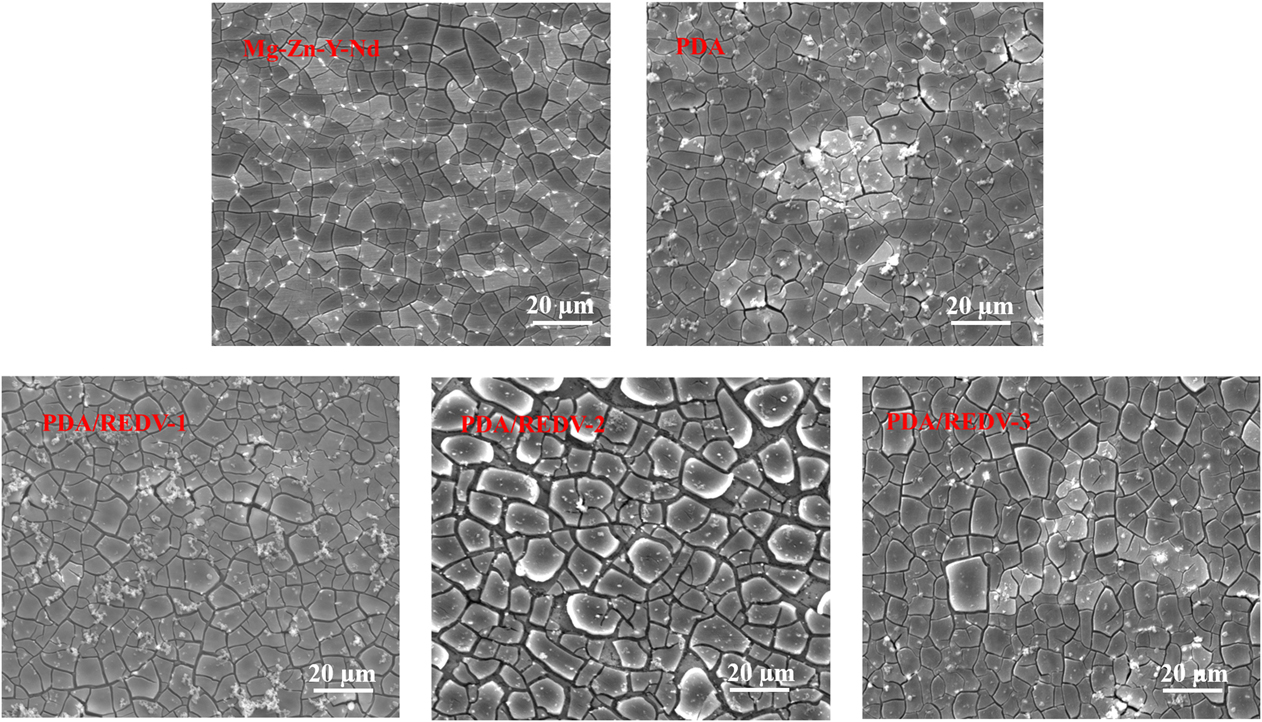
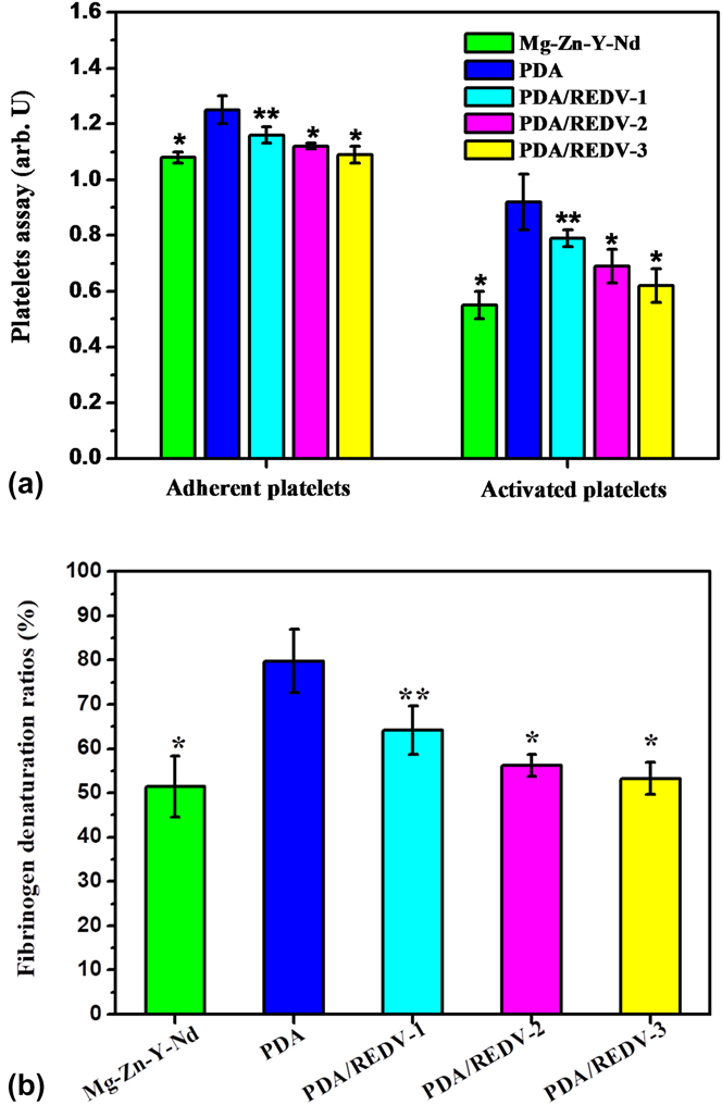
Blood flow contacts the stent material’s surface that may lead to the adhesion of the blood components, such as platelets and fibrinogen, which may cause coagulation and further restenosis.Reference Xu, Li, Yao, Li, Yang and Huang42 Thus, we conducted an in vitro dynamic blood test to evaluate the blood compatibility of the PDA/REDV coatings and the control samples. As shown in the SEM images of the adherent blood components in Fig. 4, fine polishing obviously reduced the number of platelets and the fibrinogen level in the Mg–Zn–Y–Nd substrate compared with a previous study,Reference Chen, Li, Chang, Jin, Wu, Yan, Wang and Guan30 suggesting improved blood compatibility. However, PDA film deposition increased the number of adherent platelets and the fibrinogen level, and the platelets on the PDA stretched their pseudopods and presented spreading morphology, indicating an activated phenotype. This phenomenon may be attributable to the rich amino group and unique “double-sided” adhesive properties of PDA. The PDA/REDV-1 surface showed a markedly lower platelet count and fibrinogen level than the PDA surface, but still a little more than Mg–Zn–Y–Nd, and platelets on the PDA/REDV-1 surface began to present a round or crumpled morphology, which indicated that they were regulated to the inactive phenotype. With the increasing level of the conjugated REDV, the PDA/REDV surface (PDA/REDV-2 and PDA/REDV-3) showed fewer platelets and a lower fibrinogen level, and more platelets presented the inactive phenotype. The quantitative characterization of adherent/activated platelets by LDH/GMP140 methods presented a consistent trend: Mg–Zn–Y–Nd, PDA/REDV-2, and PDA/REDV-3 < PDA/REDV-1 < PDA [Fig. 5(a)]. As a crucial evaluation of the surface blood compatibility, fibrinogen denaturation, which leads to platelet adhesion and aggregation, also showed a consistent trend with platelet adhesion/activation: Mg–Zn–Y–Nd, PDA/REDV-2, and PDA/REDV-3 < PDA/REDV-1 < PDA [Fig. 5(b)]. All these results demonstrated that PDA/REDV-2 and PDA/REDV-3 could maintain good blood compatibility of the fine polished Mg–Zn–Y–Nd substrate.
SEM images of adherent blood components (platelets and fibrinogen) on the PDA/REDV-1, PDA/REDV-2, PDA/REDV-3, PDA, and Mg–Zn–Y–Nd samples.

(a) Quantitative characterization of adherent (LDH) and activated (GMP140) platelets on the PDA/REDV-1, PDA/REDV-2, PDA/REDV-3, PDA, and Mg–Zn–Y–Nd samples (mean ± SD, *p < 0.05 compared with other samples, n = 3); (b) fibrinogen denaturation ratios of the PDA/REDV-1, PDA/REDV-2, PDA/REDV-3, PDA, and Mg–Zn–Y–Nd samples (mean ± SD, *p < 0.05 compared with other samples, **p < 0.05 compared with PDA, n = 3).

C. Growth of vascular endothelial cells
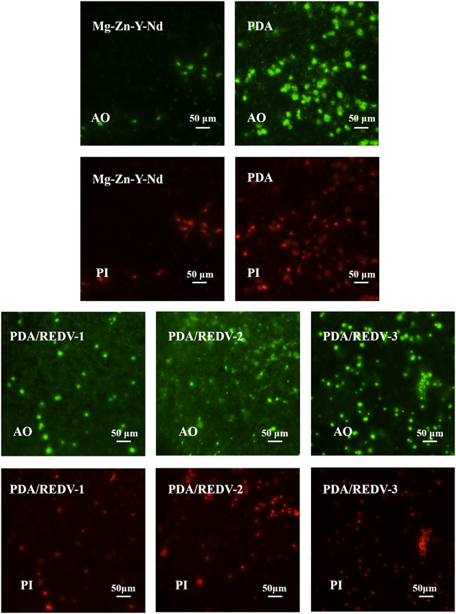
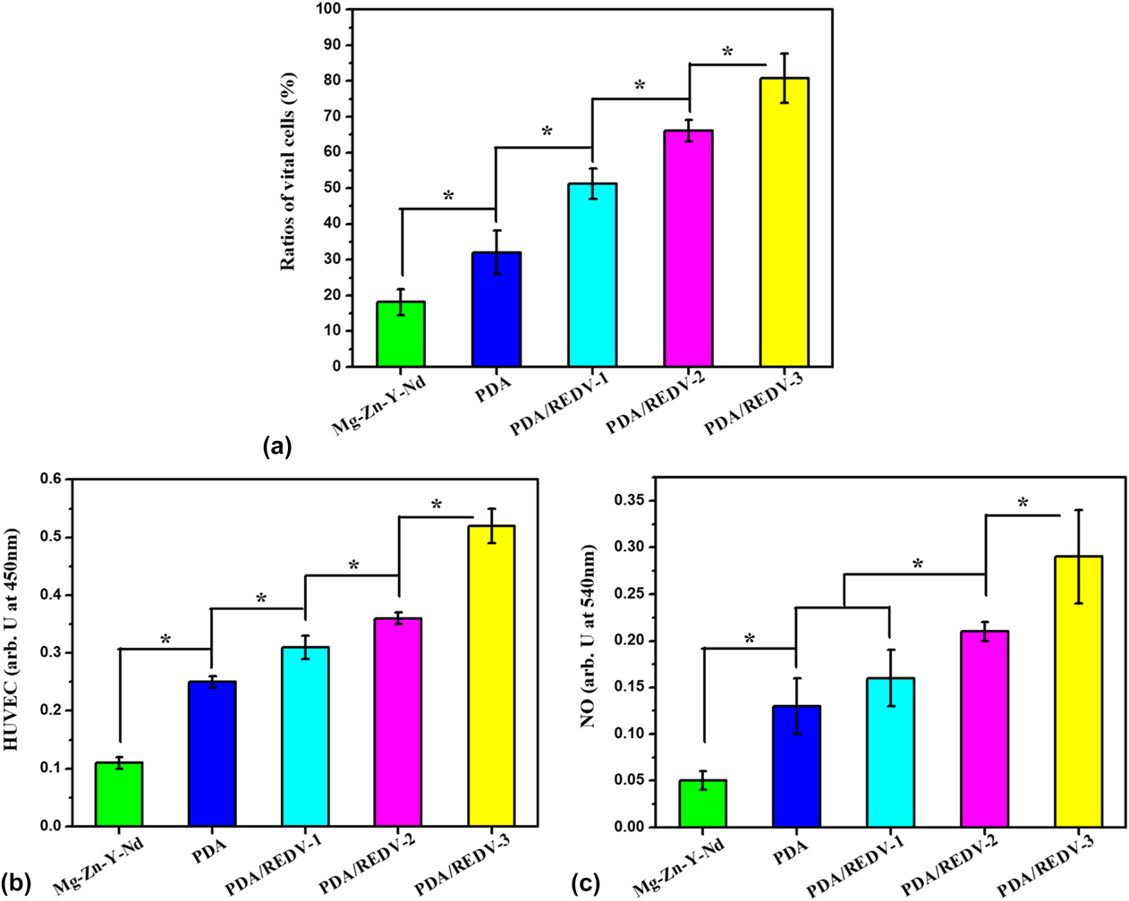
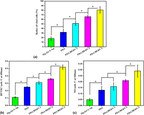
REDV has been widely reported for its adhesion to vascular endothelial cells as a modified molecule on nondegradable materials.Reference Wei, Ji, Xiao, Lin and Ji23,Reference Wei, Ji, Xiao, Lin, Xu, Ren and Ji43 However, REDV modification on Mg–Zn–Y–Nd presented a quantity-dependent phenomenon of HUVEC growth: From the fluorescence images of Fig. 6, PDA preparation obviously enhanced the number of HUVEC on the Mg–Zn–Y–Nd surface, while the number of HUVEC decreased on the PDA/REDV-1 and PDA/REDV-2 surfaces and increased again on the PDA/REDV-3 surface, which indicated that HUVEC growth on the PDA/REDV surfaces depends on the level of conjugated REDV. A new discovery was that conjugated REDV enhanced the vital ratios of HUVEC on the PDA-coated Mg–Zn–Y–Nd substrate, and this result also showed a quantity-dependent rule, i.e., a higher REDV density on the PDA/REDV coatings presented higher vital ratios of HUVEC: PDA/REDV-3 > PDA/REDV-2 > PDA/REDV-1 > PDA > Mg–Zn–Y–Nd [Fig. 7(a)]. CCK-8 assay showed a consistent result with the vital ratio calculation, which indicated that the REDV density also made the HUVEC cytoactive and further regulated their function. Then, NO as a functional factor (antiplatelets and antihyperplasia) of endothelial cells was detected, and the results showed that a higher REDV density contributed to more NO release. All the HUVEC cultures indicated that PDA/REDV-3 coatings possessed better ability to improve surface endothelialization.
HUVEC growth and viability on the PDA/REDV-1, PDA/REDV-2, PDA/REDV-3, PDA, and Mg–Zn–Y–Nd samples (green intact nucleus: vital cells; dense green areas of chromatin condensation in the nucleus: early apoptosis; dense orange areas of chromatin condensation: late apoptosis; orange intact nucleus: secondary necrosis).

(a) Ratios of vital HUVEC grown on the PDA/REDV-1, PDA/REDV-2, PDA/REDV-3, PDA, and Mg–Zn–Y–Nd samples, which were calculated from at least 15 images (*p < 0.05, mean ± SD, n = 15); (b) CCK-8 detection and (c) NO release of HUVEC on the PDA/REDV-1, PDA/REDV-2, PDA/REDV-3, PDA, and Mg–Zn–Y–Nd samples (*p < 0.05, mean ± SD, n = 3).

D. Growth of vascular smooth muscle cells
Damaged vessel wall during stent intervention will trigger the pathological proliferation and migration of smooth muscle cells from blood vessel media onto the surface of the stent, and this will lead to late hyperplasia and increase the lumen loss.Reference Li, Zhang, Yang, Wu, Chen, Zhao, Li and Huang44 Therefore, we investigated the number of HUASMC on each surface to evaluate the antihyperplasia ability of the REDV-coated Mg–Zn–Y–Nd material. DAPI staining images (Fig. S1) and CCK-8 results (Fig. S2) of HUASMC indicated that Mg–Zn–Y–Nd presented a lower number of HUASMC, compared with PDA, PDA/REDV-1, and PDA/REDV-2, while the PDA/REDV-3 surface exhibited fewer HUASMC compared with all the other samples, suggesting better antihyperplasia function. More HUASMC may be due to the degradation of the Mg–Zn–Y–Nd material, and the better corrosion resistance of PDA/REDV-3 shown in Fig. 4 and Table I may be the main reason of its better function in inhibiting HUASMC growth.
E. Adhesion and activation of macrophages
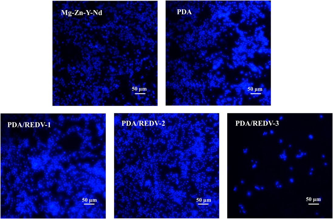
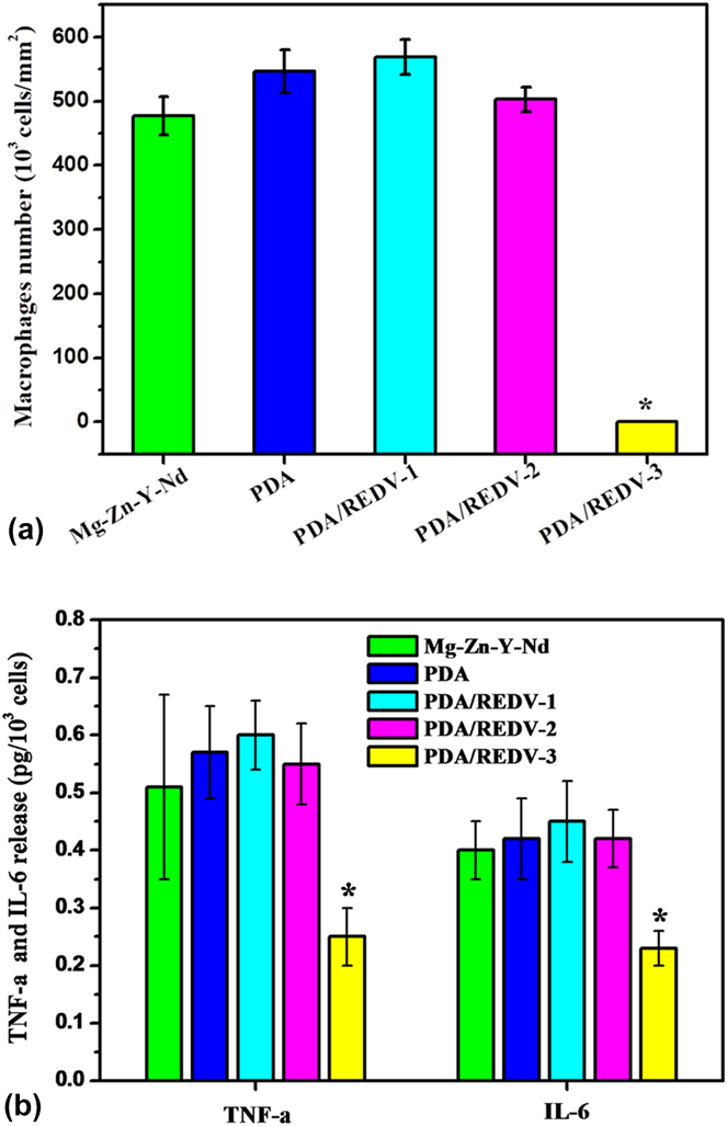
Macrophages play a crucial role during the inflammatory response after stent implantation.Reference Li, Zou, Zhang, Luo, Yang, Jing, Zhang, Cui and Huang45 Fluorescence images and counting of macrophages cultured by the DMEM culture medium immersed by the PDA/REDV-1, PDA/REDV-2, PDA/REDV-3, PDA, and Mg–Zn–Y–Nd samples are shown in Figs. 8 and 9(a). Both Figs. 8 and 9(a) show that the modification of the PDA, PDA/REDV-1, and PDA/REDV-2 significantly increased the number of attached macrophages on Mg–Zn–Y–Nd, while the modification of PDA/REDV-3 significantly reduced the number of attached macrophages on Mg–Zn–Y–Nd, and the latter presented a lower number of macrophages compared with all the other samples. The trend was consistent with the number of HUASMC: PDA/REDV-3 < Mg–Zn–Y–Nd < PDA, PDA/REDV-1, and PDA/REDV-2. Numerous macrophages in Mg–Zn–Y–Nd, PDA, PDA/REDV-1, and PDA/REDV-2 groups are significantly correlated with the excessive hydrogen enrichment and corrosion products from the degradation process, and PDA/REDV-3 coating possessed better corrosion resistance (displayed in Fig. 3 and Table I) which may prevent excessive hydrogen and corrosion products’ release.
Fluorescence images of macrophages stained with DAPI on the samples of PDA/REDV-1, PDA/REDV-2, PDA/REDV-3, PDA, and Mg–Zn–Y–Nd, respectively (blue dots were the macrophage nucleus stained with DAPI).

(a) Macrophage count of PDA/REDV-1, PDA/REDV-2, PDA/REDV-3, PDA, and Mg–Zn–Y–Nd groups, respectively (mean ± SD, *p < 0.05 compared to other samples, n = 3); (b) TNF-α and IL-6 release from macrophages of PDA/REDV-1, PDA/REDV-2, PDA/REDV-3, PDA, and Mg–Zn–Y–Nd groups, respectively (*p < 0.05 compared with all the other samples, mean ± SD, n = 3).

When the inflammation response happened, adherent and activated macrophages on the implants will release inflammatory factors, such as TNF-α and IL-6, which may aggravate the immune-related pathological process.Reference Li, Zou, Zhang, Luo, Yang, Jing, Zhang, Cui and Huang45 In this study, TNF-α and IL-6 released from the macrophages on the PDA/REDV-1, PDA/REDV-2, PDA/REDV-3, PDA, and Mg–Zn–Y–Nd samples were measured by specific kits. Figure 9(b) shows that macrophages on the PDA/REDV-3 coatings released significantly less TNF-α and IL-6 compared with the other samples, suggesting a suppressing effect on macrophage activation and better anti-inflammation function of the PDA/REDV-3 coating, and this function may also benefit from the coating’s better corrosion resistance.
IV. CONCLUSION
In this study, Mg–Zn–Y–Nd alloy substrate was coated with polydopamine (film) and then modified with Arg-Glu-Asp-Val (REDV) peptide in series of solutions (0.5 mg/mL, 1.0 mg/mL, and 2.0 mg/mL). Characterization of AFM, water contact angle measurement, and REDV quantity proved useful in the successful preparation of three PDA/REDV coatings (PDA/REDV-1: 0.5 mg/mL, PDA/REDV-2: 1.0 mg/mL, and PDA/REDV-3: 2.0 mg/mL). Polarization curves suggested that the PDA/REDV-3 coating had better corrosion resistance compared with the other samples. Dynamic blood test and static platelet/fibrinogen tests demonstrated the better blood compatibility of the Mg–Zn–Y–Nd and the PDA/REDV-3 coatings. HUVEC culture experiment showed that the all the PDA/REDV and PDA coatings significantly promoted HUVEC growth on the Mg–Zn–Y–Nd surface, wherein the PDA/REDV-3 coating possessed better pro-endothelial function. HUASMC culture experiment indicated that the PDA/REDV-3 coating inhibited smooth muscle cell growth, suggesting a strong antihyperplasia function. Macrophage culture results presented a consistent trend that the PDA/REDV-3 coating inhibited macrophage attachment and activated TNF-α and IL-6 release, suggesting a better anti-inflammation function. All the results revealed that this PDA/REDV modification with specific parameters could markedly improve surface biocompatibility, and this study may possess potential application for the surface modification of degradable magnesium alloys as cardiovascular biomaterial implants.
Supplementary Material
To view supplementary material for this article, please visit https://doi.org/10.1557/jmr.2018.410.
ACKNOWLEDGMENTS
This study was funded by the National Key Research and Development Program of China (Grant Nos. 2017YFGX090043-04 and 2016YFC1102403), Fostering Talents of National Natural Science Foundation of China and Henan Province (No. U1504310), Key Scientific Research Projects of Higher Education Institutions in Henan Province (No. 16A430030), Key Project and Special Foundation of Research, Development and Promotion in Henan Province (No. 182102310076), Young Teachers Foundation of Zhengzhou University (No. 32210475), and the Joint Fund for Fostering Talents of NCIR-MMT & HNKL-MMT (No. MMT2017-01).