INTRODUCTION
In the mammalian host, schistosomes encounter both endogenous and exogenous oxidative environments capable of damaging invading worms. However, investigations of adult Schistosoma mansoni worms have revealed the presence of a variety of potentially protective antioxidant-related molecules (Mkoji et al. Reference Mkoji, Smith and Prichard1988; Nare et al. Reference Nare, Smith and Prichard1990; Mei and LoVerde, Reference Mei and LoVerde1997), including a unique linked thioredoxin-glutathione redox system responsible for detoxification of oxidative radicals (Alger and Williams, Reference Alger and Williams2002; Salinas et al. Reference Salinas, Selkirk, Chalar, Maizels and Fernandez2004). Two key molecules in this pathway, peroxiredoxin (Prx) and thioredoxin (Trx) are involved in thiol-mediated hydroperoxide metabolism (Alger et al. Reference Alger, Sayed, Stadecker and Williams2002; Sayed and Williams, Reference Sayed and Williams2004). Both have been detected in adult S. mansoni and in egg and cercarial secretions (Kwatia et al. Reference Kwatia, Botkin and Williams2000; Williams et al. Reference Williams, Asahi, Botkin and Stadecker2001; Alger et al. Reference Alger, Sayed, Stadecker and Williams2002; Knudsen et al. Reference Knudsen, Medzihradszky, Lim, Hansell and McKerrow2005) suggesting a role for these enzymes in providing protection against parasite- or host-derived reactive oxygen species (ROS), especially hydrogen peroxide.
The capacity of the intramolluscan sporocyst stage of Schistosoma mansoni to establish a successful infection within its snail host, Biomphalaria spp., also depends on its ability to survive within a potentially hostile oxidative environment (Bayne et al. Reference Bayne, Hahn and Bender2001). The immune response of B. glabrata against recognized trematode larvae is characterized by the rapid encapsulation of sporocysts by multiple layers of circulating haemocytes resulting in the destruction of larvae usually within 24–48 h post-infection (Bayne et al. Reference Bayne, Buckley and DeWan1980; Sullivan and Richards, Reference Sullivan and Richards1981; Loker et al. Reference Loker, Bayne, Buckley and Kruse1982). Under in vitro conditions, the production of ROS, mainly hydrogen peroxide (H2O2) and nitric oxide (NO), by encapsulating haemocytes appears to be responsible for larval killing (Hahn et al. Reference Hahn, Bender and Bayne2001a, Reference Hahn, Bender and Bayneb). In addition to ROS of haemocyte origin, haemoglobin-containing plasma of B. glabrata, the initial host tissue to contact the invading miracidium, also may contribute to the oxidative stress placed on developing larvae (Bender et al. Reference Bender, Bixler, Lerner and Bayne2002). In response to these sources of potential oxidative damage, it has been proposed that S. mansoni produces protective antioxidant molecules that function to counteract the potentially damaging oxidative products generated through the parasite's own metabolism or by the snail host (Bayne et al. Reference Bayne, Hahn and Bender2001).
Previous studies have shown that S. mansoni excretory-secretory products (ESP) released from larvae during the miracidium-to-mother sporocyst transformation can inhibit superoxide production in snail haemocytes (Connors and Yoshino, Reference Connors and Yoshino1990), suggesting that larval products may provide a measure of protection from exogenous sources of oxidative stress. However, in contrast to the parasite stages found in the mammalian host, little is known regarding the expression of specific antioxidant proteins during early mother sporocyst development or their role in the snail-sporocyst relationship. Therefore, the current study was undertaken to (1) determine the mRNA expression levels of redox pathway members in larval S. mansoni during in vitro miracidium-to-mother sporocyst transformation and subsequent early sporocyst development, (2) explore the role of H2O2 in regulating antioxidant gene expression, (3) immunolocalize native Prx proteins within developing larvae, and (4) functionally characterize the metabolic/detoxifying activities that S. mansoni Prxs present in larval ESP.
MATERIALS AND METHODS
Parasite isolation and culture
Schistosoma mansoni (NMRI strain) eggs were recovered from the livers of mice at 7–8 weeks post-infection as previously described (Yoshino and Laursen, Reference Yoshino and Laursen1995). Briefly, miracidia were hatched from the eggs in sterile artificial pond water and concentrated on ice in conical polypropylene centrifuge tubes. Hatched miracidia were axenically transformed into primary sporocysts in 24-well plates and cultured at 26°C in Chernin's balanced salt solution (CBSS; Chernin, Reference Chernin1963) containing antibiotics and 1 mg/ml each of glucose and trehalose (CBSS+) for 24 h. Culture supernatants containing parasite ES products (ESP) were collected 24 h post-hatching, filtered with a 0·22 μm syringe filter (Amicon, Danvers, MA), and stored on ice until used in experiments (<2 h). For Western blotting only, ESP was concentrated 3-fold by filter centrifugation using Ultra-4 centrifugal filter units (Amicon) with a 5000 Da nominal molecular weight cut-off.
Real-time quantitative PCR (qPCR) analyses of antioxidant mRNA transcripts
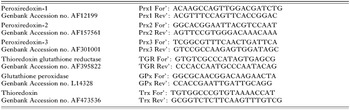
In these experiments selected antioxidant mRNA transcript levels were quantified via qPCR in cDNA populations obtained from in vitro-developing mother sporocysts cultured in CBSS+. First strand cDNA synthesis from sporocyst total RNA and subsequent qPCR amplification procedures were performed as previously described (Vermeire et al. Reference Vermeire, Taft, Hoffmann, Fitzpatrick and Yoshino2006) using a GeneAmp 7300 PCR apparatus (Applied Biosystems). The specific genes targeted and the primer sequences employed in this quantitative comparison are summarized in Table 1. Quantitative PCR data were analysed using the comparative CT method (Livak and Schmittgen, Reference Livak and Schmittgen2001) as described by Boyle et al. (Reference Boyle, Wu, Shoemaker and Yoshino2003). This method normalizes expression levels of experimental genes using a housekeeping gene, in our study, 18S ribosomal RNA (rRNA). P-values were calculated by comparing ΔCt values (N=3) for each gene using a Student's t-test order to determine if an mRNA transcript was significantly differentially expressed between the 2 developmental stages. Fold differences were calculated using the 2−ΔΔCt method (Livak and Schmittgen, Reference Livak and Schmittgen2001) to express the delta delta Ct (ΔΔCt) values in a manner suitable for graphical representation.
Primer sequences of Schistosoma mansoni antioxidant genes assayed using real-time quantitative PCR

Transcriptional activation of S. mansoni peroxiredoxins (Prxs) in response to oxidative stress
In order to determine the effect of oxidative stress on the transcriptional activation of Prx genes in in vitro-developing mother sporocysts the following experiments were performed. One-day-old mother sporocysts were obtained as described above, washed twice in CBSS+ and plated in 48-well plates at a density of approximately 3000 sporocysts per well in a total volume of 250 μl. The parasites were then exposed to a low, non-lethal level of exogenous H2O2 (10 μm; Mkoji et al. Reference Mkoji, Smith and Prichard1988; Hahn et al. Reference Hahn, Bender and Bayne2001a) and parasite populations were sampled at 0, 2, 4, and 24 h post-exposure to H2O2, and at 1 h post re-exposure to H2O2 to determine the effects of treatment on Prx mRNA transcript abundance using qPCR as described above. In order to calculate P values for H2O2-treated and control groups, delta Ct values (N=3) were compared using a one-way ANOVA and Tukey's honest post-test.
Immunolocalization of S. mansoni peroxiredoxin proteins
Localization of immunoreactive Prx was accomplished using monoclonal antibodies (mAbs) specific for Prx1, Prx2 or Prx3 kindly provided by A. Sayed and D. Williams (Sayed and Williams, Reference Sayed and Williams2004). S. mansoni miracidia and 2-day-old in vitro-developing mother sporocysts were fixed in 4% paraformaldehyde (PFA) in PBS for 1 h at 4°C using end-over-end rotation. Larvae were then washed extensively in PBS, followed by simultaneous blocking and permeabilization with 5% BSA containing 0·5% Triton X-100 (Sigma) in PBS under constant mixing at 4°C. Parasites were incubated in anti-Prx-1, -2, and -3 mAbs at a 1:100 dilution for 3 days at 4°C under constant agitation, followed by 2 washes in PBS for 2 h each and incubation in fluorescein-conjugated goat anti-mouse IgG (1:500; Promega Corp., Madison, WI) for 2 days at 4°C under constant agitation. Miracidia and sporocysts were subjected to 2 final washes (3 h each) in PBS before microscopical examination. Immunofluorescent images were collected using an inverted epifluorescent microscope (Nikon Eclipse TE 300, Melville, NY) equipped with a Synsys digital camera connected to a personal computer running Metamorph® Imaging System software version 4.12 (Universal Imaging Corporation, West Chester, PA). Confocal microscopy (Zeiss Axiovert 200M) also was performed to confirm the localization of Prx reactivity in sporocysts (Biological and Biomaterials Preparation, Imaging, and Characterization Facility, University of Wisconsin, Madison).
Western blotting of S. mansoni peroxiredoxins (Prxs)
The presence of Prx proteins (Prx1, Prx2 and Prx3) in larval ESP was assessed by SDS-PAGE using standard Western blot techniques (Sambrook et al. Reference Sambrook, Fritsch and Maniatis1989). ESP-containing transformation supernatants, derived as described above, were mixed with SDS-PAGE lysis buffer, boiled for 10 min in a water bath under reducing conditions and separated on 10% polyacrylamide gels using a Mini-PROTEAN II (Bio-Rad, Hercules, CA) apparatus. Proteins were transferred to PVDF membranes using a Hoefer TE 70 semi-dry transfer apparatus (Amersham Biosciences, Piscataway, NJ), followed by blocking with 5% bovine serum albumin (BSA) in Tris-buffered saline (TBS; pH 7·4) and incubating in the anti-Prx mAbs (1:2500) overnight at 4°C. After extensive washing with TBS, the membranes were incubated in alkaline phosphatase-conjugated goat-anti-mouse IgG secondary antibody (1:10 000). Colorometric detection of immunoreactivity was carried out using 5-bromo-4-chloro-3-indolyl phosphate (BCIP) and nitro-blue tetrazolium (NBT).
Immunoprecipitation of Prx1 and Prx2 from ESP-containing transformation supernatants
Prx1 and Prx2 proteins were removed from ESP-containing supernatants using specific anti-Prx monoclonal antibodies in order to create immunoabsorbed supernatants for use in H2O2 metabolism assays (described below). Briefly, 5 μl of both Prx1 and Prx2 mAbs were added to 1 ml of transformation supernatants and mixed by inversion for 2 h at 4°C on a rotating platform. Fifty microlitres of Protein G-conjugated Sepharose® 4B Fast Flow beads (Sigma-Aldrich, St Louis, MO) were washed with CBSS following the manufacturer's instructions and added to the supernatants. The tubes were then mixed by inversion for another 1 h at 4°C. Finally, the Protein G-sepharose bead-mAb-Prx protein complexes were removed from solution through low speed centrifugation and Western blotting used to assess the presence of Prx1/Prx2 in immunoabsorbed and unabsorbed ESP supernatants.
H2O2 metabolism by S. mansoni excretory-secretory products (ESPs)
The ability of ESP-containing transformation supernatants collected 24 h post-hatching to metabolize H2O2 was assessed using the Amplex® Red hydrogen peroxide/peroxidase assay kit (Molecular Probes, Eugene, OR) as described by Bender et al. (Reference Bender, Broderick, Goodall and Bayne2005). ESP was incubated with 10 μm H2O2 at 22°C in 96-well plates (100 μl reaction volume) and monitored over a time-course of 30 min at 5-min intervals using a Biotek Synergy™HT Multi-detection microplate reader (Biotek Instruments, Winooski, VT) to determine (1) if ES products were capable of metabolizing H2O2in vitro and (2) whether Prx1 and Prx2 contained in ESP were responsible for this activity. This horseradish peroxidase-dependent, fluorometric method uses the reagent 10-acetyl-3, 7-dihydroxyphenoxazine, which reacts with H2O2 to produce the red-fluorescent oxidation product, resorufin, in order to detect H2O2 in solution (530/590 excitation/emission wavelengths). Standard curves with values ranging from 0 to 20 μm H2O2 were produced by calculating the average fluorescence units (FU) of triplicate runs for each experiment. Control reactions without ESP (CBSS+ only) were run at each time-point to determine the rate of spontaneous breakdown of H2O2 in solution. Control values were subtracted from the FU of experimental time-points yielding the actual amount of H2O2 being metabolized by proteins in the ESP-containing supernatants. Prx1/Prx2 immunoabsorbed and unabsorbed ESP supernatants were then evaluated for their H2O2-scavenging activities in the Amplex® Red hydrogen peroxide assay.
RESULTS
Developmental regulation of mRNA abundance of antioxidant genes
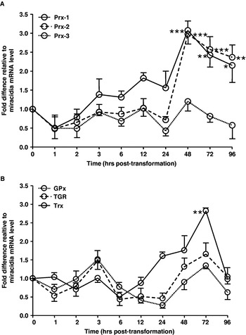
Parasite antioxidant transcripts, especially the peroxiredoxins, are expressed in a stage-associated manner during early larval development (Fig. 1). Mother sporocyst mRNA populations contained significantly more transcripts for Prx1 at 48 h (∼3-fold, P⩽0·001), 72 h (∼2·5-fold, P⩽0·01) and 96 h (∼2·25-fold, P⩽0·05) post-transformation when compared with miracidial mRNA levels (Fig. 1A). Prx2 mRNA levels also were significantly higher at 48 h (∼3-fold, P⩽0·001), 72 h (∼2·5-fold, P⩽0·001) and 96 h (∼2·5-fold, P⩽0·01) when compared to miracidial Prx2 transcripts levels. In contrast, Prx3 mRNA levels did not differ significantly between miracidia and sporocysts at any of the time-points (24, 48, 72 or 96 h) post-miracidial transformation. Also, thioredoxin (Trx) mRNA transcript abundance was significantly higher at 72 h (∼2·75-fold, P⩽0·01) for developing mother sporocysts when compared to Trx mRNA levels in miracidia (Fig. 1B). Transcript levels for other members of the redox balance pathway (GPx and TGR) were not significantly different between developmental time-points (Fig. 1B).
Real-time quantitative PCR (qPCR) determination of steady-state mRNA transcripts for peroxiredoxin 1 (Prx1), 2 (Prx2) and 3 (Prx3) (A) and glutathione peroxidase (GPx), thioredoxin glutathione reductase (TGR), thioredoxin (Trx) (B) in cDNA populations from in vitro-developing Schistosoma mansoni mother sporocysts following in vitro cultivation for 1, 2, 3, 6, 12, 24, 48, 72 and 96 h post-transformation. Cycle-threshold (Ct) values are expressed as fold-differences relative to mRNA transcript levels in miracidia (T=0 h). A one-way ANOVA and Tukey's honest post-test was utilized to calculate a P value (* P⩽0·05, ** P⩽0·01, *** P⩽0·001) for each comparison and standard deviations (N=3).

Peroxiredoxins-1 and -2 are transcriptionally activated in response to oxidative stress
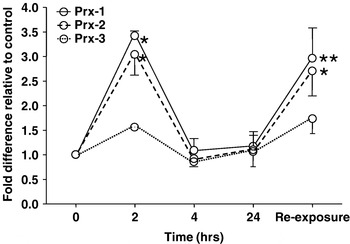
Exposure of 1-day-old, in vitro-developing mother sporocysts to exogenous H2O2 (10 μm) resulted in a rapid (<2 h) increase in steady-state mRNA levels for both Prx1 and Prx2 (Fig. 2). Transcript levels for these two H2O2-scavenging enzymes returned to baseline levels by 4 h post-exposure and continued to be expressed at levels similar to non-H2O2 exposed parasites at 24 h post-exposure. When the sporocysts were pulsed a second time with H2O2 for 1 h at 24 h post-initial exposure, mRNA levels for Prx1 and Prx2 again increased significantly in comparison to control non-H2O2 exposed parasites, suggesting a specific response to exogenous oxidative stress. In contrast, H2O2-exposure had no effect on Prx3 transcript levels (Fig. 2).
Effect of sublethal exogenous hydrogen peroxide exposure (initial concentration, 10 μm) on peroxiredoxin 1 (Prx1), 2 (Prx2) and 3 (Prx3) mRNA transcript levels in 1-day-old in vitro-developing Schistosoma mansoni mother sporocysts as determined by real-time quantitative PCR (qPCR). Differences in steady-state Prx mRNA levels in sporocyst cDNA populations were obtained at 2, 4 and 24 h post-exposure and 1 h after H2O2 re-exposure. For time-matched comparisons, untreated parasite control cDNA obtained at each time-point. A one-way ANOVA and Tukey's honest post-test was utilized to calculate a P value (* P⩽0·05, ** P⩽0·01, *** P⩽0·001) for each comparison and standard deviations (N=3).

Immunolocalization of Prx proteins in miracidia and mother sporocysts of S. mansoni
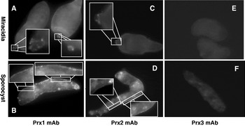
Fluorescence microscopy using anti-Prx1 and Prx2 monoclonal antibodies revealed the presence of immunoreactivity in the miracidium and 2-day-old in vitro-developing mother sporocyst (Fig. 3). Prx mAb reactivity was localized as punctate staining in the apical papilla (terebratorium) of miracidia (Fig. 3A, C) and continuously along the tegumental syncytium of sporocysts (Fig. 3B, D). Tegumental localization was confirmed with confocal microscopy of sporocysts probed with mAbs Prx1 and Prx2 (data not shown). Prx3 immunofluorescence was observed only as weak, diffuse internal staining in both miracidia and sporocysts (Fig. 3E, F). Control sporocysts probed only with FITC-conjugated secondary antibody showed minimal non-specific fluorescence.
Immunofluorescent localization of peroxiredoxins (Prx 1–3) in Schistosoma mansoni miracidia and 2-day in vitro-developing mother sporocysts. Note localization of Prx1 (A, B) and 2 (C, D) reactivity associated with the apical papilla of miracidia, and surface and interior of the tegumental syncytium of mother sporocysts. By contrast, only diffuse, low-level fluorescence was observed in miracidia and sporocysts using mAb Prx3 (E, F).

Prx1 and Prx2 proteins in the excretory-secretory products (ESP) of in vitro developing larval S. mansoni
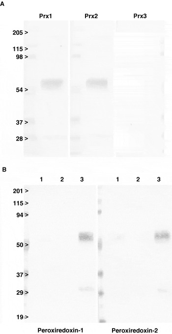
Western blot analysis of parasite ESP-containing transformation supernatants showed immunoreactivity when probed with Prx1- and Prx2-specific mAbs (Fig. 4A), but not Prx3 (Fig. 4A). Immunoreactivity with proteins of approximately 29 kDa and 60 kDa was detected in ESP samples corresponding to the predicted molecular masses of the native Prx proteins (∼26 kDa) and its naturally-occurring dimeric form. Immunoprecipitation with Prx1- and Prx2-specific antibodies successfully removed all detectable Prx protein present in ESP-containing culture supernatants (Fig. 4B) as no discernible bands were seen following ESP absorption. Enrichment of Prx1 and 2 in the Prx/anti-Prx-bound bead sample was further evidence for the specific depletion of Prxs from the ESP (Fig. 4B).
Western blot analysis of ESP-containing transformation supernatants probed with Prx1, Prx2 or Prx3 monoclonal antibodies. (A) Two immunoreactive bands (∼56 and 28 kDa) were evident in neat culture supernatants following 24-h of larval incubation in CBSS. (B) Immunoprecipitation of Prx-containing ESP culture supernatants with anti-Prx 1 and Prx 2 antibodies and Protein G-coupled agarose beads. Lane 1, neat ESP-containing culture supernatant prior to immunoprecipitation; Lane 2, neat ESP-containing culture supernatants post-immunoprecipitation; Lane 3, sample derived after extraction of the Protein G-linked Sepharose beads with SDS-PAGE lysis buffer showing enrichment of immunoprecipitated Prx proteins.

H2O2-metabolizing activity in larval ESP is due in part to Prx1 and 2
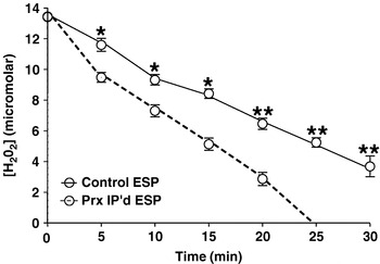
ESP-containing transformation supernatants collected 24 h post-cultivation of miracidia in CBSS were capable of scavenging exogenous H2O2in vitro (Fig. 5). Removal of Prx1 and Prx2 by specific immunoprecipitation from ESP-containing supernatants significantly inhibited their ability to scavenge H2O2 compared to controls in vitro (Fig. 5). The specific removal of Prx proteins from transformation supernatants, however, did not completely ablate their ability to break down H2O2 in solution suggesting that other factors contained in ESP also may be contributing to this action.
Metabolism of exogenous hydrogen peroxide (H2O2) by excretory-secretory product (ESP)-containing transformation supernatants collected 24 h post-miracidial transformation from cultures of in vitro-developing Schistosoma mansoni mother sporocysts. Following addition of H2O2 (∼12 μm) to wells containing ESP supernatants, hydrogen peroxide concentrations were determined at 0, 10, 15, 20, 25, and 30 min post-exposure using an Amplex Red H2O2 assay system as described in the Materials and Methods section. A paired Student's t-test (* P⩽0·05, ** P⩽0·01) was used to statistically compare relative H2O2 concentrations at the 6 time-points from replicate treatments and to calculate standard deviations (N=3).

DISCUSSION
Members of the Platyhelminthes have evolved linked glutathione (GSH) and thioredoxin (Trx) systems, made possible by a multifunctional, selenocysteine-containing enzyme, thioredoxin glutathione reductase (TGR), which possesses TrxR, GR and Grx activities responsible for recycling both Trx and GSH (reviewed by Salinas et al. Reference Salinas, Selkirk, Chalar, Maizels and Fernandez2004). In schistosomes these primary reductants maintain either GPx or Prx in a reduced state, permitting detoxification of reactive oxygen species (ROS) such as H2O2. To date, 3 schistosome Prxs have been identified in adult worm stages, and their substrate specificities and kinetics determined (Sayed and Williams, Reference Sayed and Williams2004). Because Prxs function as protective antioxidant molecules in cells through their peroxidatic activity, it has been hypothesized that such enzymes serve a protective role for the parasite, not only in the detoxification of ROS produced by their own metabolic activity, but also potential oxidative damage from the host environment (Nare et al. Reference Nare, Smith and Prichard1990; Sayed et al. Reference Sayed, Cook and Williams2006). Although many of the components of the S. mansoni redox system for the mammalian stages have been identified at the molecular level and their biochemical characteristics elucidated, little is known regarding the structure and function of the redox system in the early stages of intramolluscan schistosome development.
Upon penetration of its snail host, the miracidia enters a haemoglobin-rich oxygenated environment that may pose a potential risk of oxidative damage. In addition, in vitro encapsulation reactions by haemocytes of resistant B. glabrata snails results in larval killing via a hydroperoxide-mediated mechanism (Hahn et al. Reference Hahn, Bender and Bayne2001a). Our finding of antioxidant enzymes, Prx1 and 2, in the tegument and in vitro culture supernatants containing products released (ESP) during the miracidia-to-sporocyst transformation of S. mansoni sporocysts is consistent with a potential role of these enzymes in protecting the larval surface from H2O2 damage. Recent observations that haemocytes from resistant B. glabrata snails produce significantly more H2O2 than their susceptible counterparts (Bender et al. Reference Bender, Broderick, Goodall and Bayne2005) and that in vitro encapsulation by resistant strain haemocytes can down-regulate expression of sporocyst antioxidant transcript levels (Zelck and von Janowsky, Reference Zelck and Janowsky2004) has led to speculation that the ability of resistant B. glabrata haemocytes to produce greater quantities of H2O2 and/or to modulate larval antioxidant defences may be key elements in the destruction of sporocysts in the snail host (Bayne et al. Reference Bayne, Hahn and Bender2001).
In support of a hypothesized parasite-protective antioxidant system in larval schistosomes (Bayne et al. Reference Bayne, Hahn and Bender2001), the present study demonstrated that genes encoding the major S. mansoni redox balance pathway enzymes were expressed in miracidia and mother sporocysts during in vitro culture, suggesting an important role for these molecules in larval parasite development. Moreover, the specific induction of Prx1, Prx2 and Trx transcripts in sporocysts 48–72 h post-cultivation further implies a selective ability of sporocysts to upregulate H2O2-scavenging enzymes, possibly in response to environmental oxidative stress. Our finding that exposure of in vitro developing sporocysts to sublethal H2O2 levels significantly upregulates Prx1 and 2, but not Prx3, gene expression strongly implies that larvae possess a system for acute sensing of its oxidative environment. This result is consistent with a previous report of ROS-mediated induction of GPx transcript levels in S. mansoni sporocysts (Zelck and von Janowsky, Reference Zelck and Janowsky2004). While the mechanisms of sensing and monitoring H2O2 levels within the sporocyst's internal and external environments remain unknown, peroxide sensing systems have been proposed in other organisms. For example, the redox sensing machinery of Saccharomyces cerevisiae has been postulated to consist of receptor-initiated relay systems comprised of ‘peroxide receptors’ and ‘redox transducers’ (Orp1-Yap1) (Toledano et al. Reference Toledano, Delaunay, Monceau and Tacnet2004). It is presently unknown whether schistosomes possess homologues of the Orp1-Yap1 sensory relay, although its ability to respond to sublethal H2O2 levels suggests that such a system (or functional equivalent) likely exists.
After penetration of the snail host (mimicked during in vitro culture) the epidermal ciliated plates of the miracidia are shed through expansion along intracellular ridges and a syncytial tegument is formed (Pan, Reference Pan1980). In this paper we define excretory-secretory products (ESP) as any molecules released by the parasite either passively or actively during in vitro or in vivo miracidia-to-sporocyst transformation. Our finding of Prx 1 and 2 in larval transformation ESP also is consistent with a putative larval protective function during invasion of the molluscan host. The direct participation of ESP Prx1 and Prx 2 in the scavenging of exogenous H2O2 was clearly demonstrated by Prx mAB-specific immunoabsorption of ESP Prx proteins in Western blot analyses and H2O2 activity assays. Moreover, immunofluorescent localization of Prx1 and Prx2 at the tegumental syncytium of in vitro-developing mother sporocysts and at the apical papilla of miracidia (presumably ductal openings connecting penetration glands) (Pan, Reference Pan1980), localizes these Prx enzymes to larval compartments at the host-parasite interface and those consistent with a potential antioxidant function directed towards host ROS. The reason why penetration glands themselves revealed no anti-Prx reactivity is not known, but may be an artifact of larval fixative/detergent treatment and/or insufficient mAb incubation times. Similarly, Prx3 was predicted to be a mitochondrial protein with a probability of export to the mitochondrion at 0·85 and a cleavage site after Ala33 when analysed with the SignalP algorithm (www.cbs.dtu.dk/services/SignalP/). However, the low level diffuse staining observed also may be due to fixation artifacts and insufficient mAb penetration, or possibly suboptimum concentrations of Prx3.
Previously published reports on various trematodes suggest that antioxidant protein localization in the tegumental region or secretions may be quite commonplace. For example, tegument-enriched proteins of S. mansoni adults include Prxs, Trx-like proteins, GSH-S-transferases (GSTs) or superoxide dismutases (SODs) (Mei and LoVerde, Reference Mei and LoVerde1997; van Balkom et al. Reference van Balkom, van Gestel, Brouwers, Krijgsveld, Tielens, Heck and van Hellemond2005; Braschi et al. Reference Braschi, Curwen, Ashton, Verjovski-Almeida and Wilson2006), while Prx1, and its required reducing agent Trx, have been identified in secretions of S. mansoni eggs (Williams et al. Reference Williams, Asahi, Botkin and Stadecker2001). Additionally, a parasite-protective role has been suggested for various antioxidant proteins detected in cercarial acetabular gland secretions and adult worm ES products of S. mansoni and S. japonicum, respectively (Knudsen et al. Reference Knudsen, Medzihradszky, Lim, Hansell and McKerrow2005; Kumagai et al. Reference Kumagai, Osada and Kanazawa2006). The liver fluke, Fasciola hepatica, also possesses an antioxidant system as evidenced by the presence of Prx, TPx, SOD and GST (McGonigle et al. Reference McGonigle, Dalton and James1997; Jefferies et al. Reference Jefferies, Campbell, van Rossum, Barrett and Brophy2001). Like the schistosomes, the finding that F. hepatica egg ESP can inhibit the superoxide output of sheep and human neutrophils (Jefferies et al. Reference Jefferies, Turner and Barrett1997), further supports their involvement in anti-immune protection.
Although S. mansoni Prxs have been shown to be released during larval development, none of the Prx proteins contain canonical N-terminal signal sequences that normally allow for packaging by the ER/Golgi network, transport to the tegument surface and extracellular release. In fact both Prx1 and Prx2 of S. mansoni were predicted by SecretomeP (Bendtsen et al. Reference Bendtsen, Jensen, Blom, Von Heijne and Brunak2004; www.cbs.dtu.dk/services/SecretomeP/), a web-based, sequence-locating algorithm for proteins that lack N-terminal signal peptides, to be candidates for non-classical secretion (0·62 and 0·731, respectively, with a threshold cut-off of 0·55). This raises the question as to how these ‘non-signalling’ antioxidant proteins are packaged and transported to the outer tegumental membrane for secretion?
One possibility is that the proteins released at the time of transformation are the result of passive ‘leakage’ of molecules as ciliated epidermal plates are cast off during formation of the sporocyst tegument (Pan, Reference Pan1980). However, our present demonstration of Prx immunoreactivity as granular localizations in the tegument and subtegument of fully transformed sporocysts is highly suggestive of a secretory process. Moreover, earlier reported ultrastructural observations of sporocyst vesicular trafficking and larval secretion are consistent with this notion (Dunn and Yoshino, Reference Dunn and Yoshino1988). In this study immunoelectron microscopy was used to track immunogold-labelled vesicular contents from subtegumental perikarya to the sporocyst surface membrane, followed by vesicle-membrane fusion and extracorporeal release of their immunoreactive molecules (ESP), which now has been shown to contain Prx1 and 2. In eukaryotic cells there is substantial evidence supporting an alternative secretory process for proteins with known extracellular functions and that lack canonical signal peptides. Termed ‘non-classical’ or ‘non-conventional’ secretion, Rubaretlli and colleagues (Rubartelli and Sitia, Reference Rubartelli and Sitia1991; Rubartelli et al. Reference Rubartelli, Bajetto, Allavena, Wollman and Sitia1992) provided some of the first evidence for leaderless protein secretion in a variety of mammalian cell-types. Several potential mechanisms for non-classical translocation of leaderless proteins, including thioredoxin, have been proposed for their transport to the cell surface and extracellular secretion (review by Nickel, Reference Nickel2005). Such mechanisms may involve insertion of leaderless proteins into multivesicular bodies in the endocytic membrane system, their transport through the cytoplasm and exocytosis via fusion with the plasma membrane, as illustrated in the trafficking of small membranous vesicles from tegumental perikarya (cytons) to the surface membrane of adult (Skelly and Shoemaker, Reference Skelly and Shoemaker2000, Reference Skelly and Shoemaker2001) and larval (Dunn and Yoshino, Reference Dunn and Yoshino1988) S. mansoni stages. Other non-classical secretory processes may include membrane blebbing, in which plasma membrane-derived vesicles are shed extracellularly (Cleves and Kelly, Reference Cleves and Kelly1996; Stahl and Barbieri, Reference Stahl and Barbieri2002), or the involvement of chaperonins in mediated sorting/trafficking of leaderless proteins into vesicles destined for extracellular release. S. mansoni adult worms appear to engage in a form of membrane blebbing as they continuously replenish their outer tegumental membrane (review by Skelly and Wilson, Reference Skelly and Wilson2006). Moreover, the presence of various heat-shock proteins with known chaperonin function (including HSP60 HSP70 and HSP86) in secretions/ESP from S. mansoni cercariae (Knudsen et al. Reference Knudsen, Medzihradszky, Lim, Hansell and McKerrow2005) and larval transformation ESP (unpublished data) support indirectly a role of chaperonins in vesicular transport of Prx and other leaderless proteins.
In summary, this study reports the expression levels of antioxidant molecules for S. mansoni miracidia and mother sporocysts during early larval development under in vitro culture conditions. Two Prx proteins (Prx1 and Prx2) were localized to the tegument of developing sporocysts and were demonstrated to be present in ESP released by transforming larvae. Larval Prx in ESP possessed H2O2-detoxifying activity supporting the concept that production and active secretion of Prxs during early larval development may constitute a frontline component of the parasite's defence system, facilitating the establishment of successful infections within the snail host.
The authors would like to thank Dr David L. Williams (Illinois State University) for providing the antibodies used in this study and for helpful advice in preparation of this manuscript. This study was supported by NIH Grant AI015503.