





Introduction
Bipolar affective disorder (BD) is a severe, chronic mood disorder that ranks among the most prevalent and disabling neuropsychiatric conditions worldwide. It is a major contributor to global disease burden and is linked to significant morbidity and premature mortality (Crump, Sundquist, Winkleby, & Sundquist, Reference Crump, Sundquist, Winkleby and Sundquist2013; Grande, Berk, Birmaher, & Vieta, Reference Grande, Berk, Birmaher and Vieta2016; WHO, 2008). Although BD is known to have a strong genetic component, its exact etiology remains unclear, with growing evidence suggesting a multifactorial origin involving both genetic vulnerability and a range of environmental influences (Franco, Reyes-Resina, & Navarro, Reference Franco, Reyes-Resina and Navarro2021; Grande et al., Reference Grande, Berk, Birmaher and Vieta2016; Robinson & Bergen, Reference Robinson and Bergen2021).
Loneliness, defined as the distressing subjective experience that arises from a perceived gap between desired and actual social connections, is characterized by increased emotional vulnerability, hypervigilance, and persistent negative thinking (Hawkley & Cacioppo, Reference Hawkley and Cacioppo2010; National Academies of Sciences, Engineering, and Medicine et al., 2020). It reflects not only the absence or loss of companionship but also serves as a key marker of social isolation (Holt-Lunstad, Reference Holt-Lunstad2024; Vitale & Smith, Reference Vitale and Smith2022). Individuals with mood disorders are particularly prone to experiencing loneliness (Duggal, Fertuck, & Huprich, Reference Duggal, Fertuck, Huprich, Parra, Dagnino and Behn2021; Mann et al., Reference Mann, Wang, Pearce, Ma, Schlief, Lloyd-Evans, Ikhtabi and Johnson2022; Stein, Aguirre, & Hunt, Reference Stein, Aguirre and Hunt2013). In the context of BD, those undergoing manic episodes often feel misunderstood and socially excluded, which can gradually erode their social networks (Stein et al., Reference Stein, Aguirre and Hunt2013; Van Der Voort, Goossens, & Van Der Bijl, Reference Van Der Voort, Goossens and Van Der Bijl2009). Perceived social support has been associated with a lower risk of recurrence, whereas heightened loneliness is linked to poorer functioning and greater symptom severity in multiple domains (Cohen, Hammen, Henry, & Daley, Reference Cohen, Hammen, Henry and Daley2004; Van den Brink et al., Reference Van den Brink, Schutter, Hanssen, Elzinga, Rabeling-Keus, Stek, Comijs, Penninx and Oude Voshaar2018; Wang et al., Reference Wang, Mann, Lloyd-Evans, Ma and Johnson2018). The COVID-19 pandemic has intensified concerns about loneliness due to widespread social distancing measures, affecting individuals across all age groups. However, despite its recognized impact on mental health, research specifically exploring the relationship between loneliness and BD remains limited, and there is a notable lack of studies examining whether loneliness might serve as a precursor to newly diagnosed BD.
Evidence from preclinical studies suggests that social isolation can induce alterations in neurotransmitter systems, receptor sensitivities, and biomarker levels, though only a limited number of studies have examined its effects on specific brain regions (Cacioppo, Capitanio, & Cacioppo, Reference Cacioppo, Capitanio and Cacioppo2014; Mumtaz, Khan, Zubair, & Dehpour, Reference Mumtaz, Khan, Zubair and Dehpour2018; Tomova, Tye, & Saxe, Reference Tomova, Tye and Saxe2021; Xiong, Hong, Liu, & Zhang, Reference Xiong, Hong, Liu and Zhang2023). The subjective nature of loneliness and differences in social behavior and brain structure across species, however, make it challenging to directly translate findings from animal models to humans (Mumtaz et al., Reference Mumtaz, Khan, Zubair and Dehpour2018). In human neuroimaging research, structural abnormalities associated with loneliness have been identified in regions such as the prefrontal cortex, limbic striatum, insula, amygdala, hippocampus, and posterior superior temporal cortex (Lam et al., Reference Lam, Murray, Yu, Ramsey, Nguyen, Mishra, Martis, Thomas and Lee2021; Meeks & Jeste, Reference Meeks and Jeste2009). Despite these advances, no studies have directly investigated the neurobiological mechanisms that may link loneliness to the risk of developing BD, nor have BD-specific structural brain markers underlying this association been systematically explored.
Genetic factors are also known to play a substantial role in BD, with heritability estimates ranging from 40% to 70% (Craddock & Sklar, Reference Craddock and Sklar2013; Fabbri, Reference Fabbri2020). BD is recognized as a highly polygenic disorder, with multiple genes involved in neurotransmitter synthesis, metabolism, and signaling (Chen et al., Reference Chen, Yang, Liu, Cai, Huo, Zhang, Li, Chang and Luo2022; Wang et al., Reference Wang, Wang, Wang, Chen, Lu and Yao2021). Polygenic risk scores (PRS), which aggregate the effects of multiple risk alleles across the genome, provide a comprehensive measure of genetic susceptibility to BD (Bigdeli et al., Reference Bigdeli, Voloudakis, Barr, Gorman, Genovese, Peterson, Burstein, Velicu, Li, Gupta, Mattheisen, Tomasi, Rajeevan, Sayward, Radhakrishnan, Natarajan, Malhotra, Shi, Zhao and Roussos2022; Birmaher et al., Reference Birmaher, Hafeman, Merranko, Zwicker, Goldstein, Goldstein and Sakolsky2022; Li et al., Reference Li, He, Sun, Ma, Zhao, Wang, Hu, Feng, Li, Chu, Chen, Han, Sun, Liu, Sun and Shen2024; Musliner et al., Reference Musliner, Krebs, Albiñana, Vilhjalmsson, Agerbo, Zandi and Werge2020). PRS has proven effective in predicting BD risk and correlating with symptom severity, thus facilitating the identification of risk factors and improving understanding of the genetic architecture of the disorder (Biederman, Green, DiSalvo, & Faraone, Reference Biederman, Green, DiSalvo and Faraone2021; Birmaher et al., Reference Birmaher, Hafeman, Merranko, Zwicker, Goldstein, Goldstein and Sakolsky2022; Coombes et al., Reference Coombes, Markota, Mann, Colby, Stahl, Talati and Frye2020; Hasseris et al., Reference Hasseris, Albiñana, Vilhjalmsson, Mortensen, Østergaard and Musliner2023). Applying PRS to investigate the neurobiological relationship between loneliness and BD holds significant potential for advancing research in this field.
In this study, we utilized data from a large, nationwide UK adult cohort to prospectively examine the association between loneliness and the risk of first-episode BD. Furthermore, we aimed to assess the contributions of genetic predisposition and neurobiological indicators in mediating this association.
Methods
Study design and population
The UK Biobank recruited over 500,000 individuals aged 37–73 across the United Kingdom between 2006 and 2010 (Sudlow et al., Reference Sudlow, Gallacher, Allen, Beral, Burton, Danesh, Landray, Downey, Elliott, Green, Liu, Matthews, Ong, Pell, Silman, Young, Sprosen, Peakman and Collins2015). This ongoing, population-based cohort study enrolls participants who are registered with the UK National Health Service (NHS). All participants signed written informed consent, and the study received approval from the North West Multicenter Research Ethics Committee (no: 11/NW/0382). The current analysis was conducted using UK Biobank data under application number 713374.
Measures and outcome ascertainment
Loneliness
In the UK Biobank cohort, loneliness was evaluated through two questions administered via a touchscreen questionnaire. The first question, “Do you often feel lonely?” was scored as 1 point for ‘yes’ and 0 points for ‘no’. Participants were then categorized into two groups: those with a score of 1 were classified as experiencing loneliness, while those with a score of 0 were considered not lonely.
Outcome definition
BD diagnoses at baseline and during subsequent follow-up were confirmed through multiple data sources, including hospital admissions, primary care records, death registers, and self-reported physician diagnoses, which were further verified by nursing staff (Jongsma, Turner, Kirkbride, & Jones, Reference Jongsma, Turner, Kirkbride and Jones2019). The identification was based on ICD-10 code F31. Hospital admission data, routinely collected by the NHS, were linked to UK Biobank participants using anonymized numeric identification numbers. Death dates and causes were obtained from death certificates maintained by the NHS Information Center. Follow-up for all participants spanned from enrollment until the first occurrence of BD, death, or the cutoff date of October 30, 2022, whichever occurred first.
Phenotypic characterization of incident BD
Among participants who developed BD during follow-up, we used ICD-10 F31 subcodes from linked hospital inpatient records to classify the polarity of the first recorded BD episode as manic/hypomanic (F31.0–F31.2), depressive (F31.3–F31.5), mixed (F31.6), or remission/unspecified (F31.7–F31.9). To directly compare depressive versus manic/hypomanic onset, we restricted a secondary analysis to incident BD cases with a clearly specified first-episode type (F31.0–F31.5) and fitted a logistic regression model (depressive-onset vs. manic/hypomanic-onset) with baseline loneliness as the exposure, adjusted for age and sex. Psychiatric comorbidities were identified using ICD-10 codes for unipolar depression (F32–F33), anxiety disorders (F40–F41), psychotic disorders (F20–F29), and substance use disorders (F10–F19).
Genetic data and polygenic risk score (PRS)
The PRS for BD was obtained from the established PRS dataset available in the UK Biobank PRS Release. Detailed methodologies for PRS calculation have been described in prior publications (Lewis & Vassos, Reference Lewis and Vassos2020; Thompson et al., Reference Thompson, Wells, Selzam, Peneva, Moore, Sharp, Tarran, Beard, Riveros-Mckay, Giner-Delgado, Palmer, Seth, Harrison, Futema, McVean, Plagnol, Donnelly and Weale2024). In brief, the UK Biobank employed a standardized testing subset along with predefined criteria for disease and trait classification, utilizing Bayesian approaches to develop the PRS algorithm. This approach effectively integrated data across diverse ancestral backgrounds and relevant phenotypic characteristics. The individualized PRS was computed by summing the posterior effect sizes of genetic variants, weighted by the allele dosage across the entire genome. Participants were subsequently stratified into three categories based on their PRS distribution: low (lowest tertile), medium (middle tertile), and high (highest tertile).
Brain imaging
From the original cohort of 501,968 UK Biobank participants, 63,573 individuals underwent brain MRI scanning, with each participant receiving a single scan at designated assessment centers. Imaging was performed using a standardized Siemens Skyra 3T scanner equipped with a 32-channel head coil. The acquisition protocols and sequence parameters have been publicly available and detailed in prior publications (Thompson et al., Reference Thompson, Wells, Selzam, Peneva, Moore, Sharp, Tarran, Beard, Riveros-Mckay, Giner-Delgado, Palmer, Seth, Harrison, Futema, McVean, Plagnol, Donnelly and Weale2024). All MRI data were processed into image-derived phenotypes (IDPs) within the UK Biobank dataset. The T1-weighted images, which provide high-resolution structural representations of brain anatomy, were processed through a pipeline utilizing the FMRIB Software Library (FSL) and FreeSurfer tools. The total grey matter volume (GMV) and total white matter volume (WMV) were calculated by adjusting the raw volumes with the head size scaling factor provided by UK Biobank, ensuring these measures were normalized for head size. Additionally, cortical surfaces were reconstructed using FreeSurfer, and IDPs related to regional volumes were extracted based on surface atlases. Volumes of subcortical structures were obtained using FSL’s FIRST tool, also generating IDPs. Specifically, the FreeSurfer Desikan atlas was employed to delineate and quantify cortical and subcortical regions. To facilitate comparison and interpretation, all continuous measures of brain structure volumes were standardized using Z-scores.
Covariates
In our analysis, we accounted for a range of potential confounding variables informed by prior research, including age, gender, ethnicity, pre-tax household income, educational attainment, body mass index (BMI), residential location, alcohol consumption, and smoking status. Sociodemographic data were collected at baseline. Ethnicity was categorized into six groups within the UK Biobank: white, multiracial, Asian, Black, Chinese, and other ethnicities. Educational level was dichotomized into individuals with or without a degree or professional qualification. Household income before tax was classified as either less than £31,000 or £31,000 and above. Residential setting was determined based on population density, categorizing participants as residing in either urban or rural areas. BMI was computed from measurements of weight and height obtained by trained nurses. Smoking status was self-reported and categorized into three groups: never smokers, former smokers, and current smokers. Similarly, alcohol consumption history was self-reported and classified as never drinkers, former drinkers, or current drinkers.
Statistical analysis
Baseline characteristics were summarized using means and standard deviations (s.d.) for continuous variables, and frequencies accompanied by percentages for categorical variables. To address missing data in covariates, the fully conditional specification method was employed through multiple imputation techniques. Differences in baseline features between individuals with and without schizophrenia were evaluated using Student’s t-test, Mann–Whitney U-test, or chi-squared tests, as appropriate.
To examine the associations between loneliness and the risk of developing BD, a Cox proportional hazards model was utilized. This model incorporated time-varying annual air pollution exposure data and was structured to analyze the time from the initial loneliness assessment to the first diagnosis of BD, death, or the censoring date of October 30, 2022. All analyses initially included an unadjusted model assessing the crude association between loneliness and BD (model 1). Following this, all subsequent models were adjusted for sex and age to account for their potential confounding effects. Specifically, model 2 included only sex and age; model 3 further incorporated education level and pre-tax household income; model 4 added BMI, alcohol consumption, and smoking status; model 5 included PRS; and model 6 was fully adjusted, including all covariates.
Furthermore, logistic regression models were employed to assess the associations between loneliness and various brain structure phenotypes, such as GMV, WMV, and regional cortical and subcortical volumes. Participants with a diagnosis of BD before baseline imaging were excluded from all neuroimaging analyses. Before analysis, each phenotype was standardized to ensure comparability across measures. Additionally, stratified analyses based on genetic risk groups (low/middle/high) were performed in this context.
Besides, we performed two sensitivity analyses. First, to reduce the possibility of reverse causation, we reestimated the fully adjusted Cox models after excluding participants who received a first diagnosis of BD within the first 2 years of follow-up. Second, to further account for baseline affective symptoms that may predispose to both loneliness and BD, we fitted an additional Cox model that included the baseline Patient Health Questionnaire-4 (PHQ-4) total score (0–12, higher values indicating greater depressive/anxiety symptom burden) as a covariate, in addition to all variables in the fully adjusted model (Kroenke, Spitzer, Williams, & Löwe, Reference Kroenke, Spitzer, Williams and Löwe2009).
Results
As exhibited in Table 1, a total of 501,968 individuals were initially recruited for the study. Exclusions included 907 participants who lacked loneliness assessment data, 638 individuals with a baseline diagnosis of BD, 1,428 respondents who selected ‘prefer not to answer’, and 7,523 individuals who chose ‘do not know’, resulting in a final sample size of 491,472 participants, with 62,750 included for the further brain imaging analyses. At baseline, participants’ ages ranged from 38 to 73 years, with a mean age of 56.55 years (SD = 8.09). The cohort comprised 54.4% females and 45.6% males. Compared to individuals reporting no loneliness (n = 400,455; 81.5%), those who reported loneliness (n = 91,017; 18.5%) were more likely to be female, have a lower level of educational attainment, possess a reduced income, have a higher BMI, and have a history of smoking.
Descriptive statistics for the overall sample and by baseline loneliness and incident BD

Table 1. Long description
A complex data table with 6 columns and 24 rows. The columns are: Characteristics, Total (N = 491,472), Loneliness at baseline No (n = 400,455), Loneliness at baseline Yes (n = 91,017), Incident B D No (n = 490,156), and Incident B D Yes (n = 1,316).
Key data points include:
* Follow-up: Total mean is 13.29 years. Incident B D Yes group has a significantly lower mean of 6.64 years.
* Reported loneliness: 18.5 percent of the total sample. In the Incident B D Yes group, this rises to 42.7 percent.
* Gender: Females represent 54.4 percent of the total sample and 57.5 percent of the Incident B D Yes group.
* Age: Total mean is 56.55 years.
* Ethnicity: White participants make up 90.8 percent of the total sample.
* Education: 46.7 percent of the total sample have a degree level or professional qualification.
* Household Income: 40.9 percent earn less than 31,000 pounds, while 44.2 percent earn more than 31,000 pounds. In the Incident B D Yes group, 57.2 percent earn less than 31,000 pounds.
* B M I: Total mean is 27.43 kg m super -2.
* Smoking: 59.6 percent of the total sample have ever smoked, rising to 68.3 percent in the Incident B D Yes group.
* Drinking: 91.9 percent are current drinkers.
* B D polygenic risk score: Total mean is -0.23. The Incident B D Yes group has a higher mean score of 0.33.
Note: SD, standard deviation; BMI, body mass index (calculated as weight in kilograms divided by height in meters squared); BD, bipolar affective disorder.
a Data are presented as number (percentage) of participants unless otherwise indicated.
Over the duration of the study, which spanned ~16.58 years (mean = 13.29 years, SD = 2.03; totaling 6,531,663 person-years), 1,316 participants developed BD. Among these first-episode cases, 754 belonged to the non-lonely group, while 562 were from the lonely group. Participants diagnosed with BD were more likely to be female, possess lower income, have a higher BMI, and have a history of smoking. Additionally, they demonstrated higher BD PRS.
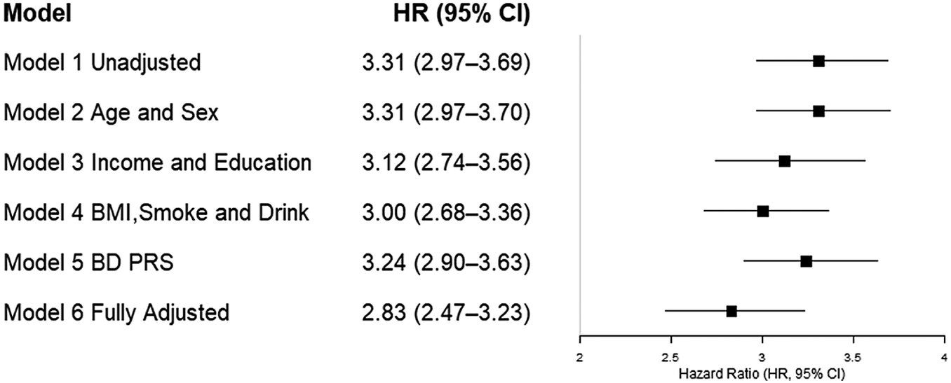
As shown in Figure 1, the unadjusted analysis demonstrated that individuals reporting loneliness had a significantly increased risk of developing BD, with a hazard ratio (HR) of 3.31 (model 1, 95% CI, 2.97–3.69). This association persisted after adjusting for age and sex (HR, 3.31; 95% CI, 2.97–3.70), indicating a robust relationship between loneliness and BD risk (model 2). Further adjustment for socioeconomic factors, including household income and education (model 3), slightly attenuated the association, but it remained statistically significant (HR, 3.12; 95% CI, 2.74–3.56). Additional adjustments for lifestyle variables, such as BMI, smoking, and alcohol consumption (model 4), resulted in a modest decrease in the HR to 3.00 (95% CI, 2.68–3.36), suggesting that behavioral factors partially mediate the relationship but do not fully account for it. The link between loneliness and BD risk persisted as significant after additional adjustment for the PRS (model 5, HR, 3.24; 95% CI: 2.90–3.63), suggesting that loneliness independently contributes to an increased risk of BD regardless of genetic predisposition. In the fully adjusted model (model 6), which included socioeconomic status, lifestyle factors, and PRS, the association between loneliness and newly diagnosed BD remained significant with an HR of ~2.83 (95% CI, 2.47–3.23). These findings indicate that loneliness is independently associated with a substantially increased risk of developing BD, even after comprehensive adjustment for potential confounders.
Association of loneliness with newly developed bipolar affective disorder (BD) in models with different sets of covariates and the fully adjusted model. Age and sex were included as covariates in models 2–6.

Figure 1. Long description
A forest plot with three columns: Model name, H R (95% C I) numerical values, and a graphical representation. The x-axis of the plot is labeled Hazard Ratio (H R, 95% C I) and ranges from 2 to 4.
* Model 1 Unadjusted: H R 3.31 (2.97–3.69). The point estimate is a black square centered at 3.31 with horizontal error bars extending from 2.97 to 3.69.
* Model 2 Age and Sex: H R 3.31 (2.97–3.70). The point estimate is at 3.31 with error bars from 2.97 to 3.70.
* Model 3 Income and Education: H R 3.12 (2.74–3.56). The point estimate is at 3.12 with error bars from 2.74 to 3.56.
* Model 4 B M I, Smoke and Drink: H R 3.00 (2.68–3.36). The point estimate is at 3.00 with error bars from 2.68 to 3.36.
* Model 5 B D P R S: H R 3.24 (2.90–3.63). The point estimate is at 3.24 with error bars from 2.90 to 3.63.
* Model 6 Fully Adjusted: H R 2.83 (2.47–3.23). The point estimate is at 2.83 with error bars from 2.47 to 3.23.
All models show a positive association as all H R values and their confidence intervals remain above the null value of 1.0.
Among participants who developed BD during follow-up, the polarity of the first recorded BD episode (based on ICD-10 F31 subcodes) was manic/hypomanic in ~9.9%, depressive in 3.9%, mixed in 1.2%, and remission/unspecified in 85.0%. These proportions were similar in participants with and without baseline loneliness. Restricting the analysis to incident BD cases with a clearly specified first-episode type (F31.0–F31.5), 28.3% were depressive-onset (F31.3–F31.5), and 71.7% were manic/hypomanic-onset (F31.0–F31.2). In a logistic regression model adjusted for age and sex, baseline loneliness was not significantly associated with depressive-onset BD relative to manic/hypomanic-onset BD (OR = 0.87, 95% CI = 0.50–1.50). Incident BD cases also exhibited substantial psychiatric comorbidity: 57.2% had a recorded diagnosis of unipolar depression, 31.8% had anxiety disorders, 17.7% had psychotic disorders, and 28.0% had substance use disorders, highlighting the high burden of depressive and anxiety morbidity in individuals who later develop BD.
The results of the logistic regression analysis examining the relationship between loneliness and brain volumes showed that GMV was significantly associated with loneliness (p = 8.46 × 10⁻⁵, z = −3.93), indicating that individuals reporting loneliness tend to have lower GMV. In contrast, WMV was not significantly related to loneliness (p = 0.118, z = 1.56), suggesting no meaningful association. Based on the regional analysis, 19 out of 34 cortical regions and 4 out of 8 subcortical regions showed significant FDR-corrected (p < 0.05) associations. All findings were negative, indicating that individuals reporting loneliness had smaller volumes in these regions. Key areas include middle frontal, fusiform, inferior parietal, inferior temporal, lateral and medial orbitofrontal, middle temporal, paracentral, pericalcarine, postcentral, posterior cingulate, precentral, rostral anterior cingulate, superior parietal, superior temporal, supramarginal, temporal pole, insula, as well as nucleus accumbens, hippocampus, putamen, and thalamus.
As illustrated in Figure 2, the stratified logistic regression results by genetic risk groups showed that in the low-risk group, only the thalamus (p = 0.034, z = −3.35) and postcentral cortex (p = 0.002, z = −4.28) remained significant after FDR correction. In the middle-risk group, only the thalamus (p = 0.022, z = −3.65) was significant. In the high-risk group, several regions – including the inferior parietal (p = 0.024, z = −3.09), medial orbitofrontal (p = 0.003, z = −4.08), inferior temporal (p = 0.003, z = −4.01), middle temporal (p = 0.020, z = −3.29), temporal pole (p = 0.020, z = −3.24), insula (p = 0.024, z = −3.12), and thalamus (p = 0.005, z = −3.77) – all survived FDR correction. All significant associations were negative, indicating smaller regional volumes among individuals reporting loneliness.
Associations between loneliness and brain imaging phenotypes. (a–c) Association between loneliness and brain imaging phenotypes stratified by genetic risk groups (low/middle/high). (d–f) The same associations that remained statistically significant after false discovery rate (FDR) correction based on p-value thresholds. The brain imaging phenotypes analyzed included cortical and subcortical volumes. Associations between loneliness and brain structures, stratified by genetic risk groups, were estimated for 62,750 participants with available neuroimaging data using logistic regression models. Results are expressed as Z-scores. P-values were adjusted using the FDR method to account for multiple comparisons, with a two-sided P-value <0.05 after FDR correction indicating statistical significance.

Figure 2. Long description
The figure consists of six panels labeled a through f. A color scale bar at the bottom of each row indicates Z-scores ranging from negative 4 in dark blue to positive 4 in dark red, with white representing zero.
Top Row: Uncorrected associations.
Panel a, Low P R S: Shows light blue and orange patches across the cortical surface and subcortical regions, indicating mixed positive and negative associations.
Panel b, Middle P R S: Displays more widespread blue regions across the cortex and subcortical structures, indicating stronger negative associations compared to the low risk group.
Panel c, High P R S: Shows the most extensive and darkest blue regions, indicating the strongest negative associations between loneliness and brain volume in the high genetic risk group.
Bottom Row: F D R corrected associations (p less than 0.05).
Panel d, Low P R S: Most cortical areas are white, with only small blue regions remaining in the subcortical structures.
Panel e, Middle P R S: Shows significant blue clusters in the temporal and frontal lobes, as well as subcortical regions.
Panel f, High P R S: Displays the largest significant blue clusters across the lateral and medial cortical surfaces and subcortical volumes, confirming that the high genetic risk group has the most robust negative associations after statistical correction.
In sensitivity analyses, excluding individuals who were diagnosed with BD within the first 2 years of follow-up yielded hazard ratios for loneliness that were very similar to those in the main analysis (fully adjusted HR, 2.89; 95% CI, 2.50–3.34). When the baseline PHQ-4 total score was additionally included in the fully adjusted Cox model, PHQ-4 was independently associated with incident BD (HR per 1-point increase, 1.23; 95% CI, 1.20–1.26), and the hazard ratio for loneliness was attenuated but remained statistically significant (HR, 1.73; 95% CI, 1.47–2.03). These findings indicate that early BD cases and baseline depressive/anxiety symptoms explain part, but not all, of the association between loneliness and subsequent BD.
Discussion
To our knowledge, this is the first large-scale, population-based study to comprehensively examine the relationship between loneliness and the risk of developing BD in adults. Our findings demonstrate that lower GMV is significantly associated with loneliness, particularly in specific brain regions such as the thalamus, postcentral cortex, and various cortical areas. Importantly, these associations are more widespread and pronounced among individuals with a higher genetic predisposition to BD, as indicated by PRS.
The existing literature on loneliness and BD remains limited, largely consisting of cross-sectional studies with small sample sizes and confounded by comorbid mental health conditions, which complicate causal interpretations. While some prior research has suggested a bidirectional relationship between loneliness and BD – where loneliness may both contribute to and result from social impairments – findings have been inconsistent (Giacco, Reference Giacco2023). For instance, studies have reported no significant association between loneliness and manic symptom severity (Johnson et al., Reference Johnson, Winett, Meyer, Greenhouse and Miller1999; Koenders et al., Reference Koenders, Giltay, Hoencamp, Elzinga, Spinhoven and Spijker2015), but subtle social cognitive deficits, such as impaired emotion recognition, have been observed even during euthymic phases of BD (Green et al., Reference Green, Wynn, Eisenberger, Horan, Lee, McCleery, Miklowitz, Reavis and Reddy2024; Lee et al., Reference Lee, Huang, Chang, Kuo, Lu, Hsieh, Chang, Yang, Chen and Tseng2022). Moreover, loneliness has been linked to poorer quality of life in older adults with mood symptoms and mediates the relationship between self-stigma, depression, and suicidality (Świtaj et al., Reference Świtaj, Grygiel, Leciak, Stefaniak, Opozda-Suder and Anczewska2025; Warner et al., Reference Warner, Holland, Lobban, Bentley, Tyler and Palmier-Claus2025). The COVID-19 pandemic further emphasized the detrimental impact of social isolation on mental health, underscoring the importance of social connectedness in vulnerable populations (Hofer et al., Reference Hofer, Kachel, Plattner, Chernova, Conca, Fronthaler, Haring, Holzner, Huber, Miller, Pardeller, Perwanger, Pycha, Schmidt, Sperner-Unterweger, Tutzer, Frajo-Apor and Marksteiner2022). Our findings extend this body of work by providing prospective evidence that loneliness increases the risk of incident BD, even after adjusting for socioeconomic, lifestyle, and genetic factors. This suggests that loneliness independently contributes to BD risk, highlighting its potential role as a modifiable environmental factor.
A key conceptual issue is the possibility of reverse causation and gene–environment correlation. Individuals with high genetic liability for BD may already exhibit subthreshold mood and behavioral changes at baseline (e.g. social withdrawal, emotional instability, and interpersonal conflict) that increase their likelihood of feeling lonely. In this scenario, loneliness may partly represent an early manifestation or consequence of emerging BD rather than a purely antecedent risk factor. To address this concern, we conducted additional sensitivity analyses. The association between loneliness and incident BD remained robust after excluding cases diagnosed within the first 2 years of follow-up and after further adjustment for baseline depressive/anxiety symptoms (PHQ-4), although it was partially attenuated in the latter analysis. These findings suggest that early BD onset and baseline psychological distress account for part, but not all, of the observed association. Nevertheless, given the observational design and single baseline assessment of loneliness, our results should be interpreted as evidence of a strong longitudinal association rather than definitive proof of a causal effect of loneliness on BD onset.
An additional question is whether our findings simply reflect depressive, rather than manic, morbidity within BD. We characterized the polarity of the first recorded BD episode among incident cases. We observed that 3.9% of incident BD cases had a depressive first episode, 9.9% had a manic/hypomanic first episode, 1.2% had a mixed episode, and 85.0% were coded as remission or unspecified at the index date, with a similar distribution in lonely and non-lonely participants. In a subset of incident BD cases with clearly specified episode type, we directly compared depressive-onset versus manic/hypomanic-onset BD and found that baseline loneliness was not associated with higher odds of depressive-onset BD (OR = 0.87, 95% CI = 0.50–1.50). These analyses indicate that the longitudinal association between loneliness and BD incidence is not driven solely by BD cases in which depressive polarity predominates at onset. At the same time, incident BD cases displayed substantial psychiatric comorbidity, with 57.2% having hospital diagnoses of unipolar depression and 31.8% having anxiety disorders, alongside psychotic and substance use disorders, consistent with the well-documented depressive and anxiety burden in BD. Taken together, these observations support the interpretation of loneliness as a transdiagnostic vulnerability factor closely linked to depressive and anxiety morbidity, but not exclusively tied to depressive-onset BD.
Regarding neurobiological mechanisms, prior studies have identified associations between loneliness and structural impairments in regions involved in socioemotional processing, including the prefrontal cortex, insula, amygdala, hippocampus, and ventral striatum (Lam et al., Reference Lam, Murray, Yu, Ramsey, Nguyen, Mishra, Martis, Thomas and Lee2021; Morr et al., Reference Morr, Liu, Hurlemann, Becker and Scheele2022; Wong et al., Reference Wong, Mabel-Kenzie, Lin, Huang, Liu, Lee and Lee2022). Our results corroborate and expand upon these findings by demonstrating that individuals reporting loneliness tend to have reduced volumes across a broad network of regions, including the middle frontal, inferior parietal, temporal, orbitofrontal, insula, hippocampus, thalamus, and basal ganglia structures. Notably, these associations are most significant in individuals at high genetic risk for BD, with multiple cortical and subcortical regions showing volume reductions. These regions are integral to emotional regulation, social cognition, reward processing, and self-referential thought – domains often disrupted in BD and linked to feelings of loneliness (Bilgili, Reference Bilgili2022; Gillissie et al., Reference Gillissie, Lui, Ceban, Miskowiak, Gok, Cao, Teopiz, Ho, Lee, McIntyre and Rosenblat2022; Marchand, Reference Marchand2012; Van Rheenen, Meyer, & Rossell, Reference Van Rheenen, Meyer and Rossell2014).
Stratification by BD PRS further revealed that the relationship between loneliness and brain structure is amplified in genetically susceptible individuals. Higher-risk groups exhibited more widespread and significant volume reductions, particularly in regions such as the inferior parietal, medial orbitofrontal, temporal pole, insula, and thalamus. Conversely, associations in low- and middle-risk groups were limited to fewer regions, such as the thalamus and postcentral cortex. These findings suggest a gene–environment interaction, where genetic vulnerability heightens susceptibility to neurostructural impairments related to social disconnection.
Collectively, these results align with neurobiological models of BD that implicate deficits in regions governing emotion regulation and social cognition. The observed interaction between genetic risk and loneliness emphasizes that genetic predisposition may sensitize brain circuits, thereby increasing vulnerability to social and emotional impairments that contribute to BD development. Recognizing these mechanisms underscores the importance of early interventions targeting social connectedness, particularly among individuals with elevated genetic risk, to potentially mitigate neurostructural vulnerabilities and reduce BD incidence.
Importantly, the neuroimaging findings should be interpreted with similar caution regarding directionality. All MRI scans were acquired at a single time point, and the observed associations between loneliness and reduced GMV are therefore cross-sectional. We cannot determine whether loneliness leads to progressive gray matter loss in BD-relevant circuits, whether preexisting structural anomalies predispose individuals both to feel lonely and to develop BD, or whether both processes co-occur. The stronger and more widespread associations we observed in individuals with high BD PRS are consistent with a gene–environment interaction framework, in which genetic vulnerability may sensitize neural circuits to the effects of social disconnection. However, they are also compatible with gene–environment correlation, whereby shared genetic factors contribute simultaneously to BD liability, brain structural alterations, and behavioral tendencies (e.g. social withdrawal and negative social cognitions) that increase the likelihood of loneliness. Furthermore, although we excluded individuals with a recorded diagnosis of BD at baseline, some participants may have been in an early or prodromal phase of BD, such that the structural differences reflect emerging BD-related pathology that co-occurs with loneliness. Longitudinal neuroimaging studies with repeated measurements of both brain structure and social experiences will be crucial to establish whether loneliness exerts a causal neurobiological impact on BD-related circuits, or primarily indexes an already altered neurodevelopmental trajectory.
While loneliness is often conceptualized as a modifiable social risk factor, its reduction in practice can be challenging, especially in individuals with mood disorders or high BD liability. The previous study identified social interactions as a coping strategy and pre-pandemic social functioning and loneliness as modifiable correlates of changes in well-being and psychopathology during the COVID-19 pandemic, but also showed that these operate within a network of nonmodifiable and contextual constraints, including country income, COVID-19-related restrictions, urbanicity, physical health and disability, socioeconomic status, and prior mental disorder history (Solmi et al., Reference Solmi, Thompson, Cortese, Estradé, Agorastos, Radua, Dragioti, Vancampfort, Thygesen, Aschauer, Schlögelhofer, Aschauer, Schneeberger, Huber, Hasler, Conus, Cuénod, von Känel, Arrondo and Correll2025). In BD, severe depressive or mixed states can reduce motivation and energy to engage with others or make social contact feel aversive, while manic or hypomanic episodes may lead to interpersonal conflict and stigma that erode social networks (Fowler & Dooley, Reference Fowler and Dooley2023; Gilbert & Gruber, Reference Gilbert and Gruber2014). Physical impairments, chronic medical conditions, caregiving responsibilities, geographical isolation, and structural barriers further limit the feasibility of face-to-face interaction (Nuara et al., Reference Nuara, Fabbri-Destro, Scalona, Lenzi, Rizzolatti and Avanzini2022). These considerations temper the notion of loneliness as a straightforwardly modifiable factor and suggest that effective strategies will need to be multimodal, targeting both objective social isolation and subjective appraisals of relationships – for example, through psychological therapies addressing maladaptive social beliefs and hypervigilance, interventions to enhance social skills and self-efficacy, digital or remote modalities to facilitate connection despite constraints, and public health policies that reduce socioeconomic and environmental barriers. Our findings, particularly the stronger neurostructural associations in individuals with higher genetic risk for BD, underscore the need to tailor interventions to individual motivational, cognitive, physical, and contextual capacities rather than assuming that increasing social contact alone will reliably alleviate loneliness or prevent BD onset.
Strengths and limitations
The primary strengths of this study include its large sample size and consequently high statistical power, the extended follow-up period, and the comprehensive range of covariates incorporated to account for relevant risk factors. Additionally, the independent ascertainment of diagnoses through health records enhances the validity of our findings. A novel aspect of this research is the investigation of the combined effects of loneliness, genetic predisposition, and BD risk, supported by neurobiological evidence – an approach that has not been previously explored in the literature.
However, the study also has several limitations. First, because loneliness and BD-related behavioral changes may mutually influence each other, and loneliness was only measured once at baseline, we cannot exclude the possibility of reverse causation or gene–environment correlation. Our sensitivity analyses excluding BD cases occurring within 2 years of baseline and additionally adjusting for baseline PHQ-4 scores reduced, but did not eliminate, this concern. Thus, the present findings should be viewed as demonstrating a robust longitudinal association between loneliness and BD risk rather than proving a causal relationship. Second, our neuroimaging analyses were based on a single cross-sectional MRI scan per participant. As a result, the observed associations between loneliness and reduced GMV in BD-relevant regions cannot be interpreted as causal effects of loneliness on brain structure. It remains possible that preexisting structural differences predispose individuals to both loneliness and BD, or that shared genetic factors influence neural architecture and social experiences in parallel. Third, although the UK Biobank’s recruitment process was efficient and facilitated the inclusion of ~500,000 participants, it was associated with a low response rate of around 5.5%, which may introduce selection bias (Schoeler et al., Reference Schoeler, Speed, Porcu, Pirastu, Pingault and Kutalik2023). Fourth, despite adjusting for numerous potential confounders, residual confounding from unmeasured or unknown factors cannot be entirely excluded. Fifth, the reliance on registry-based ICD-10 codes means that we cannot fully characterize BD subtypes (e.g. type I vs. type II), lifetime predominant polarity, or the detailed course and phenomenology of mixed states; most index F31 diagnoses in our cohort were coded as remission or unspecified. Finally, the reliance on a single measurement of loneliness at baseline precluded an examination of potential time-varying associations and dynamic changes in loneliness over the follow-up period.
Conclusions
In conclusion, this large-scale study found that loneliness is strongly and prospectively associated with the subsequent onset of BD. Neuroimaging evidence revealed that loneliness is associated with widespread reductions in gray matter volume, especially in regions involved in emotion and social processing, with stronger effects in those at higher genetic risk. These findings highlight loneliness as a potentially modifiable social factor linked to BD risk and neurobiological vulnerability, rather than as a simple or easily alterable risk factor, and suggest that addressing social disconnection may help prevent neurobiological vulnerabilities and reduce BD incidence.
Data availability statement
The data could be accessed from the following link: https://www.ukbiobank.ac.uk.
Author contribution
Designed and performed research: Jianing Wang and Zhongpeng Dai. Analyzed data: Jianing Wang and Zhongpeng Dai. Wrote and revised the paper: Jianing Wang and Zhongpeng Dai. Supervision: Jianing Wang and Zhongpeng Dai.
Funding statement
Not applicable.
Competing interests
The authors declare none.
Ethical standards
This ongoing population-based cohort study involves participants registered with the UK National Health Service, all of whom provided written informed consent. The study has been approved by the North West Multicenter Research Ethics Committee (no: 11/NW/0382). The current analysis utilizes data from the UK Biobank under application number 713374, ensuring adherence to ethical guidelines throughout the research process.



 Open access

