Pulmonary artery sling is a congenital disease in which the left pulmonary artery abnormally originates from the right pulmonary artery instead of the main pulmonary artery.Reference Berdon, Baker and Wung 1 , Reference Backer, Idriss, Holinger and Mavroudis 2 Because of the aberrant left pulmonary artery, the trachea and right bronchus are typically compressed and induce respiratory symptoms after birth, such as wheezing, lung infection, and other severe respiratory distress.Reference Potts, Holinger and Rosenblum 3 – Reference Cohen and Landing 5 Infant pulmonary artery sling requires surgical intervention to relieve symptoms, especially tracheobronchial stenosis. The surgical procedure includes left pulmonary artery re-implantation and tracheobronchial tracheoplasty. The latter involves post-operative complications. Better treatment has yet to be developed.Reference Hong, Zhou, Liu, Liu, Wang and Feng 6
Three-dimensional printing provides customized three-dimensional objects of various materials.Reference Rankin, Giovinco, Cucher, Watts, Hurwitz and Armstrong 7 , Reference Schmauss, Juchem, Weber, Gerber, Hagl and Sodian 8 It provides novel and effective ways to resolve medical problems. Our team developed a patient-customised three-dimensional printed splint.
Here, we report a rare and complex case of pulmonary artery sling associated with severe bilateral bronchus stenosis, left pulmonary artery stenosis, atrial septal defect, and patent ductus arteriosus. We successfully treated bronchus stenosis in an infant with a three-dimensional printed splint. The infant underwent an uneventful recovery and was discharged from our hospital 10 days later.
Case report
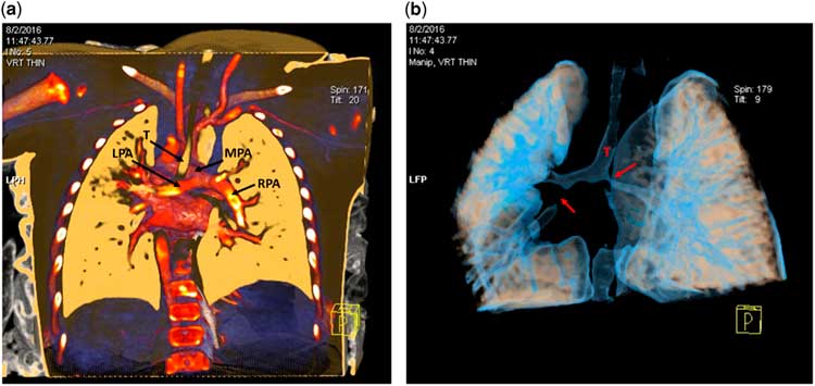
A 5-month-old boy was admitted with complaints of recurrent wheezing, stridor, and respiratory distress. An echocardiogram revealed an aberrant left pulmonary artery rising from the right pulmonary artery associated with left pulmonary artery stenosis, atrial septal defect, and patent ductus arteriosus. Three-dimensional CT angiography of the chest identified pulmonary artery sling associated with a severe stenosis of the right main bronchus and the left lower bronchus.
Surgery was conducted to restore the normal location of the left pulmonary artery and relieve bronchus compression. The surgery also protected the bronchus from compression by highly oedematous vessels and tissues, post-operatively. We used a three-dimensional printed splint to treat this life-threatening condition.
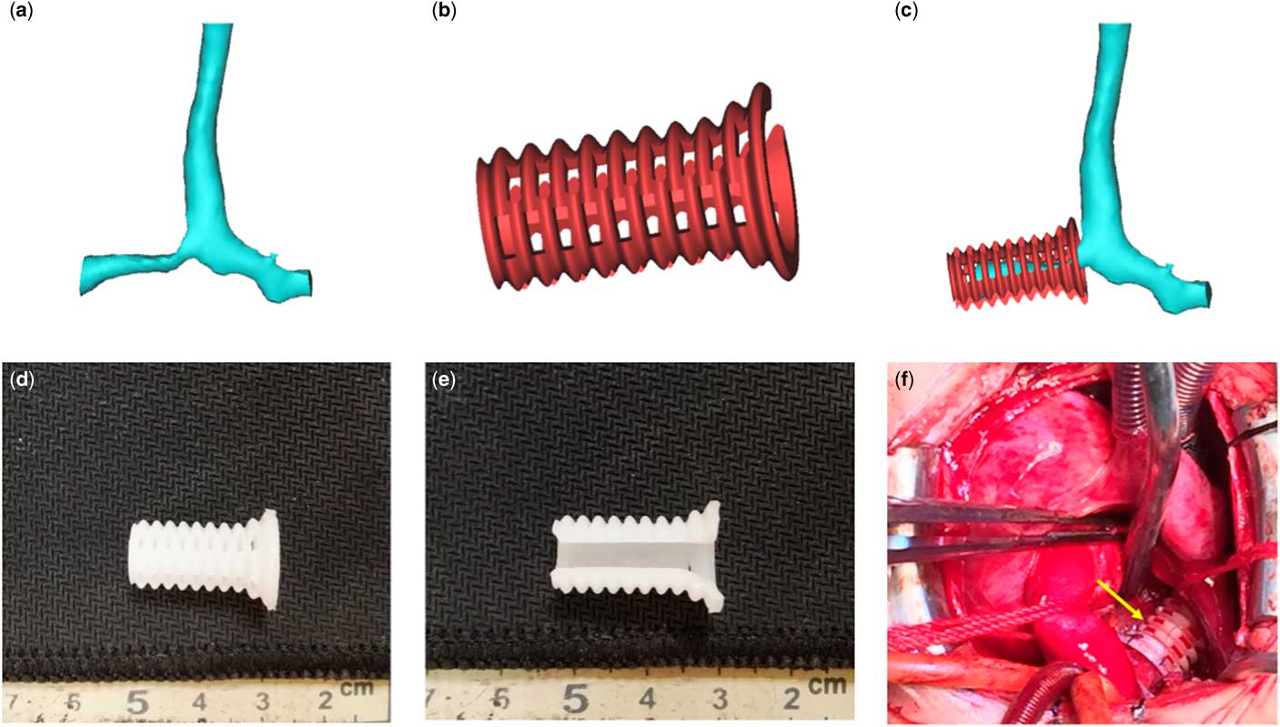
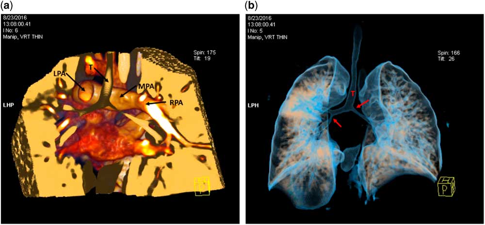
A median sternotomy approach was used and a normothermic cardiopulmonary bypass was placed. After ligation and dissection of the patent ductus arteriosus, the aberrant left pulmonary artery was divided at its origin off the right pulmonary artery and re-implanted anterior to the trachea into the main pulmonary artery with an end-to-side anastomosis. Then, we placed the three-dimensional printed splint and sutured around the circumference of the stenotic malacic bronchus. We tied the interstices of the splint and expanded the bronchus. Subsequently, the atrial septal defect was repaired. Early extubation is critical to the post-operative recovery of a patient. To achieve early extubation, the cardiopulmonary bypass was shortened to 35 minutes during left pulmonary artery re-implantation, which may post-operatively reduce bronchus secretions and make extubation easier. The surgery was successful. Compared with preoperative data, the arterial blood oxygen partial pressure increased from 50 to 90 mmHg, the arterial blood oxygen saturation increased from 88 to 100 mmHg, and the carbon dioxide partial pressure in venous blood decreased from 80 to 42 mmHg. The infant was post-operatively extubated for 8 hours. Echocardiograms and three-dimensional CT angiography at 5 days after the operation indicated that the recovery progressed well. The patient was discharged after 10 days.
Discussion
Pulmonary artery sling is a congenital vascular disorder first described in 1897 by Glaevecke and Doehle.Reference Backer, Russell, Kaushal, Rastatter, Rigsby and Holinger 9 Patients usually present with respiratory symptoms, including recurrent chest infection and other respiratory distress.Reference Cohen and Landing 5 , Reference Procacci, Residori, Bertocco, Di Benedetto, Andreis and D’Attoma 10 , Reference Loukanov, Sebening, Springer, Hagl, Karck and Gorenflo 11 Three-dimensional reconstructions of CT scans and two-dimensional echocardiography are useful tools to establish pulmonary artery sling diagnoses.Reference Lovett, Entrikin, Ungerleider and Ootaki 12 Once pulmonary artery sling is diagnosed, a patient must undergo a surgical procedure.Reference Healey, Ron, Hromada and Chhabra 13 After the left pulmonary artery has been reconnected, the trachea needs to be repaired. In recent years, many different types of tracheoplasties have been undertaken. However, none of these procedures are satisfactory, and post-operative complications remain a great challenge, which include tracheobronchomalacia, anastomosis leakage, and granulation tissue formation.Reference Yamaguchi, Ohashi, Hosokawa, Oshima, Tsugawa and Kimura 14 Stenting is an important therapeutic method for stenotic tracheobronchial. However, stenting is equally unsatisfactory owing to problems of fragmentation, extrusion, and penetration into neighbouring structures.Reference Dutau, Maldonado, Laroumagne and Astoul 15 , Reference Bolliger, Probst, Tschopp and Perruchoud 16
In our case study, the tracheobronchial lumen diameter of the infant was relatively small compared with adults, and had a high degree of stenosis. All repair methods mentioned above were not suitable for our patient. Cardiopulmonary bypass can greatly damage the circulatory system and induce oedemas of the heart and ambient vessels and tissues. Because it is important to provide external support for stenotic bronchial tubes, we needed to find a way to relieve bronchus stenosis and prevent ambient compression.
Three-dimensional printing could provide customised replicas according to patient-specific data in a variety of materials.Reference Frame and Leach 17 Zopf et alReference Zopf, Hollister, Nelson, Ohye and Green 18 treated a tracheal stenosis by using a three-dimensional printed splint. Our team found that a customised degradable splint may be the appropriate treatment for our patient. This splint provided resistance against collapse and space to grow.
We coordinated with engineering specialists and developed a customised, degradable three-dimensional printed bronchus splint. We added new features according to specific circumstances of the patient:
(1) The splint was made using polycaprolactone, which is compatible with biological cells. Normal cells can be grown in the base frame, and polycaprolactone can be degraded into CO2 and H2O.Reference Jurgens, Kroeze, Bank, Ritt and Helder 19 This splint can provide sufficient strength to resist external compression. We modulated the degradation rate of polycaprolactone to 2 years by adjusting its molecular weight.
(2) On the basis of the normal diameter of a bronchus of a 2-year-old child and the space required for bronchial growth, the splint inner diameter was set to 6 mm.
(3) As limited by the technique difficulty, the shape of a conventional splint is straight and has only been applied for median sections of stenotic bronchi. Our team overcame the difficulties and designed one end of the splint in a trumpet-like shape owing to the location of the stenotic bronchus.
The surgery was successful, and the patient was discharged from our hospital 10 days later. To our knowledge, this is a novel and successful case in which a three-dimensional printed splint was used in a complex congenital cardiovascular disease associated with severe bronchus stenosis. The post-operative recovery period was particularly superior to typical tracheobronchial stenosis recovery periods. We can expect three-dimensional printing to enter more aspects of medical treatment.
In conclusion, the effect of using three-dimensional printed splint treatments in infant pulmonary artery sling associated with severe bilateral bronchus stenosis is significant. Owing to the special physiological makeup of infants and complicated structure of complex congenital heart diseases, traditional tracheobronchial stenosis treatment could not satisfy our clinical needs. We suggested the use of a three-dimensional printed splint. The prospect of three-dimensional printing of degradable material for treating infant illnesses is worthwhile to explore (Figs 1–3).
Posterior view of three-dimensional CT angiogram reconstruction preoperatively. Panel a shows aberrant left pulmonary artery origin from the right pulmonary artery. It circles around the right main bronchus and travels along the left main bronchus and enters the hilum of the left lung. Panel b shows severe stenosis of the right main bronchus and left lower bronchus (arrow), and left lower lobe atelectasis. MPA=main pulmonary artery, T=trachea.

Panels a–c show the three-dimensional reconstruction models of stegnotic bronchus and splint. Panels d and e show the patient-specific three-dimensional printed degradable splint with one trumpet-like shape end. Panel f shows intraoperative placement of the splint (yellow arrow) overlying the stegnotic malacic right main bronchus segment.

Posterior view of the three-dimensional CT angiogram reconstruction at 5 days post-operatively. Panel a shows a normal origin of the left pulmonary artery and effective release for the stegnotic bronchus. Panel b shows airway after placement of the splint.

Informed Consent
Informed consent was obtained from all individual participants included in the study.
Financial Support
This research received no specific grant from any funding agency or from commercial or not-for-profit sectors.
Conflicts of Interest
None.
Ethical Standards
Ethical approval: All procedures performed in studies involving human participants were in accordance with the ethical standards of the institution.