Introduction
Complete atrioventricular septal defect represents a complex congenital cardiac malformation characterised by a common atrioventricular valve, a primum atrial septal defect, and an inlet ventricular septal defect. Reference Jacobs, Burke, Quintessenza and Mavroudis1,Reference Atz, Hawkins and Lu2 Over recent decades, primary biventricular repair has evolved into the predominant surgical strategy, achieving satisfactory outcomes, Reference Ginde, Lam and Hill3,Reference Schleiger, Miera and Peters4 long-term prognosis remains influenced by post-operative left atrioventricular valve regurgitation, left ventricular outflow tract obstruction, or residual intracardiac shunts. Reference Ginde, Lam and Hill3–Reference Habermann, Meza and Dischinger5
Progressive left atrioventricular valve regurgitation constitutes the primary indication for reoperation, with a reported incidence ranging from 6% to 15%. Reference Ramgren, Nozohoor and Zindovic6–Reference Malik and Khalid Nuri9 Despite substantial advances in medical technologies and perioperative care in current clinical practice, significant heterogeneity in surgical outcomes still exists across clinical centres. However, contemporary clinical data regarding biventricular repair for complete atrioventricular septal defect remain insufficient. The present study aimed to report the latest clinical outcomes based on a large-scale patient cohort.
Materials and methods
Study population
This retrospective cohort study was approved by the Institutional Research Ethics Committee (Approval No. 2025-2653) and conducted in strict accordance with the principles of the Declaration of Helsinki. We systematically reviewed the medical records of all patients diagnosed with complete atrioventricular septal defect at our medical centre between January 2010 and April 2025. All paediatric patients diagnosed with complete atrioventricular septal defect who underwent primary biventricular repair at our institution were enrolled in this cohort. Exclusion criteria: (1) staged or single-ventricle palliative procedures; (2) non-cardiac malformations with severe adverse prognostic implications; and (3) loss to follow-up or severely incomplete data.
Definitions
Complex complete atrioventricular septal defect: complete atrioventricular septal defect associated with double-outlet right ventricle, tetralogy of Fallot, or unroofed coronary sinus syndrome.
Pre-operative left atrioventricular valve regurgitation > moderate: The last echocardiogram assessment before the operation showed that the degree of left atrioventricular valve regurgitation was greater than moderate.
Early post-operative left atrioventricular valve regurgitation > moderate: At least one echocardiogram performed within 1 month after the operation showed that the degree of left atrioventricular valve regurgitation was greater than moderate.
Surgical technique
All procedures were performed via median sternotomy using aorto-bicaval cannulation, moderate hypothermia, and antegrade cardioplegia. For patients with complete atrioventricular septal defect, either the modified single-patch or double-patch technique was used according to ventricular septal defect size and surgeon preference. Double-patch technique is preferentially indicated for patients with a large or deep ventricular septal defect, complex chordal attachments to the ventricular septum, parachute mitral valve, or concomitant complex cardiac anomalies. Reference Bakhtiary, Takacs and Cho10,Reference Mery, Zea-Vera, Chacon-Portillo, Zhu and Kyle11 A small subset of patients underwent novel repair methods, such as the V-shaped double-layer patch repair and edge-to-edge valve repair. Reference Sun, Sun and Huang12,Reference Sideris, Burri and Prinzing13
When leaflet tension was acceptable, complete cleft closure was performed whenever possible. Reference Buratto, Lui and Hu14 Partial cleft closure is adopted in patients with hypoplastic left atrioventricular annulus, in whom complete cleft closure would induce left atrioventricular annular stenosis or compromise the left ventricular outflow tract. In cases of insufficient leaflet tissue, autologous pericardium was used for leaflet augmentation. Intraoperative saline testing was performed repeatedly during the procedure. When central regurgitation was identified on testing, left atrioventricular valve annuloplasty was routinely performed. Reference Fong, Betts and Ayer15
Post-operative management included individualised inotropic support tailored to the post-operative haemodynamic status, with early endotracheal extubation performed as clinically feasible. Pharmacological agents were routinely administered to reduce afterload. All patients received adequate post-operative analgesia. Post-operative pulmonary arterial pressure was closely monitored and actively controlled. For patients with pulmonary hypertension, management strategies included adequate sedation, inhaled nitric oxide therapy, and maintenance of continuous mechanical ventilation. Reference Meller, Grinenco and Aiello16
Statistical analysis
Continuous variables are presented as mean ± standard deviation for normally distributed data or median with interquartile range for non-normally distributed data. Categorical variables were presented as n (%). Survival curves were constructed using the Kaplan–Meier method. Univariable Cox regression was used to screen potential predictors. Variables with P < 0.10 were entered into the multivariable Cox model. The proportional hazards assumption was verified using Schoenfeld residuals. Fine–Gray competing-risk analysis was performed to evaluate reoperation and mortality risk. A two-sided P < 0.05 was considered statistically significant. All analyses were performed using SPSS 27.0.1 (IBM) and R 4.3.1 (R Foundation for Statistical Computing).
Results
Baseline patient characteristics
A total of 401 patients with a confirmed diagnosis of complete atrioventricular septal defect were initially enrolled. Sixteen patients performed staged or palliative procedures, 43 patients were lost to follow-up or had insufficient data, and 342 patients who underwent primary biventricular repair were included in the final analysis. During the study period, 16 patients died after the primary repair, 36 patients underwent reoperation, among whom 6 died after the operation, and 320 patients were still alive at the time of the latest follow-up (Figure 1).
Flowchart of the study cohort.

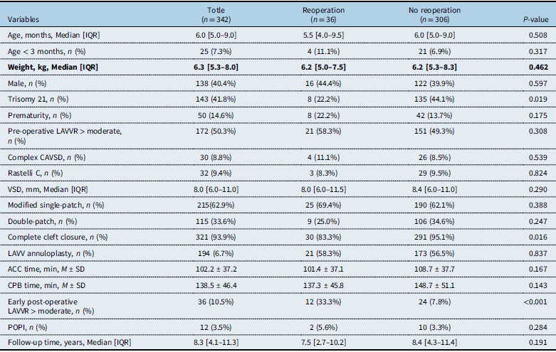
In our cohort, the median age at surgical repair was 6.0 months (interquartile range 5.0–9.0), the median weight at surgical repair was 6.3 kg (interquartile range 5.3–8.0), and the median follow-up time was 8.3 (interquartile range 4.1–11.3) years. Hundred and thirty-eight (40.4%) patients were male, and 143 (41.8%) patients had trisomy 21. Complex complete atrioventricular septal defect was present in 30 (8.8%) patients. Baseline demographic, anatomic, and surgical characteristics, stratified according to reoperation and mortality during follow-up, are detailed in Table 1 and Table S1.
.Baseline patient characteristics stratified by reoperation

Table 1. Long description
A table with 17 rows and 6 columns comparing baseline patient characteristics stratified by reoperation. The columns are labeled Totle (n = 342), Reoperation (n = 36), No reoperation (n = 306), and P-value. The rows include variables such as Age, months, Median [IQR], Age < 3 months, n (%), Weight, kg, Median [IQR], Male, n (%), Trisomy 21, n (%), Prematurity, n (%), Pre-operative LAWR > moderate, n (%), Complex CAVSD, n (%), Rastelli C, n (%), VSD, mm, Median [IQR], Modified single-patch, n (%), Double-patch, n (%), Complete cleft closure, n (%), LAVV annuloplasty, n (%), ACC time, min, M ± SD, CPB time, min, M ± SD, Early post-operative LAWR > moderate, n (%), POPI, n (%), and Follow-up time, years, Median [IQR]. Each row provides specific data points for each variable across the different columns.
IQR = interquartile range; LAVVR = left atrioventricular valve regurgitation; CAVSD = complete atrioventricular septal defect; VSD = ventricular septal defect; LAVV = left atrioventricular valve; ACC = aortic cross-clamp; M ± SD = mean ± standard deviation; CPB = cardiopulmonary bypass; POPI = post-operative pacemaker implantation.
Reoperation
Of the 36 patients who underwent reoperation, the indications were severe left atrioventricular valve regurgitation alone in 29 cases (80.6%), severe left atrioventricular valve regurgitation with residual ventricular septal defect in 5 cases (13.9%), and left atrioventricular valve regurgitation with concomitant left ventricular outflow tract obstruction in 2 cases (5.6%). These patients exhibited a significantly lower prevalence of trisomy 21 (22.2% vs. 44.1%, P = 0.019) and a lower rate of complete cleft closure (83.3% vs. 95.1%, P = 0.016). Most notably, the incidence of early post-operative left atrioventricular valve regurgitation > moderate was significantly higher in the reoperation group (33.3% vs. 7.8%, P < 0.001).
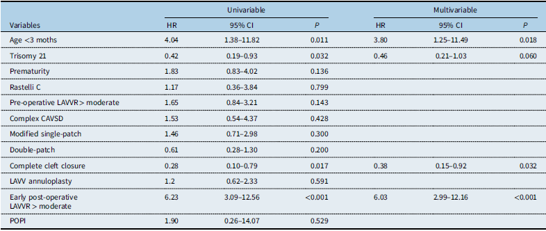
Univariable Cox regression analysis identified four candidate variables associated with reoperation (P < 0.10), including age < 3 months, trisomy 21, complete cleft closure, and early post-operative left atrioventricular valve regurgitation > moderate. In the multivariable Cox proportional hazards model, three independent risk factors for reoperation were identified: age < 3 months at repair (hazard ratio 3.80, 95% CI 1.25–11.49; P = 0.018), complete cleft closure (hazard ratio 0.38, 95% CI 0.15–0.92; P = 0.032), and early post-operative left atrioventricular valve regurgitation > moderate (hazard ratio 6.03, 95% CI 2.99–12.16; P < 0.001), while trisomy 21 showed a borderline association (hazard ratio 0.46, 95% CI 0.21–1.03, P = 0.060) (Table 2). In the Fine–Gray analysis model, trisomy 21 was associated with a significantly decreased sub-distribution hazard of reoperation (Fine–Gray, P = 0.015) (Figure 2 a ).
Long-term outcomes according to trisomy 21 and complex anatomy status. ( a ) Freedom from reoperation in patients with and without trisomy 21 (Fine–Gray, P = 0.015). ( b ) Overall survival in patients with complex complete atrioventricular septal defect (CAVSD) versus non-complex CAVSD (Fine–Gray, P = 0.068).

Figure 2. Long description
Two line graphs compare freedom from reoperation and survival rates over time for different patient groups. Panel A: The line graph shows freedom from reoperation by Trisomy 21 status. The x-axis represents follow-up time in years, ranging from 0 to 18 years. The y-axis represents the percentage of freedom from reoperation, ranging from 70 percent to 100 percent. Two lines are plotted: one for Non-Trisomy 21 (blue) and one for Trisomy 21 (red). The blue line shows a decline in freedom from reoperation over time, while the red line remains relatively stable. The P value is 0.015. Panel B: The line graph shows overall survival according to complex CAVSD status. The x-axis represents follow-up time in years, ranging from 0 to 18 years. The y-axis represents the overall survival percentage, ranging from 70 percent to 100 percent. Two lines are plotted: one for Complex CAVSD (black) and one for Non-Complex CAVSD (blue). The black line shows a decline in survival over time, while the blue line remains relatively stable. The P value is 0.068.
Risk factors for reoperation after CAVSD repair

Table 2. Long description
A table comparing risk factors for reoperation after CAVSD repair using univariable and multivariable analysis. The table has 13 rows and 6 columns. The columns are labeled Variables, Univariable HR, Univariable 95% CI, Univariable P, Multivariable HR, Multivariable 95% CI, and Multivariable P. Row 1: Age <3 moths, 4.04, 1.38–11.82, 0.011, 3.80, 1.25–11.49, 0.018. Row 2: Trisomy 21, 0.42, 0.19–0.93, 0.032, 0.46, 0.21–1.03, 0.060. Row 3: Prematurity, 1.83, 0.83–4.02, 0.136,, , . Row 4: Rastelli C, 1.17, 0.36–3.84, 0.799,, , . Row 5: Pre-operative LAWR > moderate, 1.65, 0.84–3.21, 0.143,, , . Row 6: Complex CAVSD, 1.53, 0.54–4.37, 0.428,, , . Row 7: Modified single-patch, 1.46, 0.71–2.98, 0.300,, , . Row 8: Double-patch, 0.61, 0.28–1.30, 0.200,, , . Row 9: Complete cleft closure, 0.28, 0.10–0.79, 0.017, 0.38, 0.15–0.92, 0.032. Row 10: LAVV annuloplasty, 1.2, 0.62–2.33, 0.591,, , . Row 11: Early post-operative LAWR > moderate, 6.23, 3.09–12.56, <0.001, 6.03, 2.99–12.16, <0.001. Row 12: POPI, 1.90, 0.26–14.07, 0.529,, , .
HR = hazard ratio; CI = confidence Interval; LAVV = left atrioventricular valve; LAVVR = left atrioventricular valve regurgitation; CAVSD = complete atrioventricular septal defect; POPI = post-operative pacemaker implantation.
The Kaplan–Meier curve for freedom from reoperation after complete atrioventricular septal defect repair is presented in Figure 3 a . Estimated freedom from reoperation rates were 91% at 5 years (95% CI 88–94%), 89% at 10 years (95% CI 85–92%), and 87% at 15 years (95% CI 82–91%) (Figure 3 a ).
Long-term outcomes following primary biventricular repair for complete atrioventricular septal defect (CAVSD). ( a ) Freedom from reoperation during the follow-up period. ( b ) Overall survival rate during the follow-up period.

Mortality
During the follow-up period, 22 of 342 (6.4%) patients died. Patients who died were characterised by a significantly higher proportion of age < 3 months at repair (27.3% vs. 5.9%, P = 0.003), longer aortic cross-clamp time (126.5 ± 52.6 vs. 100.5 ± 35.4 minutes, P = 0.013), and longer cardiopulmonary bypass time (179.6 ± 74.0 vs. 135.6 ± 42.6 minutes, P = 0.001). The rate of early post-operative left atrioventricular valve regurgitation > moderate was markedly higher in the death group (54.5% vs. 7.5%, P < 0.001). Other information is detailed in Table S1.
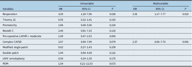
Univariable Cox regression analysis identified reoperation (hazard ratio 3.09, 95% CI 1.20–7.96, P = 0.020) and complex complete atrioventricular septal defect (hazard ratio 2.67, 95% CI 0.89–7.99, P = 0.079) as potential risk factors for mortality. In the multivariable Cox proportional hazards model, reoperation remained an independent risk factor for mortality (hazard ratio 3.01, 95% CI 1.17–7.77, P = 0.023), while complex complete atrioventricular septal defect showed a borderline association (hazard ratio 2.57, 95% CI 0.86–7.70, P = 0.091) (Table 3). To further evaluate the impact of complex complete atrioventricular septal defect on mortality while accounting for competing risks, Fine–Gray analysis was performed. Patients with complex complete atrioventricular septal defect had numerically lower overall survival compared with those with non-complex complete atrioventricular septal defect, though the difference did not reach statistical significance (P = 0.068) (Figure 2 b ).
The Kaplan–Meier curve for overall survival following complete atrioventricular septal defect repair is presented in Figure 3 b . Estimated overall survival rates were 93% at 5 years (95% CI 90–95%), 91% at 10 years (95% CI 87–93%), and 89% at 15 years (95% CI 84–92%) (Figure 3 b ).
The proportional hazards assumption of the Cox regression models was tested and verified for all included variables in both the reoperation and mortality models, confirming the validity of the Cox model results (Table S2).
Discussion
Primary biventricular repair has become the standard treatment for complete atrioventricular septal defect, and its long-term clinical outcomes have been stable and satisfactory over recent decades. Reference Ginde, Lam and Hill3,Reference Schleiger, Miera and Peters4 Although the quality of healthcare and paediatric cardiac surgery has advanced remarkably in East Asia over the same period, large, contemporary, long-term outcome studies focusing on local children with complete atrioventricular septal defect remain scarce. The present study was therefore designed to address these gaps by reporting contemporary long-term outcomes, identifying predictors of reoperation and mortality, and validating these associations using advanced statistical approaches in a large East Asian cohort.
The principal findings of this study can be summarised as follows. First, primary biventricular repair for complete atrioventricular septal defect yields satisfactory long-term outcomes, with 15-year freedom from reoperation of 87% and overall survival of 89%, which are comparable to those reported in other regions worldwide. Second, severe left atrioventricular valve regurgitation was the predominant indication for reoperation. Early post-operative left atrioventricular valve regurgitation > moderate, age < 3 months at primary repair, and incomplete cleft closure were independent risk factors for reoperation. Notably, after Fine–Gray analysis, without trisomy 21 was associated with a risk of reoperation, which is in line with several recent international studies. Third, reoperation was an independent predictor of late mortality, whereas complex complete atrioventricular septal defect was not associated with a significantly elevated risk of death. Fourth, all variables satisfied the proportional hazards assumption, confirming the robustness of our multivariable Cox regression results.
The overall survival and freedom from reoperation curves demonstrated nearly parallel trajectories over time, reflecting a robust temporal association between mortality and reoperation following primary complete atrioventricular septal defect repair. Reference Malhotra, Lacour-Gayet and Mitchell17 The two curves showed the most pronounced decline within the first post-operative year, suggesting that early short-term reoperation failed to achieve satisfactory outcomes and was a major contributor to early mortality in paediatric patients. Adverse events continued to accumulate notably from 3 to 5 years, which emerged as another high-risk phase dominated by progressive left atrioventricular valve regurgitation that greatly increased the likelihood of reoperation. Both curves entered distinct plateau phases during long-term follow-up: overall survival stabilised after 10 years, while freedom from reoperation levelled off at 12 years. These findings indicate that patients surviving beyond this high-risk window maintain favourable long-term cardiac and valvular stability. Reference Schumacher, Marin Cuartas and Meier8,Reference Bakhtiary, Takacs and Cho10,Reference Cai, Chen and Chen18 We believed that a risk-stratified individualised surveillance strategy is recommended. Intensive clinical follow-up and serial echocardiographic monitoring should be strictly implemented during the initial high-risk 5 years, particularly for vulnerable children, including those aged <3 months at primary repair and those with early post-operative left atrioventricular valve regurgitation > moderate. For patients who remain stable beyond 10 years post-operatively with a low likelihood of late adverse events, an individualised and less intensive follow-up regimen is clinically reasonable.
In our cohort, severe left atrioventricular valve regurgitation was the predominant indication (80.6%) for reoperation. Such adverse valvular events were predominantly observed in patients with congenital malformations of the left atrioventricular valve, including papillary muscle dysplasia and parachute valve malformation, which were consistent with the findings of international reports. Reference Selcuk, Spurney and Ozturk19 Early post-operative left atrioventricular valve regurgitation > moderate was identified as the strongest independent predictor of reoperation (P < 0.001), conferring an approximately sixfold higher risk. This finding is highly consistent with large Western cohorts, reinforcing that early post-operative valvular function serves as the cornerstone of long-term repair durability. Reference Hoashi, Hirano and Hosoda20 In terms of surgical strategy, neither the modified single-patch technique nor the double-patch technique demonstrated distinct superiority in protecting left atrioventricular valve function. A small proportion of patients received adjunctive innovative techniques in our cohort, such as edge-to-edge suture, V-shaped double-layer patch, and pericardial patch reinforcement. Owing to the limited sample size of these subgroups, separate comparative analyses of these novel approaches were not performed in our study.
Globally, the optimal surgical window for complete atrioventricular septal defect repair is universally recognised as 3–6 months of age. However, due to rapid progressive heart failure and haemodynamic deterioration, some infants require urgent surgical intervention. Currently, clinical studies for infants under 3 months of age are still insufficient worldwide. Reference Ma, He and Dou21 In our study, only 25 infants underwent complete atrioventricular septal defect repair before the age of 3 months, with a median follow-up time of 4.5 years (range 0–15 years). Most patients in this subgroup underwent emergency surgery due to uncontrollable pulmonary hypertension or worsening heart failure during the clinical observation period. Four (16.0%) patients received early reoperation, and two (50.0%) died in the hospital due to post-operative left ventricular dysfunction. Up to the last follow-up, a total of six deaths were documented in this subgroup: five died within 6 months after the primary repair, and one death occurred at 3.3 years post-operatively. These findings confirm that age < 3 months is a prominent risk factor for reoperation and mortality. Young infants have miniature cardiac anatomy, delicate valvular tissue, and poor physiological tolerance to surgical stress, which collectively increase the technical difficulty of precise valve reconstruction. Moreover, emergent surgery for progressive haemodynamic compromise further increases perioperative uncertainty and mortality risk. There remains ongoing controversy regarding whether pulmonary artery banding or primary biventricular repair should be performed in young infants at an early stage. All patients enrolled in our study underwent primary biventricular repair. The overall survival rate was 93% at 5 years (95% CI 90–95%), 91% at 10 years (95% CI 87–93%), and 89% at 15 years (95% CI 84–92%), and the freedom from reoperation rates were 91% at 5 years (95% CI 88–94%), 89% at 10 years (95% CI 85–92%), and 87% at 15 years (95% CI 82–91%). We conclude that early primary biventricular repair is safe and reliable for patients with stable haemodynamics and acceptable ventricular development.
A notable finding is that in the Fine–Gray competing-risk model, complete atrioventricular septal defect children without trisomy 21 syndrome have a risk of requiring reoperation after surgery (Fine–Gray, P = 0.015). This result is consistent with recent studies. Reference Wang, Verrocchi, Liew and Zentner22 The high reoperation risk observed in patients without trisomy 21 may be attributed to specific histological and anatomical features. Patients with trisomy 21 typically exhibit redundant left atrioventricular valve tissue, which facilitates superior leaflet coaptation and improves the durability of primary repair. Furthermore, these patients often have more favourable valve morphology and less severe leaflet distortion, allowing more effective cleft closure and valvular reconstruction. Reference Azzab, Samy and Singab23 These intrinsic advantages lower the incidence of progressive left atrioventricular valve regurgitation and thereby reduce the need for reoperation.
There is no doubt that reoperation was identified as an independent risk factor for all mortality (P = 0.023). Combined with our multivariate risk analysis of reoperation, individualised perioperative management strategies are strongly recommended. Sufficient pre-operative physical maturation should be guaranteed to ensure adequate valvular tissue area; meticulous anatomical reconstruction of the atrioventricular valve leaflets should be performed during surgery; and post-operative haemodynamic monitoring and blood pressure regulation should be strictly implemented.
We also investigated the reoperation and mortality risks for patients with complex complete atrioventricular septal defect. The definition of complex complete atrioventricular septal defect is provided in the previous text. A total of 30 patients were classified as having complex complete atrioventricular septal defect in our cohort, including 10 with double-outlet right ventricle, 8 with tetralogy of Fallot, 8 with unroofed coronary sinus syndrome, 1 with tetralogy of Fallot + unroofed coronary sinus syndrome, and 3 with double-outlet right ventricle + unroofed coronary sinus syndrome. Although patients with complex complete atrioventricular septal defect had longer aortic cross-clamp time and cardiopulmonary bypass time, complex complete atrioventricular septal defect was not a significant risk factor for reoperation (P = 0.428) or mortality (P = 0.091). These findings were further validated by Fine–Gray competing-risk analysis, which confirmed no significant association between complex complete atrioventricular septal defect and mortality (Fine–Gray, P = 0.068). Some clinicians may question this result, as it is generally believed that prolonged aortic cross-clamp and cardiopulmonary bypass time increase the risk of post-operative death. Our team offers the following explanations. First, there is no correlation between complex complete atrioventricular septal defect and reoperation. This is because reoperation after complete atrioventricular septal defect primary repair is mainly caused by left atrioventricular valve regurgitation, and this condition is largely independent of whether complete atrioventricular septal defect is complex or simple. Second, regarding the mortality, although the P = 0.068 did not reach a statistically significant level, the limited number of death events in this study may introduce statistical uncertainty. Therefore, we believe it is necessary to conduct long-term continuous follow-up and expand the sample size to clarify this issue. Within the limitations of this study, we conclude that complex complete atrioventricular septal defect is not a significant independent risk factor for mortality.
Limitations
This study has several limitations that warrant consideration when interpreting its findings. First, the retrospective single-centre design inherently confers a risk of selection bias. Second, surgical techniques and perioperative management evolved over the 15-year study period, which may influence early and late outcomes. Third, the number of deaths in our study was relatively small, and we cannot exclude the possibility of a high mortality rate among patients lost to follow-up.
Conclusion
This East Asian complete atrioventricular septal defect cohort employed Fine–Gray competing-risk analysis for accurate assessment. Favourable long-term outcomes were achieved after primary repair. Early progressive left atrioventricular valve regurgitation and young age independently increased the reoperation risk, while incomplete cleft closure and absence of trisomy 21 are also risk factors. Reoperation was a strong predictor of mortality. This study provides essential regional evidence to optimise surgical strategies and personalised management for paediatric complete atrioventricular septal defect.
Risk factors for mortality after CAVSD repair

Table 3. Long description
A table comparing risk factors for mortality after CAVSD repair. The table has 11 rows and 6 columns. The columns are labeled Variables, Univariable HR, Univariable 95% CI, Univariable P, Multivariable HR, Multivariable 95% CI, and Multivariable P. The rows list different variables such as Reoperation, Trisomy 21, Prematurity, Rastelli C, Pre-operative LAVVR > moderate, Complex CAVSD, Modified single-patch, Double-patch, LAVV annuloplasty, and POPI. Each row provides the hazard ratio (HR), 95% confidence interval (CI), and P-value for both univariable and multivariable analyses.
HR = hazard ratio; CI = confidence Interval; LAVV = left atrioventricular valve; LAVVR = left atrioventricular valve regurgitation; CAVSD = complete atrioventricular septal defect; POPI = post-operative pacemaker implantation.
Supplementary material
The supplementary material for this article can be found at https://doi.org/10.1017/S1047951126123944.
Data availability statement
The data that support the findings of this study are available from the first author upon reasonable request.
Acknowledgements
We acknowledge the roles of our colleagues, perfusionists, nurses, and others involved in the care of the study participants. The article was written without using any artificial intelligence tools.
Author contributions
Honghao Fu and Zhangwei Wang: Conceptualisation; Data curation; Formal analysis; Investigation; Methodology; Software; Validation; Writing—original draft; Shoujun Li: Writing—review and editing. All authors reviewed the results and approved the final version of the manuscript. Honghao Fu and Zhangwei Wang contributed equally to this work.
Financial support
This research is supported by the following funds:
-
1. Noncommunicable Chronic Disease-National Science and Technology Major Project of China (No.2024ZD0527001).
-
2. National Natural Science Foundation of China (No.82470318).
Competing interests
The authors declare no conflicts of interest to report regarding the present study.
Ethical standard
This study was approved by the Ethics Committee of Fuwai Hospital, Chinese Academy of Medical Science, and Peking Union Medical College (approval no. 2025-2653). This study was conducted in accordance with the Declaration of Helsinki.