INTRODUCTION
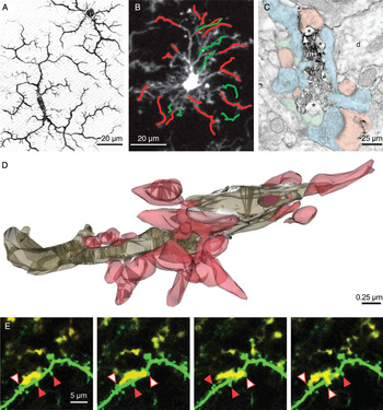
Microglia were discovered almost a century ago by Pio del Rio-Hortega. From his light microscopic observations of silver carbonate impregnated microglia in rabbit, cat and human brain in situ (1919–27; see Fig. 1A), del Rio-Hortega proposed that microglia have a mesodermal origin, colonize the brain during embryonic development, become evenly distributed, display little morphological variation in the mature brain, but respond to pathological insults by transforming their morphology and acquiring the capacity to migrate, proliferate and phagocytose (Del Rio-Hortega, Reference Del Rio-Hortega and Penfield1932; reviewed in Kettenmann et al., Reference Kettenmann, Hanisch, Noda and Verkhratsky2011). Since his formulation of these visionary postulates, which are still valid today, the changes in microglial morphology, gene expression and functional behavior during infection, trauma, ischemia and various neuropsychiatric and neurodegenerative diseases have been extensively studied. In all these situations, where normal homeostasis is impaired, activated microglia were shown to perform a variety of roles essential to the immune and inflammatory response of the central nervous system (CNS), including the release of numerous factors and compounds, presentation of antigens to T cells and phagocytosis of tissue debris, damaged cells and microbes (reviewed in Kreutzberg, Reference Kreutzberg1996; Hanisch and Kettenmann, Reference Hanisch and Kettenmann2007; Ransohoff and Perry, Reference Ransohoff and Perry2009; Rivest, Reference Rivest2009; Graeber, Reference Graeber2010; Prinz et al., Reference Prinz, Priller, Sisodia and Ransohoff2011).
Evolving views of resting microglia, from the pioneer observation of silver carbonate impregnated microglia in situ to two-photon imaging of microglia-synapse dynamics in vivo. (A) Drawing by Pio del Rio-Hortega of ramified microglial morphology (modified from Del Rio-Hortega, Reference Del Rio-Hortega1919). (B) Two-photon micrograph showing microglial process extensions (green) and retractions (red) in a CX3CR1-GFP mouse over the course of 20 min (reproduced from Nimmerjahn et al., Reference Nimmerjahn, Kirchhoff and Helmchen2005). (C) Electron micrograph showing direct contacts of an Iba1-immunostained microglial process with dendritic spines (pink), axon terminals (blue), synaptic cleft (arrow) and perisynaptic astrocytic processes (green). Note the extracellular space pockets surrounding the microglia (asterisks). (D) 3D reconstruction of proximal (cut in transverse) and distal (longitudinal) microglial processes uncovering simultaneous contacts with dendritic spines varying in shape and size (pink). Extracellular space pockets are displayed in white, and a phagocytic inclusion within the proximal microglial process is in purple. (E) Two-photon time-lapse micrographs showing a dynamic microglial process (yellow for presentation purposes) transiently interacting with dendritic spines (green) in a CX3CR1-GFP/Thy1-YFP mouse over the course of 20 min. Each frame was captured 5 min apart. Red arrowheads indicate non-targeted spines, and white arrowheads targeted ones. (C) to (E) reproduced from Tremblay et al. (Reference Tremblay, Lowery and Majewska2010b).

The roles of ‘resting’ or immunologically quiescent microglia have remained relatively unknown (also see Tremblay et al., Reference Tremblay, Stevens, Sierra, Wake, Bessis and Nimmerjahn2011). This is largely due to the difficulties of studying microglia in their non-activated state. Microglia respond promptly to any changes occurring in their environment, and therefore experimental ex vivo and in vitro preparations inevitably result in transformation of their normally prevailing behavior. Recently, the development of in vivo two-photon laser scanning microscopy (or two-photon) has for the first time allowed the study of resting microglia in the intact brain of living animals (Davalos et al., Reference Davalos, Grutzendler, Yang, Kim, Zuo and Jung2005; Nimmerjahn et al., Reference Nimmerjahn, Kirchhoff and Helmchen2005), using CX3CR1-GFP transgenic mice in which microglia are fluorescently labeled (Jung et al., Reference Jung, Aliberti, Graemmel, Sunshine, Kreutzberg and Sher2000; see Section 1 for details on these important mice). When imaged through the skull, microglia in the superficial layers of somatosensory and motor cortices of adult animals displayed small cell bodies with highly ramified thin processes extending radially and were distributed homogeneously in three dimensions (3D), as anticipated from previous in situ observations (see Del Rio-Hortega, Reference Del Rio-Hortega and Penfield1932). But quite unexpectedly, microglia were also extremely dynamic, continuously extending and retracting processes at an average velocity of ~1.5 µm min−1 (~2.2 µm min−1 for terminal protrusions) and maximal velocity of ~4 µm min−1, thereby leading to comprehensive changes in cellular morphology on a time scale of minutes (Davalos et al., Reference Davalos, Grutzendler, Yang, Kim, Zuo and Jung2005; Nimmerjahn et al., Reference Nimmerjahn, Kirchhoff and Helmchen2005; Fig. 1B). This dynamic remodeling contrasts both to neurons and other types of glial cells imaged in vivo in the cerebral cortex of juvenile and adult mice, which showed no comparable restructuring of processes. Astrocytic processes showed no motility at all over 1 h (Eom et al., Reference Eom, Stanco, Weimer, Stabingas, Sibrack and Gukassyan2011), neuron–glial antigen 2 (NG2)-positive glial cells remodeled their processes over several days (Hughes et al. (Reference Hughes, Fukaya, Kang and Bergles2011); Society for Neuroscience abstract No. 548.12), while the most motile dendritic spines only reached an average velocity of ~0.02 µm min−1 (Majewska and Sur, Reference Majewska and Sur2003). Following these observations, ‘resting’ microglia have emerged as the most structurally dynamic cells of the mature CNS discovered so far.
This unexpected behavior suggested that resting or surveillant microglia may continuously survey the brain parenchyma as part of their immune function, which would justify the substantial expenditure of energy required to continuously maintain microglial dynamics in the normal brain, without excluding the possibility of an additional, distinct contribution to normal brain physiology. Although the field of research addressing the roles of microglia under non-pathological conditions is still very young and comprises only a few studies, for the purpose of this special issue of Neuron Glia Biology, I will discuss these emerging roles of microglia at synapses in the mammalian CNS. Within this limited scope, I will focus on non- or minimally invasive in situ and in vivo imaging approaches, and discuss recent observations describing the structural interactions between microglia and synaptic elements. I will also examine the evidence supporting a regulation of microglial behavior by neurotransmission, neuronal activity and sensory experience, and conversely, for an involvement of microglia in the elimination of synaptic structures during experience-dependent plasticity.
Characterization of microglial interactions with synaptic elements
Electron microscopy (EM) enables the investigator to accurately visualize the fine structural intricacies between all the cells and their various compartments, and the extracellular matrices binding them together, thereby uncovering their immensely complex array of possible functional interactions at the local level within any tissue, at the best resolution (approximately 1 nm) so far achieved by a morphological technique. Over the last 60 years, ultrastructural examinations of the CNS have provided unprecedented insights into the functional relationships of neurons and glial cells (astrocytes, NG2-positive cells, oligodendrocytes and microglia in most regions of the mature CNS) under various conditions of health and disease. For example, EM contributed to the establishment of astrocytes as synaptic partners modulating neurotransmission (see Ventura and Harris, Reference Ventura and Harris1999; reviewed in Theodosis et al., Reference Theodosis, Poulain and Oliet2008) and to the recent discovery of synaptic interactions between NG2-positve cells and neurons (reviewed in Bergles et al., Reference Bergles, Jabs and Steinhäuser2010). Some 40 years ago, a possible but still controversial role of activated microglia in synaptic stripping also emerged from EM observations showing that microglial processes structurally intervene between pre-synaptic axon terminals and post-synaptic dendrites or neuronal cell bodies under pathological conditions (Blinzinger and Kreutzberg, Reference Blinzinger and Kreutzberg1968; reviewed in Graeber, Reference Graeber2010; Perry and O'Connor, Reference Perry and O'Connor2010; see also Trapp et al., Reference Trapp, Wujek, Criste, Jalabi, Yin and Kidd2007).
Microglial cell bodies under healthy, non-pathological conditions were also described with EM just as long ago (Herndon, Reference Herndon1964; Mori and Leblond, Reference Mori and Leblond1969), but since microglial processes are rarely contiguous to their cell bodies in single ultrathin sections, and these processes have few distinctive ultrastructural features, their discrimination from other subcellular elements (such as small unmyelinated axons, dendrites and other small glial processes) required the development of selective labeling approaches. In particular, an enzymatic staining for thiamine pyrophosphatase (TPPase) allowed examination of microglial processes in situ for the first time 30 years ago, revealing their direct apposition with excitatory synapses in the cerebral cortex of mature rats (Murabe and Sano, Reference Murabe and Sano1982). Since TPPase labels not only microglia, but also neurons, astrocytes and oligodendrocytes (Kumamoto et al., Reference Kumamoto, Nakagawa, Yata and Shimizu1976; Castellano et al., Reference Castellano, Gonzales and Palacios1989), a more specific approach was needed to validate these observations. To this end, immunocytochemical EM with a specific antibody directed against ionized calcium binding adaptor molecule 1 (Iba1), a protein only expressed by microglia within the healthy CNS (Ito et al., Reference Ito, Imai, Ohsawa, Nakajima, Fukuuchi and Kohsaka1998), was recently performed, showing that microglial processes can interact with both pre-synaptic axon terminals and post-synaptic dendritic spines in the somatosensory cortex of adult mice (Wake et al., Reference Wake, Moorhouse, Jinno, Kohsaka and Nabekura2009). Contrary to these interactions with excitatory synapses, the existence of microglial interactions with inhibitory synapses under normal physiological conditions remains yet unknown.
Extending this characterization, Tremblay et al. (Reference Tremblay, Lowery and Majewska2010b) revealed with a similar immunocytochemical EM approach (see Tremblay et al. (Reference Tremblay, Riad and Majewska2010a) for methodological details) that almost all microglial processes localize to the vicinity of excitatory synapses in the superficial layers of juvenile mouse visual cortex: ~94% of microglial processes directly contacted terminals, spines, perisynaptic astrocytic processes and synaptic clefts, in decreasing order of frequency, and ~68% of these processes contacted more than one synaptic element (Fig. 1C). Conversely, the proportion of each of those synaptic elements contacted by microglial processes could not be determined, because not all microglial processes were positively identified, since only a subset were stained for Iba1 under these stringent immunocytochemical conditions. Recently, microglial interactions with terminals, spines, astrocytic processes and synaptic clefts were also confirmed throughout adulthood and normal aging, in the superficial layers of mouse visual and auditory cortices (Tremblay et al., Reference Tremblay, Zettel, Ison, Allen and Majewska2012). Serial section EM (SSEM) with 3D reconstruction further revealed the geometry of various contacts between a single microglial process and numerous excitatory synapses (Fig. 1D) in juvenile animals (Tremblay et al., Reference Tremblay, Lowery and Majewska2010b). While microglial contacts generally occurred en passant, one of the contacts displayed morphological specialization in the form of finger-like protrusions wrapping around a spine, suggesting immediate and functional interactions. Clathrin-coated pits were also observed at interfaces between microglial processes and neuronal or astrocytic elements of synapses, on either side of the interaction, suggesting reciprocal exchange of molecular signals through clathrin-mediated endocytosis of membrane-bound receptors and their ligands (see Le Roy and Wrana (Reference Le Roy and Wrana2005) for details on clathrin-mediated endocytosis). Even though the nature of the exchanged signals remains to be determined, these two types of morphological specializations (finger-like protrusions and clathrin-coated pits) indicate that surveillant microglia can functionally interact with synaptic structures under non-pathological conditions.
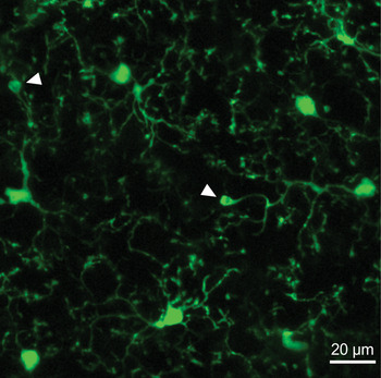
Inspired by the extreme dynamism of surveillant microglia in the intact CNS (Davalos et al., Reference Davalos, Grutzendler, Yang, Kim, Zuo and Jung2005; Nimmerjahn et al., Reference Nimmerjahn, Kirchhoff and Helmchen2005), recent studies have additionally explored the dynamic nature of microglial interactions with synaptic structures using two-photon in vivo imaging. The imaging of microglia and synaptic structures required the development of transgenic mice in which both microglia and neurons are fluorescently labeled. For this purpose, Wake et al. (Reference Wake, Moorhouse, Jinno, Kohsaka and Nabekura2009) crossed Iba1-GFP (Hirasawa et al., Reference Hirasawa, Ohsawa, Imai, Ondo, Akazawa and Uchino2005) with Thy1-GFP mice (Feng et al., Reference Feng, Mellor, Bernstein, Keller-Peck, Nguyen and Wallace2000) for visualizing microglia and neuronal elements including axons, dendrites, terminals and spines in the same color (green). More recently, other investigators crossed the CX3CR1-GFP mice from Jung et al. (Reference Jung, Aliberti, Graemmel, Sunshine, Kreutzberg and Sher2000), which provide exceptional in vivo visualization of microglial morphology including the distal processes and fine protrusions (Fig. 2), with Thy1-YFP mice (Feng et al., Reference Feng, Mellor, Bernstein, Keller-Peck, Nguyen and Wallace2000 or Hirrlinger et al., Reference Hirrlinger, Scheller, Braun, Quintela-Schneider, Fuss and Hirrlinger2005), enabling simultaneous visualization of microglia and neurons in two different colors (green and yellow respectively, see Fuhrmann et al. (Reference Fuhrmann, Bittner, Jung, Burgold, Page and Mitteregger2010); Tremblay et al. (Reference Tremblay, Lowery and Majewska2010b) and Dibaj et al. (Reference Dibaj, Nadrigny, Steffens, Scheller, Hirrlinger and Schomburg2010) for examples in cerebral cortex and spinal cord). It should be noted that CX3CR1-GFP heterozygous mice generally used for imaging may be partially deficient in fractalkine signaling, a pathway that was previously shown to modulate microglial activation and cytokine release, neuron–microglia communication and neuronal survival in different contexts of disease and CNS regions (for example see Harrison et al., Reference Harrison, Jiang, Chen, Xia, Maciejewski and McNamara1998; Cardona et al., Reference Cardona, Pioro, Sasse, Kostenko, Cardona and Dijkstra2006; Fuhrmann et al., Reference Fuhrmann, Bittner, Jung, Burgold, Page and Mitteregger2010; Cho et al., Reference Cho, Sun, Zhou, Kauppinen, Halabisky and Wes2011; Donnelly et al., Reference Donnelly, Longbrake, Shawler, Kigerl, Lai and Tovar2011) due to the replacement of one of the two alleles encoding microglial CX3CR1 with a GFP reporter. Nevertheless, many aspects of microglial morphology, process motility (Nimmerjahn et al., Reference Nimmerjahn, Kirchhoff and Helmchen2005; Wake et al., Reference Wake, Moorhouse, Jinno, Kohsaka and Nabekura2009) and interactions with synaptic elements (Wake et al., Reference Wake, Moorhouse, Jinno, Kohsaka and Nabekura2009; Tremblay et al., Reference Tremblay, Lowery and Majewska2010b) that were examined in vivo were comparable between the CX3CR1-GFP heterozygous mice and Iba1-GFP mice from Hirasawa et al. (Reference Hirasawa, Ohsawa, Imai, Ondo, Akazawa and Uchino2005) where CX3CR1 is not deleted.
Imaging of surveillant microglia in vivo with the CX3CR1-GFP mice from Jung et al. (Reference Jung, Aliberti, Graemmel, Sunshine, Kreutzberg and Sher2000). Two-photon micrograph (maximum z-projection of 20 slices captured 1 µm apart) showing the density and morphology of microglial cell bodies, processes and terminal protrusions, with or without bulbous endings (shown by arrowheads), when imaged through a thinned skull in somatosensory cortex.

Using Iba1-GFP/Thy1-GFP mice and CX3CR1-GFP/Thy1-YFP mice, respectively, Wake et al. (Reference Wake, Moorhouse, Jinno, Kohsaka and Nabekura2009) and Tremblay et al. (Reference Tremblay, Lowery and Majewska2010b) recently explored microglial interactions with axon terminals and dendritic spines from subsets of cortical layer V pyramidal neurons in vivo. With great caution, a non-invasive thinned-skull preparation was performed to prevent brain injury and microglial activation (see Tremblay et al. (Reference Tremblay, Lowery and Majewska2010b), Yang et al. (Reference Yang, Pan, Parkhurst, Grutzendler and Gan2010) and Marker et al. (Reference Marker, Tremblay, Lu, Majewska and Gelbard2010) for methodological details). In this manner, Wake et al. (Reference Wake, Moorhouse, Jinno, Kohsaka and Nabekura2009) first showed that microglial processes transiently interact with terminals and spines, in the somatosensory and visual cortices of adult animals. Microglial contacts with terminals generally lasted ~5 min and occurred at a frequency of ~1 per hour and per terminal. During the contacts, microglial processes displayed enlarged extremities or bulbous endings (Wake et al., Reference Wake, Moorhouse, Jinno, Kohsaka and Nabekura2009) resembling the morphological specializations described with SSEM and 3D reconstruction in situ (Tremblay et al., Reference Tremblay, Lowery and Majewska2010b). Additionally, Tremblay et al. (Reference Tremblay, Lowery and Majewska2010b) observed microglial contacts with terminals and spines that varied in duration between 5 and 30 min in the visual cortex of juvenile animals, using time-lapse imaging every 5 min, thereby adding a new dimension to the modalities of microglial interactions with synaptic elements occurring in vivo under non-pathological conditions.
In the mature healthy CNS, neuronal networks are continuously remodeled through the formation, modification and elimination of synaptic structures (see Fortin et al. (Reference Fortin, Srivastava and Soderling2011) for molecular mechanisms of structural plasticity) in relation with behavioral and sensory experience. In particular, two-photon in vivo imaging in Thy1-YFP or Thy1-GFP mice revealed that spines, especially small ones, are structurally dynamic and transient in mouse visual, somatosensory, motor and frontal cortices (Trachtenberg et al., Reference Trachtenberg, Chen, Knott, Feng, Sanes and Welker2002; Zuo et al., Reference Zuo, Lin, Chang and Gan2005; De Paola et al., Reference De Paola, Holtmaat, Knott, Song, Wilbrecht and Caroni2006; Majewska et al., Reference Majewska, Newton and Sur2006; also see Alvarez and Sabatini, Reference Alvarez and Sabatini2007; Holtmaat and Svoboda, Reference Holtmaat and Svoboda2009). To determine a possible role of surveillant microglia in the structural remodeling of synaptic structures under normal physiological conditions, Tremblay et al. (Reference Tremblay, Lowery and Majewska2010b) also examined the size changes of spines and terminals before, during and after microglial contacts. Spines contacted by microglial processes during imaging (30–120 min sessions) were found to be smaller initially than those which remained non-contacted. Spines, but not terminals, also underwent transient increases in size during microglial contact, with smaller spines showing the most pronounced changes. Surprisingly, chronic imaging over 2 days further revealed a statistically significant difference in the elimination rate of microglia-contacted spines: spines contacted by microglia were more frequently eliminated than non-contacted spines (~24 versus ~7%; P ~ 0.05), and in all cases, only the small spines were seen to disappear. These observations suggest that despite an apparently random sampling of the parenchyma, microglial processes specifically target a subset of small, structurally dynamic and transient dendritic spines.
Altogether, these immunocytochemical EM, SSEM with 3D reconstruction and two-photon in vivo imaging observations indicate that as part of their normal surveillance, microglial processes frequently interact with a subset of synaptic structures. This specificity raises the intriguing possibility that microglia could perceive neuronal activity or structural changes at individual synapses.
Regulation of microglial behavior by neurotransmission, neuronal activity and sensory experience
Cultured microglia were shown to express various receptors for acetylcholine, glutamate, GABA, serotonin, dopamine, noradrenaline, ATP, cannabinoids, opioids and neuropeptides among other neurotransmitters. Each of these neurotransmitters induced changes in the electrophysiological properties of microglia (reviewed in Biber et al. (Reference Biber, Neumann, Inoue and Boddeke2007), Pocock and Kettenmann (Reference Pocock and Kettenmann2007), Kettenmann et al. (Reference Kettenmann, Hanisch, Noda and Verkhratsky2011); and see Liu et al. (Reference Liu, Wang, Liu, Wang, Zhao and Hao2011) for details and receptors involved) while ATP and glutamate also influenced microglial motility in vitro (Honda et al., Reference Honda, Sasaki, Ohsawa, Imai, Nakamura and Inoue2001; Haynes et al., Reference Haynes, Hollopter, Yang, Kurpius, Dailey and Gan2006; Liu et al., Reference Liu, Nagarajah, Banati and Bennett2009; but also see Wu and Zhuo, Reference Wu and Zhuo2008; Chen et al., Reference Chen, Koga, Li and Zhuo2010). But importantly, since microglial resting state is believed to differ between in vitro and in vivo environments, given the absence of in situ evidence it remains uncertain whether surveillant microglia also express receptors for these neurotransmitters in the intact CNS (aside from ATP, see below). Therefore, how these demanding immune cells normally respond to the various neurotransmitters released by neurons and glial cells remains to be seen.
To determine whether surveillant microglia can detect neuronal and synaptic activity under physiological conditions, recent studies investigated microglial response to neurotransmission, neuronal activity and sensory experience in the intact CNS. Specifically, microglial sampling of the parenchyma, basal velocity of their processes and dynamic interactions with axon terminals and dendritic spines were examined.
NEUROTRANSMISSION
Even though it remains unknown whether changes in neurotransmission at individual synapses can influence microglia in the intact CNS, two imaging studies have revealed consequences of globally modulating glutamatergic, GABAergic or purinergic neurotransmission on microglial morphology and dynamic behavior in vivo or ex vivo (Davalos et al., Reference Davalos, Grutzendler, Yang, Kim, Zuo and Jung2005; Fontainhas et al., Reference Fontainhas, Wang, Liang, Chen, Mettu and Damani2011).
The first evidence for a regulation of surveillant microglia by neurotransmission in vivo was provided by the pioneer study from Davalos et al. (Reference Davalos, Grutzendler, Yang, Kim, Zuo and Jung2005). In this study, most of the imaging was performed through a thinned skull, but the application of pharmacological agents onto the cortical surface required a more invasive procedure, i.e. the removal of the skull and dura mater. Nonetheless, microglial morphology and motility seemed unaffected by the procedure at the cortical depths that were imaged. In this manner, cortical surface application of apyrase which catalyzes the hydrolysis of extracellular ATP and ADP reduced the basal velocity (length changes over time) of microglial processes. In contrast, application of carbenoxolone (an inhibitor of connexin and pannexin-1) or flufenamic acid (an inhibitor of connexin) nearly abolished their velocity within 30 min (Davalos et al., Reference Davalos, Grutzendler, Yang, Kim, Zuo and Jung2005), suggesting that ATP released from astrocytes via connexin channels, such as forming gap junctions, or hemichannels could mediate the effects of extracellular purines on microglial process velocity (see Fields and Burnstock, Reference Fields and Burnstock2006; Inoue et al., Reference Inoue, Koizumi and Tsuda2007; Kang et al., Reference Kang, Kang, Lovatt, Torres, Zhao and Lin2008; Halassa and Haydon, Reference Halassa and Haydon2010 for details; also see discussion below).
Building on these observations, Fontainhas et al. (Reference Fontainhas, Wang, Liang, Chen, Mettu and Damani2011) recently discovered a modulation of microglial dynamics by glutamatergic, GABAergic and purinergic neurotransmission ex vivo in adult CX3CR1-GFP mice acute retinal explants, a preparation that minimizes CNS injury because the retina is imaged as a whole. In this preparation, application of glutamate receptor agonists (AMPA and kainate) into the recording chamber increased the size (area and length), complexity and basal velocity of microglial processes, whereas GABA or GABAA receptor antagonist bicuculline (BCC) had opposite effects. ATP also increased the size, complexity and velocity of microglial processes within a few minutes of application, while opposite changes were observed with apyrase, suramin (a purinergic P2 receptor antagonist) or probenecid (a pannexin-1 inhibitor; Silverman et al., Reference Silverman, Locovei and Dahl2008), suggesting ATP release from pannexin-1 channels, which are expressed by excitatory neurons, inhibitory neurons and astrocytes under non-pathological conditions (see Inoue et al., Reference Inoue, Koizumi and Tsuda2007; MacVicar and Thompson, Reference MacVicar and Thompson2010; also see Wong et al., Reference Wong, Wang and Li2011). Additionally, voltage-clamp recordings failed to reveal any inward or outward currents in microglia during local application of AMPA or GABA, although application of ATP did induce large inward currents (Fontainhas et al., Reference Fontainhas, Wang, Liang, Chen, Mettu and Damani2011).
During brain injury, purinergic signaling was shown to regulate microglial process dynamics in vivo, with ATP released from the damage tissue and/or the surrounding cells inducing process extension in surveillant microglia via P2Y12 microglial receptors, and adenosine (produced from the extracellular degradation of ATP into ADP, AMP and adenosine notably by microglial ectonucleotidases; see Kettenmann et al., Reference Kettenmann, Hanisch, Noda and Verkhratsky2011) inducing process retraction in activated microglia via A2A receptors (Davalos et al., Reference Davalos, Grutzendler, Yang, Kim, Zuo and Jung2005; Haynes et al., Reference Haynes, Hollopter, Yang, Kurpius, Dailey and Gan2006; Orr et al., Reference Orr, Orr, Li, Gross and Traynelis2009). Under normal physiological conditions, since pre-synaptic release of glutamate and GABA is regulated by neuronal activity, and ATP is released from connexin and pannexin-1 channels in an activity-dependent manner (see Fields and Burnstock, Reference Fields and Burnstock2006; Inoue et al., Reference Inoue, Koizumi and Tsuda2007; MacVicar and Thompson, Reference MacVicar and Thompson2010), ATP could additionally represent an indirect mechanistic link between glutamatergic and GABAergic neurotransmission and microglial process dynamics. The particular subtypes of microglial purinergic receptors recruited by ATP in these conditions are yet to be determined, but P2Y12 receptor is probably not involved since surveillant microglia displayed a normal basal process velocity when imaged through the skull in the cerebral cortex of P2Y12 receptor-deficient adult CX3CR1-GFP mice in vivo (Haynes et al., Reference Haynes, Hollopter, Yang, Kurpius, Dailey and Gan2006).
NEURONAL ACTIVITY
Two additional in vivo studies correlated the changes in spontaneous or evoked neuronal activity, induced by pharmacological manipulations of neuronal excitability or neurotransmission, eye enucleation or reduction of body temperature and monitored by electrophysiological recording or calcium imaging, with the changes in microglial sampling, process velocity and dynamic interactions with synaptic elements in the intact CNS (Nimmerjahn et al., Reference Nimmerjahn, Kirchhoff and Helmchen2005; Wake et al., Reference Wake, Moorhouse, Jinno, Kohsaka and Nabekura2009). Contrary to the previously described modulations of neurotransmission in vivo or ex vivo, these manipulations influenced excitatory and inhibitory neurons simultaneously within extensive networks.
In particular, Nimmerjahn et al. (Reference Nimmerjahn, Kirchhoff and Helmchen2005) examined the changes in microglial dynamics during pharmacological manipulations of spontaneous neuronal activity in vivo. With a methodology analogous to Davalos et al. (Reference Davalos, Grutzendler, Yang, Kim, Zuo and Jung2005), enhancing excitatory neuronal activity by surface application of BCC increased microglial sampling of the parenchyma over 1 h, as evidenced by differences in fluorescence intensity between the outer (distal microglial processes) and inner rings (proximal processes) surrounding individual microglial cell bodies. However, silencing excitatory and inhibitory neuronal activity with TTX produced no significant outcome over 1 h, suggesting that microglial sampling proceeds at a basal rate that is unaffected by neuronal activity (Nimmerjahn et al., Reference Nimmerjahn, Kirchhoff and Helmchen2005). In both cases, the changes in neuronal activity were confirmed by electrocorticogram recording using minimally invasive electrodes. Moreover, both BCC and TTX were ineffective at modifying the basal velocity of microglial processes (Nimmerjahn et al., Reference Nimmerjahn, Kirchhoff and Helmchen2005), suggesting that neuronal activity increases the rate of branching or turnover of individual microglial processes without influencing their basal velocity. The discrepancy in BCC-induced changes in microglial process velocity between Nimmerjahn et al. (Reference Nimmerjahn, Kirchhoff and Helmchen2005) and Fontainhas et al. (Reference Fontainhas, Wang, Liang, Chen, Mettu and Damani2011) is unclear and could result from differences between CNS regions or between in vivo and ex vivo environments, as microglial process basal velocity was approximately two-fold higher in retina ex vivo than in cerebral cortex or spinal cord in vivo (Liang et al., Reference Liang, Lee, Wang, Ma, Fontainhas and Fariss2009 versus Nimmerjahn et al., Reference Nimmerjahn, Kirchhoff and Helmchen2005; Wake et al., Reference Wake, Moorhouse, Jinno, Kohsaka and Nabekura2009; Dibaj et al., Reference Dibaj, Nadrigny, Steffens, Scheller, Hirrlinger and Schomburg2010).
Wake et al. (Reference Wake, Moorhouse, Jinno, Kohsaka and Nabekura2009) further examined the influence of sensory-evoked and spontaneous neuronal activity on microglial interactions with terminals in vivo. In this study, binocular enucleation, injection of TTX into both eyes and reduction of body temperature from 37 to 32°C each reduced neuronal activity over 2 h, which was confirmed by calcium imaging in separate experiments, and subsequently induced a retraction of microglial processes over 4–6 h (eye enucleation), while also halving the frequency of microglial contacts with individual terminals (TTX and body temperature). These observations provided the first evidence for a modulation of microglial interactions with synaptic structures by neuronal activity. Furthermore, microglial process basal velocity remained unchanged during these manipulations (Wake et al., Reference Wake, Moorhouse, Jinno, Kohsaka and Nabekura2009), in line with the observations from Nimmerjahn et al. (Reference Nimmerjahn, Kirchhoff and Helmchen2005) during cortical surface application of TTX. The discrepancy between the other effects of TTX reported by these two studies, i.e. a retraction of microglial processes (Wake et al., Reference Wake, Moorhouse, Jinno, Kohsaka and Nabekura2009) versus an unchanged microglial sampling (Nimmerjahn et al., Reference Nimmerjahn, Kirchhoff and Helmchen2005), could result from differences in experimental paradigms, such as the application of TTX onto the brain surface versus into the eyes, differences in the anesthetics used for in vivo imaging (isoflurane for Nimmerjahn et al. (Reference Nimmerjahn, Kirchhoff and Helmchen2005) versus ketamine and xylazine for Wake et al. (Reference Wake, Moorhouse, Jinno, Kohsaka and Nabekura2009)), and in the durations of the experiments and imaging sessions during which the resting state of microglia could vary (~2 h for Nimmerjahn et al. (Reference Nimmerjahn, Kirchhoff and Helmchen2005) versus ~8 h for Wake et al. (Reference Wake, Moorhouse, Jinno, Kohsaka and Nabekura2009)).
SENSORY EXPERIENCE
In addition to these manipulations of neurotransmission and neuronal activity, the effects of environmental experience on microglial interactions with synaptic elements were recently examined in vivo, during binocular light deprivation induced by housing the animals in complete darkness throughout their fourth postnatal week (Tremblay et al., Reference Tremblay, Lowery and Majewska2010b). This time point coincides with the beginning to the peak of the critical period for experience-dependent plasticity in the visual system, where neuronal circuit remodeling in relation with visual experience is most pronounced (see Hooks and Chen, Reference Hooks and Chen2007). Binocular deprivation is known to increase spine motility and turnover in the visual cortex of juvenile and adult mice (Majewska and Sur, Reference Majewska and Sur2003; Keck et al., Reference Keck, Mrsic-Flogel, Vaz Afonso, Eysel, Bonhoeffer and Hübener2008), making this paradigm ideal for correlating microglial behavior with the experience-dependent modification of synaptic structures under non-pathological conditions.
In this manner, comparing differences in microglial morphology over time revealed a reduced microglial sampling of the parenchyma that was accompanied by a sparsening and thickening of microglial processes during visual deprivation, suggesting a relationship between microglial morphology and surveillance behavior. Even though microglial contacts with spines were not significantly altered in duration and frequency during visual deprivation, microglial processes were found to change their preference of contact among the small and structurally dynamic spines, from transiently growing spines during normal experience, to persistently shrinking spines during deprivation. All these changes reversed during reexposure to a normal 12-h light/dark cycle for 2 days (Tremblay et al., Reference Tremblay, Lowery and Majewska2010b). These observations indicate that microglial interactions with dendritic spines are regulated by sensory experience in the healthy CNS. Since microglial processes preferentially interact with a subset of small, structurally dynamic and frequently eliminated spines during normal sensory experience (Tremblay et al., Reference Tremblay, Lowery and Majewska2010b), and small and shrinking spines, such as those contacted by microglia during visual deprivation, have been shown to gradually disappear over a span of days in the somatosensory cortex of adult mice in vivo (Holtmaat et al., Reference Holtmaat, Wilbrecht, Knott, Welker and Svoboda2006), these findings suggest that microglial contacts could facilitate the elimination of small and structurally dynamic spines. Furthermore, since Knott et al. (Reference Knott, Holtmaat, Wilbrecht, Welker and Svoboda2006) revealed that small and recently formed spines generally lack synaptic contacts in the somatosensory cortex of adult mice in situ, using two-photon in vivo imaging over 1 month combined with retrospective SSEM with 3D reconstruction, it also remains uncertain whether the small spines contacted by microglia receive synaptic contacts.
Altogether, by examining different CNS regions and stages of the lifespan, using different experimental paradigms for modulating neurotransmission, neuronal activity or sensory experience, and different methodologies for assessing the changes in microglial dynamics in all these conditions, these studies have revealed that microglia do not behave randomly in the absence of pathological insults. In light of the combined observations, resting microglia can now be viewed as continuously and differentially modulating their morphology, basal process velocity, sampling of the parenchyma and dynamic interactions with synaptic structures under normal physiological conditions.
Microglial elimination of synaptic structures
The specificity of microglial interactions with structurally dynamic and frequently eliminated dendritic spines raises the intriguing likelihood that surveillant microglia may facilitate the elimination of dendritic spines, with or without synaptic contacts, thereby preventing the formation of new synapses or promoting the elimination of existing ones. In this way, they could contribute to the experience-dependent remodeling of neuronal networks that is required for learning and memory storage throughout the lifespan. Mechanisms by which surveillant microglia may eliminate synaptic structures were recently proposed and will be described here.
MICROGLIAL PHAGOCYTOSIS OF AXON TERMINALS AND DENDRITIC SPINES
Like macrophages, activated microglia contribute to preserving normal homeostasis by their phagocytosis and clearance of cellular debris (see Neumann et al. (Reference Neumann, Kotter and Franklin2009) and Napoli and Neumann (Reference Napoli and Neumann2009) for details on the mechanisms of microglial phagocytosis in different contexts). Under normal physiological conditions, Nimmerjahn et al. (Reference Nimmerjahn, Kirchhoff and Helmchen2005) observed a few examples of microglial processes engulfing unidentified tissue components which were subsequently transported to the cell bodies in vivo. More recently, Tremblay et al. (Reference Tremblay, Lowery and Majewska2010b) also noticed cellular inclusions with ultrastructural features of axon terminals and dendritic spines, i.e. synaptic vesicles and post-synaptic densities, respectively, within microglial processes and cell bodies in juvenile mouse somatosensory cortex in situ. These putative terminals and spines occasionally displayed synaptic contacts, and invariably presented ultrastructural features of healthy cellular elements such as an electron-lucent cytoplasm (see Schmechel (Reference Schmechel, Hannun and Boustany1999) for additional features of apoptotic and necrotic cells), unlike apoptotic cells that are phagocytosed by resting microglia during adult neurogenesis (Sierra et al., Reference Sierra, Encinas, Deudero, Chancey, Enikolopov and Overstreet-Wadiche2010). In vivo, microglial processes with phagocytic specializations were also observed, sometimes encircling neuronal elements, but since these structures were stable throughout the 30–120 min imaging sessions, episodes of microglial engulfment followed by a full disappearance of the synaptic elements were not completely visualized. Both in situ and in vivo, microglial phagocytic structures became more prevalent during light deprivation, and still persisted after reexposure to light for 2 days (Tremblay et al., Reference Tremblay, Lowery and Majewska2010b), in a context of sensory experience where dendritic spine remodeling and elimination is also increased (Majewska and Sur, Reference Majewska and Sur2003; Keck et al., Reference Keck, Mrsic-Flogel, Vaz Afonso, Eysel, Bonhoeffer and Hübener2008).
Additionally, Tremblay et al. (Reference Tremblay, Zettel, Ison, Allen and Majewska2012) recently observed phagocytic inclusions within microglial cell bodies and processes throughout adulthood and normal aging, using immunocytochemical EM. This study revealed that microglial cell bodies and processes accumulate large amounts of phagocytic inclusions, which often resemble terminals and spines, with or without an electron-lucent cytoplasm, but also lysosomal lipopigments, large vesicles, vacuoles and lipid droplets, in the visual and auditory cortices of two stains of mice normally undergoing complementary age-related loss of vision (CBA/CaJ mice) or hearing (C57Bl/6J mice) during their second year of life. Surprisingly, this accumulation was exacerbated by the age-related loss of visual or auditory function, with nearly all microglia containing phagocytic inclusions and ~20% of microglial cells becoming almost completely filled by inclusions, akin to fat granule cells or gitter cells, in this context of sensory deprivation. Also, microglial processes extending between axon terminals and dendritic branches were encountered, suggesting an early stage of phagocytosis or an evidence of synaptic stripping under non-pathological conditions. Together, these observations during adolescence, adulthood and normal aging suggest that microglial phagocytosis is regulated in an experience-dependent manner in the mature CNS.
In line with these observations, activated microglia have been shown to be involved in the apoptotic and phagocytic elimination of excess neurons during early postnatal development (see Bessis et al. (Reference Bessis, Bechade, Bernard and Roumier2007) and Pont-Lezica et al. (Reference Pont-Lezica, Béchade, Belarif-Cantaut, Pascual and Bessis2011) for details and additional roles during development), and recently, Paolicelli et al. (Reference Paolicelli, Bolasco, Pagani, Maggi, Scianni and Panzanelli2011) showed that microglia can also eliminate synaptic elements during the second and third postnatal weeks in CX3CR1-GFP mouse hippocampus in situ. In that study, puncta immunopositive for either PSD95 (marker of dendritic spines) or SNAP25 (axon terminals) were found to be colocalized at nanometer resolution within GFP-positive microglial processes using stimulated emission depletion microscopy (see Willig et al. (Reference Willig, Kellner, Medda, Hein, Jakobs and Hell2006) for technical details). Microglial colocalization with PSD95 was more prevalent in homozygous CX3CR1-GFP mice, where fractalkine signaling is completely eliminated (Jung et al. (Reference Jung, Aliberti, Graemmel, Sunshine, Kreutzberg and Sher2000); see Section 1 for details about these mice), and was accompanied by a reduced density of microglial cells and an increased density of PSD95 puncta with or without microglial colocalization, suggesting that reduced microglial numbers could impact the pruning of supernumerary spines (Paolicelli et al., Reference Paolicelli, Bolasco, Pagani, Maggi, Scianni and Panzanelli2011). Providing additional molecular insights, recent observations from Schafer et al. (Reference Schafer, Lehrman, Kautzman, Koyama, Mardinly and Greenberg2011; Society for Neuroscience abstract No. 663.03) also revealed that microglia can phagocytose terminals in a complement C3-dependent manner (Stevens et al., Reference Stevens, Allen, Vazquez, Howell, Christopherson and Nouri2007) during early postnatal development in mouse thalamus in situ.
Altogether, these studies propose a novel physiological role for microglial phagocytosis, in the elimination of individual synaptic structures and quite likely of entire excitatory synapses, throughout the lifespan.
REGULATION OF THE PERISYNAPTIC EXTRACELLULAR SPACE
Another proposed mechanism by which surveillant microglia could eliminate dendritic spines was suggested by the same study of Tremblay et al. (Reference Tremblay, Lowery and Majewska2010b). Immunocytochemical EM and SSEM with 3D reconstruction revealed that microglial cell bodies, proximal and distal processes are distinctively surrounded by pockets of extracellular space, which consists of interstitial fluid supplemented with various extracellular matrix proteins (see Syková and Nicholson, Reference Syková and Nicholson2008), in stark contrast to all other cellular and subcellular elements in the visual cortex of juvenile mice in situ. This tight association indicates that microglial processes could dynamically influence the volume and geometry of the extracellular space, and thus the concentration of neurotransmitters and signaling molecules in the vicinity of synapses. These pockets of extracellular space were regulated in an experience-dependent manner, undergoing expansion during light deprivation and shrinkage during light reexposure. More recently, pockets of extracellular space juxtaposing microglial cell bodies and processes were also observed in mouse auditory and visual cortices throughout adulthood and normal aging (Tremblay et al., Reference Tremblay, Zettel, Ison, Allen and Majewska2012). Interestingly, more than 40 years ago, Herndon (Reference Herndon1964) mentioned that a ‘small amount of extracellular space may be seen in a few places about the cell, particularly about its thick processes’, when describing for the first time microglial cell bodies and contiguous, proximal processes at the ultrastructural level under normal physiological conditions in adult rat cerebellum. Whether microglia create these extracellular spaces remains unknown.
Among candidate molecules for microglial involvement, the polysialylated form of neural cell adhesion molecule (PSA-NCAM) increases the size of the extracellular space (Yang et al., Reference Yang, Yin and Rutishauser1992; see Bonfanti, Reference Bonfanti2006; Syková and Nicholson, Reference Syková and Nicholson2008) and was recently found to be expressed by microglia in vitro (Wang and Neumann, Reference Wang and Neumann2010). Proteases degrading extracellular matrix proteins which are expressed by activated microglia in vitro might also be involved (see Nakanishi, Reference Nakanishi2003; Choi et al., Reference Choi, Cho, Shin, Ko, Kwon and Shin2010). Application of proteases in vitro and in vivo has been shown to promote spine motility and pruning, as well as activity-dependent and experience-dependent plasticity in the visual cortex of adult mice (Pizzorusso et al., Reference Pizzorusso, Medini, Berardi, Chierzi, Fawcett and Maffei2002; Mataga et al., Reference Mataga, Mizuguchi and Hensch2004; Oray et al., Reference Oray, Majewska and Sur2004), thereby suggesting a possible link between the regulation of the extracellular space and the elimination of the small and structurally dynamic spines preferentially contacted by microglial processes in vivo (Tremblay et al., Reference Tremblay, Lowery and Majewska2010b).
SUMMARY AND CONCLUSION
Over the last century, the development of selective staining approaches and concomitant refinement of imaging techniques, from conventional light and electron microscopy to two-photon laser scanning microscopy, have provided unprecedented insights into microglial morphology, dynamic behavior and interactions with excitatory synapses in the intact CNS. In particular, recent imaging studies have revealed that microglia are extremely dynamic in their presumed ‘resting’ state, continuously surveying their local environment with highly motile processes which transiently contact subsets of synaptic structures in vivo, especially the small, dynamic and frequently eliminated dendritic spines. Surveillant microglia also perceive ongoing changes in neurotransmission, neuronal activity and sensory experience, and respond by modulating their behavior including their interactions with dendritic spines, their phagocytic elimination of axon terminals and dendritic spines and their remodeling of the perisynaptic extracellular space. As resident immune cells, microglia are not directly bound by the constraints imposed by the continuous flow of information processing in the brain, and may uniquely contribute to remodeling neuronal circuits in relation with behavioral and sensory experience in the mature CNS. Together, these observations have opened many functional possibilities beyond immune surveillance for these once presumed ‘resting’ cells.
At this very early stage of investigation in this field, our knowledge about surveillant microglia almost exclusively comes from observing the superficial layers of cerebral cortex and cannot be confidently extrapolated to the entire CNS. Since the recent studies have barely scratched the surface (of the brain in this case), the modalities of microglial interactions with excitatory and inhibitory synapses throughout the CNS, much as their functional significance and particular cellular and molecular mechanisms still remain undetermined. For example, in which contexts do quiescent microglia directly phagocytose axon terminals and dendritic spines, use other mechanisms such as proteolytic remodeling of the extracellular space, or refrain from intervening? How do surveillant microglia recognize and respond to the various molecular signals in their environment, including dynamic changes in neurotransmission and neuronal activity at individual synapses? How do these immune cells cooperate with other glial cells, as well as peripheral myeloid cells, in maintaining or shaping neuronal architecture and activity? And, as in the case of microglial memory of past immune challenges (see Bilbo et al., Reference Bilbo, Smith and Schwarz2012), do surveillant microglia somehow remember their previous behavioral states, the flux of information processing in the brain, or the structural changes of synaptic elements in recent and not so recent windows of intervention?
Even though most essential questions remain unanswered at present, the novel insights about microglia have established integrative in vivo and in situ imaging approaches as a valuable methodological way, alongside genetics, electrophysiology and animal behavior, for studying these demanding immune cells in the healthy CNS. Promising and increasingly integrative approaches for modulating or monitoring cellular activity in a non-invasive way are now emerging, with the ongoing development of optogenetics and imaging of functional markers, sophistication of transgenic mice in the postgenomic era and improvement in the spatiotemporal resolution of microscopy techniques. Therefore, in addition to these innovative recent observations, we can optimistically envision an explosion of discoveries regarding the roles of surveillant microglia across the various CNS regions including the cerebellum and spinal cord, stages of the lifespan and states of normal brain physiology, in awake and normally behaving animals. These discoveries and their underlying technical approaches will facilitate a broader understanding of microglial involvement in various neuropsychiatric and neurodegenerative diseases in the near future.
ACKNOWLEDGEMENTS
I am extremely grateful to Andreas Buchmann, Harris A. Gelbard, Ania K. Majewska, Richard M. Ransohoff and Cynthia D. Rittenhouse for their comments and suggestions. This work was supported by Fonds de la Recherche en Santé du Québec (FRSQ) and Canadian Institutes of Health Research (CIHR) postdoctoral fellowships.
Statement of interest
None.