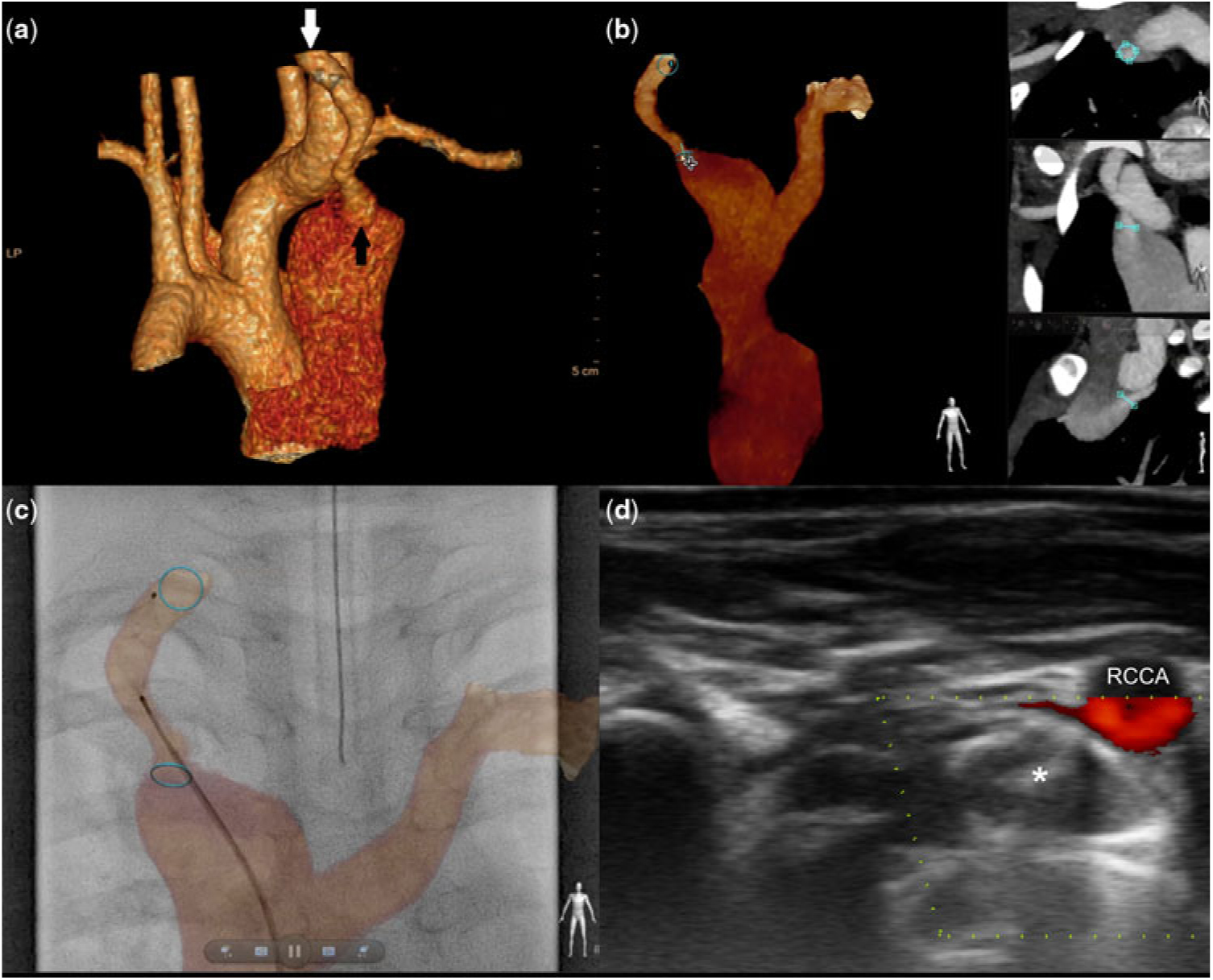
A 6-year-old patient was accidently found with a loud continuous murmur at the second right intercostal space. Echocardiography showed a 4-mm atrial septal defect with left-to-right flow and a mild biventricular enlargement. In the suprasternal notch, a continuous high-velocity flow was recorded suggesting arteriovenous fistula. Subsequent contrast computed tomography (Fig 1a) confirmed communication (minimum and maximum diameters of 4 and 8 mm, respectively) between the right common carotid artery and the enlarged superior caval vein. Before patient’s arrival to the catheterisation laboratory, the three-dimensional data set was manipulated with fusion software (VesselNavigator, Philips Healthcare, Best, Netherlands) to guide percutaneous occlusion of the fistula (Fig 1b).Reference Goreczny, Dryzek, Morgan, Lukaszewski, Moll and Moszura 1 The target vessel was manually exposed, and additional ring markers were placed to highlight the origin and the site of drainage of the connecting vessel. At the commencement of the procedure, bony structures of the neck and the upper chest were used to register the two-dimensional fluoroscopy images with the three-dimensional reconstruction (2D–3D registration). An 8-mm plug was deployed in the fistula under the sole guidance of the three-dimensional roadmap without contrast administration (Fig 1c). Before the end of the procedure, an ultrasound study confirmed unobstructed flow in the right common carotid artery with trivial residual flow through the device placed in the fistula (Fig 1d).
A contrast computed tomography shows communication between the right common carotid artery ( a , white arrow) and the enlarged superior caval vein ( a , black arrow). The 3D data set was manipulated with fusion software to highlight the origin and the site of drainage of the connecting vessel ( b ). A plug was deployed in the fistula under the sole guidance of the 3D roadmap without contrast administration ( c ). An ultrasound study confirmed unobstructed flow in the right common carotid artery with trivial residual flow through the device ( d , white asterisk). 3D = three-dimensional.

Non-invasive three-dimensional imaging may be used for direct guidance of percutaneous intervention abolishing the need for diagnostic angiograms.Reference Goreczny, Dryzek, Morgan, Lukaszewski, Moll and Moszura 1 Application of Doppler ultrasound, instead of control angiography, allows elimination of contrast administration and further reduction of radiation exposure during cardiac catheterisation.
Acknowledgements
The authors thank Dr. Katarzyna Ostrowska (Department of Cardiology, Polish Mother’s Memorial Hospital) for contribution to this patient’s care and providing constructive thoughts for the manuscript.
Financial Support
This research received no specific grant from any funding agency, commercial or not-for-profit sectors.
Conflicts of Interest
None