Aortic valve stenosis is a common lesion of the spectrum of congenital left ventricular outflow tract obstruction malformationsReference St Louis and Jaggers1 and represents about 3–6% of all congenital cardiac diseases.Reference Mack and Silberbach2 The male-to-female ratio of this disorder is about 4:1.Reference Mack and Silberbach2 The most frequent type in aortic valve stenosis is bicuspid,Reference Reich3 accounting for 70–85% of paediatric aortic valve stenosisReference Mack and Silberbach2 and 50% of adult severe aortic valve stenosis.Reference Roberts and Ko4 Tricuspid valve is seen in 25% of infants and in 40% of older patients who require treatment.Reference Reich3
Previous studies and our recent study have both suggested that genetic aetiologies may play a role in the development of aortic valve stenosis.Reference Godden, Sandhu and Kerr5–Reference Zhao, Wang and Shen8 McBride et alReference McBride, Riley and Zender7 reported that mutations in NOTCH1 gene, encoded on chromosome 9q34.3, are associated with the spectrum of left ventricular outflow tract obstruction defects, which indicates that bicuspid and tricuspid aortic valve stenosis, coarctation of the aorta, and hypoplastic left heart syndrome share a common pathogenetic mechanism at the molecular level. Previous studies have shown that vascular endothelial growth factor signalling system plays a critical role in the formation of endocardial cushions, the primordia of heart valves.Reference Armstrong and Bischoff9, Reference Wagner and Siddiqui10 Our recent study first reported that mutations in vascular endothelial growth factor-A, encoded on chromosome 6p12, may be associated with congenital left ventricular outflow tract obstruction malformations.Reference Zhao, Wang and Shen8 Of our reported three mutations, c.454C > T (p.Arg152X) nonsense mutation is related to bicuspid aortic valve stenosis.Reference Zhao, Wang and Shen8 Mutation in vascular endothelial growth factor-A gene, therefore, is another genetic factor for bicuspid aortic valve stenosis.
The question whether vascular endothelial growth factor-A variations may play a role in the aetiology of congenital tricuspid aortic valve stenosis was brought to our attention. However, to the best of our knowledge, to date, no groups have explored this question. In this study, we report for the first time that a c.973C > T (p.Arg325X) heterozygous nonsense variation in the vascular endothelial growth factor-A gene, which was previously submitted as a coding single-nucleotide polymorphism (rs45533131), occurred in a Han Chinese patient with an isolated tricuspid aortic valve stenosis. Our study suggests that the p.Arg325X nonsense variation may be associated with congenital tricuspid aortic valve stenosis.
Materials and methods
Definition
Aortic valve stenosis is an obstruction of the left ventricle outflow tract and can be categorised as mild, moderate, or severe based on the findings of transthoracic echocardiography. The peak pressure gradient across the aortic valve in the three groups is less than 40 millimetres of mercury, up to 75 millimetres of mercury, and greater than 75 millimetres of mercury, respectively, and the mean pressure gradient across the aortic valve in the three groups is less than 20 millimetres of mercury, up to 50 millimetres of mercury, and greater than 50 millimetres of mercury, respectively.Reference Nguyen11
Study population
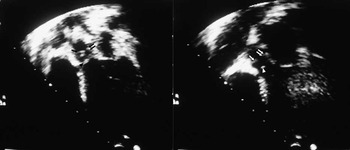
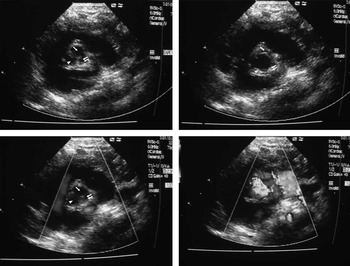
From October, 2008 to January, 2010, a total of 32 unrelated Han Chinese patients with congenital tricuspid aortic valve stenosis were enrolled. Patients with chromosomal anomalies were excluded from the study. There were 26 boys and 6 girls, aged between 2 months and 15 years with a median age of 4 years. Of the 32 patients, 15 (46.9%) had one or more other congenital cardiovascular malformations, including patent ductus arteriosus in 10 cases, subaortic stenosis (Fig 1) in six cases, coarctation of the aorta in four cases, secundum atrial septal defect in two cases, ventricular septal defect in two cases, and pulmonary valve stenosis in one case. The remaining 17 patients (53.1%) had an isolated tricuspid aortic valve stenosis (Fig 2). No family history of congenital cardiac disease was found in the study population.
Tricuspid aortic valve stenosis coexisting with subaortic stenosis. Two-dimensional apical five-chamber view showed the position of right-coronary cusp (arrow), left-coronary cusp (double arrow), and subaortic fibrous ridge (arrowhead). Left: diastole; right: systole.

Parasternal short-axis view of the aortic valve showing thickened tricuspid aortic valve with limited opening. Upper left and upper right represented the two-dimensional echocardiographic images in diastole and systole, respectively. Lower left and lower right represented the colour flow Doppler images in diastole and systole, respectively. Right-coronary, left-coronary, and non-coronary cusps are indicated by an arrow, a double arrow, and an arrowhead, respectively.

A total of 300 unrelated healthy children with no cardiovascular abnormalities were enrolled as normal controls, who underwent transthoracic echocardiography before the study. A total of 103 unrelated patients with congenital conotruncal defects, including tetralogy of Fallot in 75 cases, double-outlet right ventricle in 21 cases, and transposition of the great arteries in 7 cases, were enrolled as disease controls based on the following two reasons: one is that aortic valve stenosis is the most common type of left ventricular outflow tract obstruction,Reference St Louis and Jaggers1 which is a different category of congenital cardiac defects compared with conotruncal defects; the other is that genetic variation in vascular endothelial growth factor does not contribute significantly to the risk of non-syndromic tetralogy of Fallot as suggested by a recent study.Reference Griffin, Hall and Topf12 All cases of conotruncal defects were confirmed by surgical inspection. Subjects and controls were both racially and ethnically matched.
We obtained written informed consent from parents of each patient and control for deoxyribonucleic acid sequencing according to a protocol approved by the Medical Ethics Committee of Shanghai Children's Medical Center, Shanghai Jiaotong University School of Medicine. Blood samples were collected into tubes containing sodium citrate.
Screening for vascular endothelial growth factor-A variations
Genomic deoxyribonucleic acid was extracted from the blood samples using a standard phenol–chloroform extraction protocol. Oligonucleotide primers, which amplify all coding exons and exon–intron boundaries of the human vascular endothelial growth factor-A gene, were designed from the genomic sequence – GenBank accession number NG_008732.1 – using online Primer3 software (http://frodo.wi.mit.edu/cgi-bin/primer3/primer3.cgi) and synthesised by Invitrogen (Shanghai, China). The sequences of polymerase chain reaction primers and cycling conditions have been described in our recent study.Reference Zhao, Wang and Shen8 All polymerase chain reaction products were purified from agarose gel using a QIAquick Gel Extraction Kit (Qiagen GmbH, Hilden, Germany) and subsequently sequenced using the dideoxy chain termination method on an ABI3130XL sequencer (Applied Biosystems, Foster City, California, United States of America). Sequencing results were aligned with the reference sequence using the GenBank BLAST program. For genetic variations identified in patients with tricuspid aortic valve stenosis, the corresponding exons and exon–intron boundaries of all controls were amplified and sequenced as above.
For the patient with variations in the vascular endothelial growth factor-A gene, all available family members were screened by transthoracic echocardiography. Blood samples were collected from these individuals for corresponding sequence analysis.
Results
Classification of aortic valve stenosis severity
Among the 32 cases of congenital tricuspid aortic valve stenosis, there were 10 cases of mild stenosis, 11 cases of moderate stenosis, and 11 cases of severe stenosis. For the 10 cases of mild aortic valve stenosis, the peak pressure gradient ranged from 22.5 to 37.0 millimetres of mercury (median 34.5 millimetres of mercury) and the mean pressure gradient ranged from 13.7 to 19.9 millimetres of mercury (median 18.6 millimetres of mercury). For the 11 cases of moderate aortic valve stenosis, the peak pressure gradient ranged from 41.6 to 74.0 millimetres of mercury (median 61.3 millimetres of mercury) and the mean pressure gradient ranged from 26.1 to 43.9 millimetres of mercury (median 36.7 millimetres of mercury). For the 11 cases of severe aortic valve stenosis, the peak pressure gradient ranged from 79.2 to 149.3 millimetres of mercury (median 94.1 millimetres of mercury) and the mean pressure gradient ranged from 51.0 to 79.2 millimetres of mercury (median 57.9 millimetres of mercury).
Vascular endothelial growth factor-A gene variation
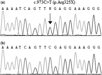
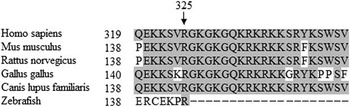
We identified a c.973C > T heterozygous nonsense variation in exon 6 of the vascular endothelial growth factor-A gene in one of the 32 unrelated patients with congenital tricuspid aortic valve stenosis, which was absent in the chromosomes of all controls. The total c.973C > T variation frequency of the vascular endothelial growth factor-A gene in this study was 3.1% (1 out of 32). The c.973C > T nonsense variation changes an arginine at amino acid 325 to a stop codon (p.Arg325X), which is predicted to produce a prematurely truncated vascular endothelial growth factor-A protein of 324 residues instead of the normal-sized product of 412 amino acids (Fig 3). Moreover, the residue involved in the p.Arg325X variant is highly conserved in different species including zebrafish (Fig 4).
Sequence of vascular endothelial growth factor-A in index patient and control subjects. (a) The heterozygous nonsense variation c.973C > T in the vascular endothelial growth factor-A gene, which was absent in 300 normal controls and 103 disease controls, was identified in a patient with an isolated tricuspid aortic valve stenosis. (b) Corresponding normal sequence in the controls. The vertical arrow indicates the position of the c.973C > T nonsense variation. All sequences are in the sense direction.

A multiple sequence alignment of the known vascular endothelial growth factor-A proteins. The arginine residue at position 325 is highly conserved among different species, including Human (NP_001020537), Mus musculus (NP_001020421), Rattus norvegicus (NP_114024), Gallus gallus (NP_990373), Canis lupus familiaris (NP_001003175), and Zebrafish (NP_001103819). The conserved Arg325 residue is indicated by an arrow.

Proband and pedigree characteristics
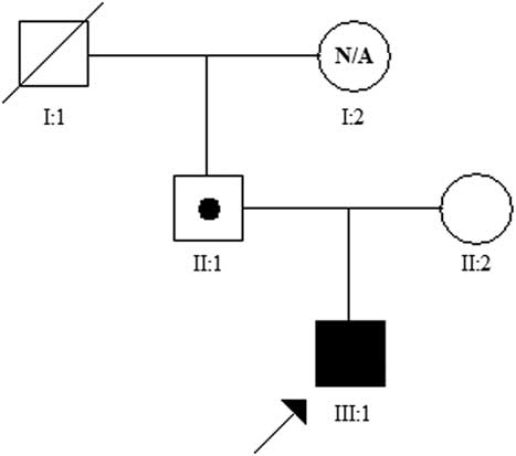
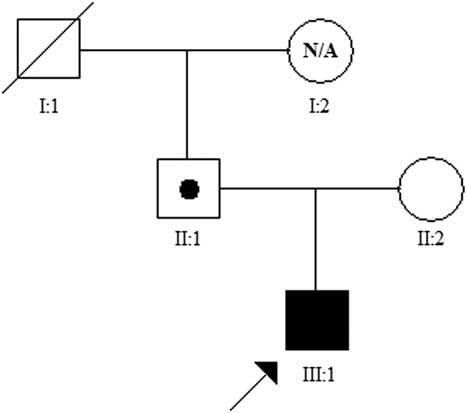
The proband, a 5-year-old boy, was referred to our hospital 2 years ago for cardiac murmur, which was discovered accidentally. Transthoracic echocardiography showed an isolated tricuspid aortic valve stenosis, with a peak systolic pressure gradient of 64 millimetres of mercury at that time. The peak systolic pressure gradient slightly increased to 69 millimetres of mercury at the time of enrolment. The proband was asymptomatic with normal cardiac function by echocardiography, and his New York Heart Association functional class was class I. He was the first and only child of healthy non-consanguineous parents with no family history of congenital cardiac disease. Sequencing results of the parents revealed that the p.Arg325X variant was carried by the father (II:1). Echocardiography of the parents showed no evidence of cardiovascular abnormalities. In the p.Arg325X variant-carrying father, the aortic valve was tricuspid, and the dimensions of the aortic annulus, the sinuses of Valsalva, the sino-tubular junction, and the proximal ascending aorta – measured 1 centimetre from the sino-tubular junction – were 2.27 centimetres, 3.09 centimetres, 2.29 centimetres, and 3.03 centimetres, respectively. The proband's grandfather (I:1) was deceased, and grandmother (I:2) was not available for the phenotype and genotype analysis (Fig 5).
Pedigree of family with the proband. Squares represent male family members; circles, female family members; blackened square, clinically and genetically affected patient; open symbol, unaffected member with normal genotype; a black dot in a square, carrier; arrow, proband; N/A, not available; and symbol with a slash, deceased member.
Discussion
In this study, to our knowledge, we identified for the first time that a c.973C > T (p.Arg325X) nonsense variation of the vascular endothelial growth factor-A gene occurred in a paediatric patient with an isolated tricuspid aortic valve stenosis. Although the c.973C > T variation of the vascular endothelial growth factor-A gene was previously submitted as a coding single-nucleotide polymorphism (rs45533131), it has not yet been validated, and its clinical association was also unknown at the time of submission (http://www.ncbi.nlm.nih.gov/SNP/snp_ref.cgi?rs=45533131).
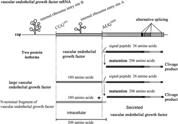
Previous studies have demonstrated that the human vascular endothelial growth factor messenger ribonucliec acid contains two internal ribosome entry sites – internal ribosome entry site A and internal ribosome entry site B – and two initiation codons – AUG1039 and CUG499. The AUG1039 is a classical initiation codon, whereas the CUG499, located in a 1038-nucleotide-long 5′-untranslated region upstream from the AUG1039 codon, is a non-canonical initiation codon.Reference Huez, Bornes, Bresson, Créancier and Prats13–Reference Tee and Jaffe15 The 5′-untranslated region of the human vascular endothelial growth factor messenger ribonucliec acid is highly conserved in mammals and contains a large open reading frame.Reference Huez, Bornes, Bresson, Créancier and Prats13, Reference Meiron, Anunu, Scheinman, Hashmueli and Levi14 A large vascular endothelial growth factor protein is generated by alternative translation initiation from the CUG499 codon and subsequently cleaved into an N-terminal fragment with 206 amino acids and a C-terminal fragment with 206 amino acids, which is identical to the classical AUG1039-initiated vascular endothelial growth factor protein (Fig 6).Reference Huez, Bornes, Bresson, Créancier and Prats13–Reference Tee and Jaffe15 Using the CUG499 as the start codon is very important, as the large vascular endothelial growth factor protein represents as much as 50% of precursors for active vascular endothelial growth factor protein.Reference Huez, Bornes, Bresson, Créancier and Prats13, Reference Tee and Jaffe15
Alternative translation initiation of the vascular endothelial growth factor-A messenger ribonucleic acid. The vascular endothelial growth factor messenger ribonucleic acid is schematised with the two active internal ribosome entry sites – internal ribosome entry site A and internal ribosome entry site B – and the two initiation codons – AUG1039 and CUG499. Beneath the messenger ribonucleic acid, the two protein isoforms, large vascular endothelial growth factor and vascular endothelial growth factor are represented together with their maturation products and their localisation. N-terminal fragment of vascular endothelial growth factor corresponding to the 180-amino acid N-terminal extension plus the signal peptide (26 amino acids).

The vascular endothelial growth factor-A is commonly referred to as vascular endothelial growth factor.Reference Dowlati16 The vascular endothelial growth factor-A gene is composed of eight exons. In humans, alternative splicing of a single vascular endothelial growth factor-A messenger ribonucliec acid results in the generation of at least eight vascular endothelial growth factor isoforms, including vascular endothelial growth factor 121 (NP_001020541.2), vascular endothelial growth factor 145 (NP_001191314.1), vascular endothelial growth factor 148 (NP_001020540.2), vascular endothelial growth factor 165 (NP_001020539.2), vascular endothelial growth factor 165b (NP_001028928.1), vascular endothelial growth factor 183 (NP_001020538.2), vascular endothelial growth factor 189 (NP_003367.4), and vascular endothelial growth factor 206 (NP_001020537.2).Reference Ribeiro, Bacci, Seren, Tamanini and Forni17 The domains, N-terminal vascular endothelial growth factor receptor-binding region, encoded by exons 1–5 are highly conserved in all vascular endothelial growth factor isoforms.Reference Tischer, Mitchell and Hartman18 The vascular endothelial growth factor isoforms are distinguished by the presence or absence of the peptides encoded by exons 6a, 6b, 7a, and 7b. These four exons encode peptides of 24, 17, 32, and 12 amino acid residues, respectively.Reference Ribeiro, Bacci, Seren, Tamanini and Forni17 The vascular endothelial growth factor 121 isoform lacks the peptides encoded by all these four exons, the vascular endothelial growth factor 165 isoform lacks the peptides encoded by exons 6a and 6b, the vascular endothelial growth factor 189 isoform lacks the peptide encoded by exon 6b,Reference Tischer, Mitchell and Hartman18 and the vascular endothelial growth factor 145 isoform lacks the peptides encoded by exons 6b, 7a, and 7b.Reference Poltorak, Cohen and Sivan19 The vascular endothelial growth factor 148 isoform lacks the peptides encoded by exons 6a, 6b, and 7b.Reference Whittle, Gillespie, Harrison, Mathieson and Harper20 The vascular endothelial growth factor 165b isoform is an inhibitor splice variant of vascular endothelial growth factor 165.Reference Bates, Cui and Doughty21 The vascular endothelial growth factor 183 isoform is only six amino acids shorter than vascular endothelial growth factor 189.Reference Lei, Jiang and Pei22 The vascular endothelial growth factor 206 isoform is the full-length form.Reference Houck, Ferrara, Winer, Cachianes, Li and Leung23 Of the eight vascular endothelial growth factor isoforms, only vascular endothelial growth factor 206, vascular endothelial growth factor 189, vascular endothelial growth factor 183, and vascular endothelial growth factor 145 contain the peptide encoded by exon 6a of the vascular endothelial growth factor-A gene.Reference Ribeiro, Bacci, Seren, Tamanini and Forni17
In our study, the p.Arg325X variation in exon 6 of the vascular endothelial growth factor-A gene introduces a premature translation termination codon. As a result of this nonsense variation, both large vascular endothelial growth factor and vascular endothelial growth factor proteins are predicted to be truncated in the peptide produced by translation of this variant transcript. Of all vascular endothelial growth factor isoforms, vascular endothelial growth factor 206, vascular endothelial growth factor 189, vascular endothelial growth factor 183, and vascular endothelial growth factor 145 are theoretically predicted not to be formed, as the Arg325 residue codon (CGA), the fourth codon located in exon 6a of the vascular endothelial growth factor-A gene, was substituted by a stop codon (TGA). Although the single-nucleotide polymorphism rs4553313 gives numerous transcripts in the single-nucleotide polymorphism database, only three main transcripts associated with c.973C > T of the vascular endothelial growth factor-A gene were submitted, that is, NM_001025366.2:c.973C > T, NM_003376.5:c.973C > T, and NM_001025367.2:c.973C > T. The vascular endothelial growth factor 206 isoform, vascular endothelial growth factor 189 isoform, and vascular endothelial growth factor 183 isoform are encoded by the above three transcript variants, respectively. The vascular endothelial growth factor 145 isoform is encoded by the transcript of NM_001204385.1; however, to date, transcript of NM_001204385.1:c.973C > T has not been reported and not been submitted to the single-nucleotide polymorphism database.
Researchers have demonstrated that messenger ribonucliec acid with a truncated open reading frame would be degraded through nonsense-mediated messenger ribonucliec acid decay.Reference Nicholson, Yepiskoposyan, Metze, Zamudio Orozco, Kleinschmidt and Mühlemann24 This mechanism would effectively prevent translation of the large vascular endothelial growth factor and vascular endothelial growth factor proteins and result in both deficiencies in patient, which is equivalent to gene knockout. Our finding therefore indicates that haploinsufficiency of the vascular endothelial growth factor-A gene may be a developmental mechanism for congenital tricuspid aortic valve stenosis. Through pedigree analysis, we found that the proband's parents (II:1 and II:2) had no phenotypic abnormalities; however, his father (II:1) was a carrier of the p.Arg325X variant, which indicates that the p.Arg325X variant is inherited and incompletely penetrant (Fig 5). Furthermore, previous numerous studies have implicated that vascular endothelial growth factor is involved in the development of endocardial cushion.Reference Armstrong and Bischoff9, Reference Wagner and Siddiqui10 Lastly, we reported in our recent study that vascular endothelial growth factor-A mutation may be associated with bicuspid aortic valve stenosis.Reference Zhao, Wang and Shen8 Taken together, our finding suggests that the p.Arg325X variant in the vascular endothelial growth factor-A gene may play a role in the aetiology of congenital tricuspid aortic valve stenosis.
In conclusion, we report for the first time that the p.Arg325X nonsense variation in the vascular endothelial growth factor-A gene may be associated with congenital tricuspid aortic valve stenosis. Nevertheless, more normal controls are needed to confirm the result of this study.
Acknowledgements
We thank our patients and their parents for agreeing to participate in the study. We also thank all staff of the Heart Center of Shanghai Children's Medical Center for their assistance in case collection.