Highlights
-
This study addresses this gap through a meta-analysis of existing data, evaluating the influence of probiotics on gut microbiota in diabetic patients.
-
It also aims to identify the optimal target population and conditions for probiotic use.
-
We assessed the effects of probiotics on clinical outcomes in diabetic patients, providing robust evidence for their role in the clinical prevention and treatment of diabetes.
Introduction
Diabetes is a common metabolic disorder with a significant impact on human health. In 2021, 537 million adults worldwide (10.5 %) were living with diabetes, and this number is projected to increase to 783 million (12.2 %) by 2045(Reference Magliano and Boyko1). Over the past 30 years, the prevalence of diabetes in China has steadily risen, making China the country with the largest diabetic population(Reference Sun, Saeedi and Karuranga2). Among adults aged 18 and older across 31 provinces, the prevalence is 11.2 %, with type 2 diabetes mellitus (T2DM) accounting for over 90 % of cases(3). Complications from diabetes are leading causes of disability and death(4). In 2021, approximately 6.7 million adult deaths were attributed to diabetes and its complications, representing over 12.2 % of global all-cause mortality, or one death every five seconds(Reference Magliano and Boyko1). The need for effective strategies to prevent and treat diabetes and its complications is urgent. Current management mainly involves pharmacotherapy and lifestyle modifications. However, the complexity of T2DM and the side effects of pharmacological treatments make it difficult for many patients to achieve optimal metabolic control(Reference Pearson5). Therefore, it is essential to explore more effective treatment strategies for T2DM.
Studies suggest that type 2 diabetes mellitus (T2DM) results from inadequate insulin secretion or insulin resistance. Increasing evidence links abnormal gut microbiota to chronic low-grade inflammation, which can lead to pancreatic β-cell destruction, apoptosis, and insulin resistance. Changes in the microbial ecosystem may also disrupt both systemic and local immune responses. The gut microbiota is closely associated with T2DM development(Reference Tsai, Lin and Chang6,Reference Gurung, Li and You7) . Modulating the gut microbiota offers a promising strategy for preventing and treating T2DM(Reference Okazaki, Zang and Nakayama8). Several studies(Reference Naseri, Saadati and Ashtary-Larky9–Reference Zhang, Jiang and Wang11) have demonstrated that probiotics and synbiotics affect glucose metabolism in individuals with prediabetes and T2DM. A meta-analysis of 46 randomized controlled trials (RCTs) involving 3067 participants found that probiotic and synbiotic intake significantly reduced fasting plasma glucose (FPG), glycated hemoglobin (HbA1c), fasting insulin, and HOMA-IR levels(Reference Naseri, Saadati and Ashtary-Larky9). Research by Tao Y(Reference Tao, Gu and Mao12) and Ruan Y(Reference Ruan, Sun and He13) further confirmed that probiotic therapy lowers HbA1c, FPG, and insulin resistance in T2DM patients.
Previous studies have primarily focused on the effects of probiotics on clinical symptoms and specific metabolic parameters of diabetes, with limited attention to their impact on gut microbiota. This study addresses this gap through a meta-analysis of existing data, evaluating the influence of probiotics on gut microbiota in diabetic patients. It also aims to identify the optimal target population and conditions for probiotic use. Furthermore, we assessed the effects of probiotics on clinical outcomes in diabetic patients, providing robust evidence for their role in the clinical prevention and treatment of diabetes.
Materials and methods
Protocol and registration
This meta-analysis is registered in the International Prospective Register of Systematic Reviews (PROSPERO) under registration number CRD42024555813. We adhered to the recommendations provided in the Preferred Reporting Items for Systematic Reviews and Meta-Analyses (PRISMA) and its extensions to report our results(Reference Hutton, Salanti and Caldwell14).
Search methods
A comprehensive search was performed in various databases, such as PubMed, Web of Science, Cochrane Library, Embase, CNKI (China National Knowledge Infrastructure), and Wanfang, covering all periods from inception to the current date. This search specifically focused on randomized controlled trials (RCTs) evaluating probiotics for the treatment of diabetes. The Chinese search terms included ‘Diabetes & Prediabetes,’ ‘Probiotics,’ ‘Gut Microbiota,’ and ‘RCTs and Clinical Trials.’ The English search terms were ‘Diabetes Mellitus,’ ‘Probiotics,’ ‘Gastrointestinal Microbiome,’ and ‘random.’ No restrictions on language or region were imposed. Additional relevant studies were identified by manually reviewing the references of the included articles. The detailed search strategy is provided in online Supplementary Figure 1.
Study selection
Studies were chosen according to the following inclusion and exclusion criteria after reviewing titles, abstracts, and full texts:
Inclusion Criteria: (a) Participants: There were no restrictions on gender or ethnicity. The studies involved patients with a clinical diagnosis of Diabetes Mellitus (DM) or Prediabetes. (b) Intervention: The intervention group was administered probiotic supplementation. (c) Control: The control group received either standard care, hypoglycemic medications, or a placebo. (d) Outcomes: The studies assessed changes in gut microbiota markers before and after treatment. (e) Study Design: Randomized controlled trials (RCTs) were included. Exclusion Criteria: (a) Studies involving pregnant or breastfeeding individuals. (b) Non-randomized trials, retrospective studies, animal research, and review articles. (c) Studies where participants took herbal medicines or other supplements, such as micronutrients or specific dietary components. (d) Outcomes: Studies with inaccurate or incomplete outcome data or where it was not possible to obtain original data from the authors. (e) Duplicate publications were excluded.
Data extraction
Two reviewers (Lifen Hong and Yan Zheng) independently performed the literature search and extracted data according to the inclusion criteria. Any disagreements between the reviewers were settled by a third reviewer. The extracted data included population characteristics (such as gender, age, and region), intervention details (including type, dosage, and duration of probiotics or placebo), study design, and outcomes (such as gut microbiota markers and clinically relevant indicators).
The researchers independently applied search strategies and inclusion criteria to gather data. Any disagreements were addressed by a third researcher. The extracted data included the following: (a) Publication Details: Title, first author, and year of publication. (b) Study Characteristics: Design and duration of treatment. (c) Participant Characteristics: Sample size, age, gender, and region. (d) Intervention Details: For the control group, treatment type, dosage, frequency, and route of administration; for the intervention group, probiotic type, dosage, frequency, route of administration, and intervention duration. (e) Outcomes: Primary outcomes included gut microbiota markers, while secondary outcomes focused on glucose metabolism profiles. Means and standard deviations were extracted for continuous variables, whereas categorical data included the number of events and total counts.
Critical appraisal
Two researchers, Lifen Hong and Yan Zheng, independently evaluated the quality of the included RCTs using the Cochrane risk of bias tool(Reference Higgins, Altman and Gøtzsche15). Their assessments were cross-checked for consistency. The evaluation examined seven domains: random sequence generation, allocation concealment, blinding of participants and personnel, blinding of outcome assessment, completeness of outcome data, selective reporting, and other potential biases. Based on methodological quality, studies were categorized into three groups: ‘high risk of bias,’ ‘low risk of bias,’ and ‘unclear risk of bias.’ Any disagreements were resolved by a third reviewer, Zhaoyang Yang, who made the final decision.
Statistical analysis
Literature management was conducted using EndNote 9.0, while data organization was handled with Excel, and statistical analyses were performed using RevMan 5.4.1 and Stata 16.0. The risk ratio (RR) was applied for binary outcomes, whereas the standardized mean difference (SMD) was used for continuous outcomes. Heterogeneity was evaluated using 95 % confidence intervals (CI), the I2 statistic, and Cochran’s Q test. Non-significant heterogeneity was defined as P ≥ 0.05 and I2 < 50 %. A random-effects model was employed for all analyses, with a significance threshold set at α = 0.05. Sensitivity analyses utilizing the leave-one-out method assessed the stability of each measure. Publication bias was evaluated through funnel plots and Egger’s test. To investigate the sources of heterogeneity, both subgroup and sensitivity analyses were conducted. Subgroup analyses examined factors such as intervention duration (> 3 months/≤ 3 months), region (Asia/Europe/America), population (prediabetes/diabetes), and age (> 60 years/≤ 60 years).
Results
Research and selection
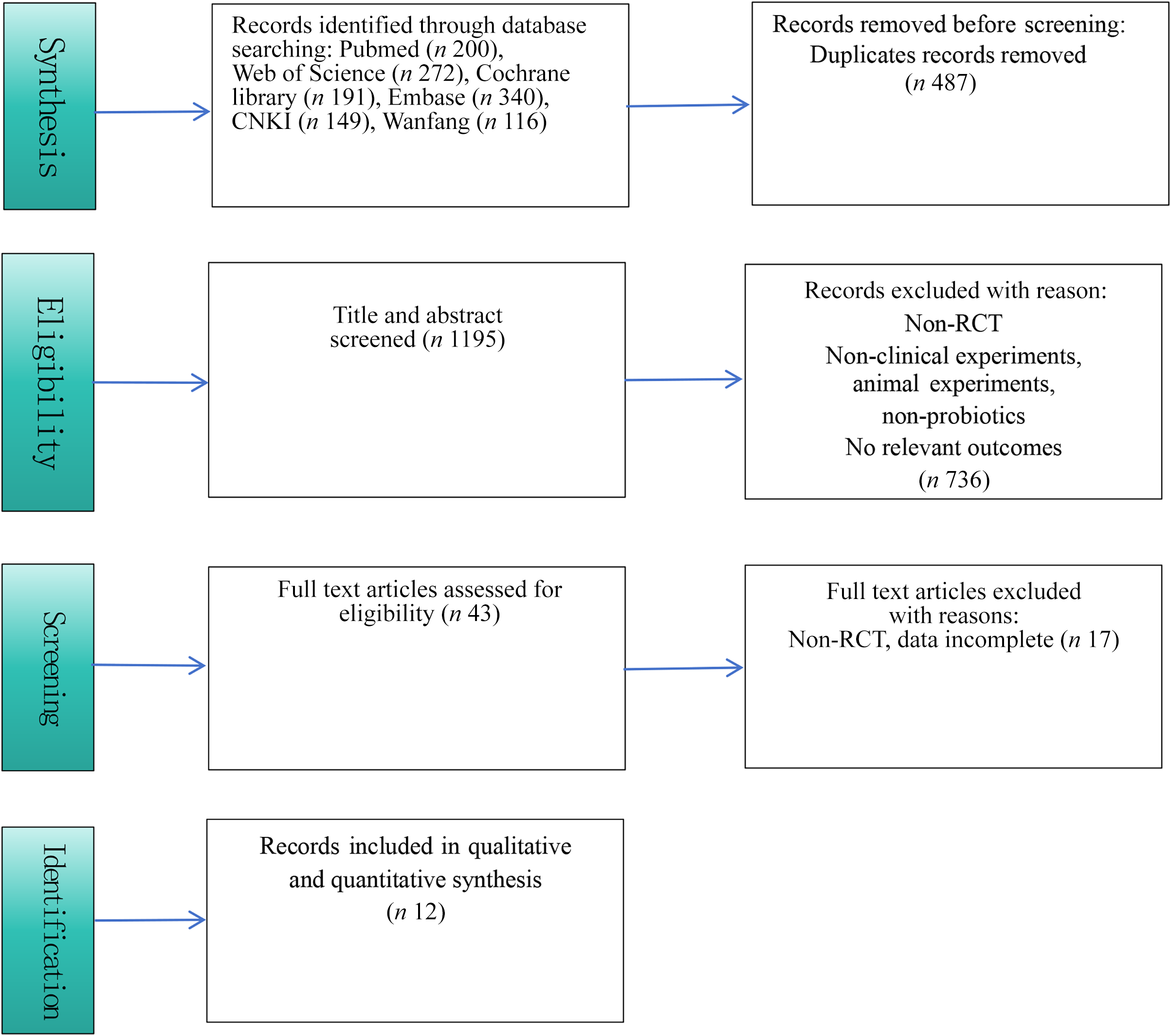
A total of 1268 studies were retrieved from the database searches. After removing 487 duplicates, 782 studies underwent title and abstract screening, which led to the exclusion of 736. Out of the remaining 46 studies, 12 were selected for inclusion after a full-text review(Reference Kassaian, Feizi and Rostami16–Reference Donghui26). The selection process is illustrated in Figure 1.
Screening flow chart.

Study characteristics
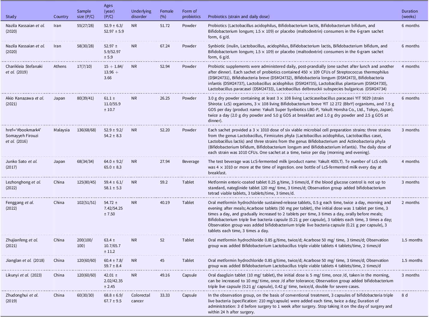
Table 1 presents the characteristics of the included studies. Eleven studies were conducted in Asia, specifically in China(Reference Zhonghong, Tingting and Feifei21–Reference Donghui26), Malaysia(Reference Firouzi, Majid and Ismail19), Iran(Reference Kassaian, Feizi and Rostami16) and Japan(Reference Kanazawa, Aida and Yoshida18,Reference Sato, Kanazawa and Azuma20) . One study was carried out in Europe (Athens)(Reference Stefanaki, Michos and Mastorakos17). The 12 randomized controlled trials involved a total of 1113 participants, allocated into probiotic (n 586) and control (n 527) groups. The mean age of participants in the intervention group ranged from 15.0 to 68.8 years, while the control group’s mean age varied from 13.96 to 67.7 years. Control interventions comprised standard treatment, hypoglycemic agents, or a placebo. Participants in the intervention group received probiotics in addition to the control treatments. Among the 12 trials, one utilized single-strain probiotics, nine employed dual or multi-strain probiotics, and two involved multi-strain probiotics combined with prebiotics. Probiotics were delivered through fermented dairy in one study, whereas the others used powder (n 5), capsules (n 3), or tablets (n 4). Daily dosages ranged from 4.0 × 10⁷ to 9.0 × 10¹¹ CFU. The median duration of probiotic intervention was 12 weeks, with a range extending from 8 d to 24 weeks.
Characteristics of included studies

Bias risk assessment results
The quality of the studies included was assessed using the Cochrane risk of bias tool. Of the 12 studies, 10 used random number tables for allocation, and 2 employed computer-generated random numbers, all categorized as low risk. One study did not conceal its allocation method, resulting in a high-risk rating. Three studies lacked details on allocation concealment and were classified as unclear risk(Reference Kanazawa, Aida and Yoshida18,Reference Zhonghong, Tingting and Feifei21,Reference Jianfeng and Ying23) . Eight studies did not report on blinding and were also considered to have an unclear risk of bias(Reference Stefanaki, Michos and Mastorakos17,Reference Kanazawa, Aida and Yoshida18,Reference Sato, Kanazawa and Azuma20–Reference Kunyi and Ge25) . All 12 studies(Reference Kassaian, Feizi and Rostami16–Reference Donghui26) provided complete outcome data and were judged as low risk. Since all studies were registered without selective reporting, they were rated as low risk. No other sources of bias were detected, leading to a low-risk rating for all studies (show in online Supplementary Figure S1a and S1b).
Effects of probiotics intervention on gastrointestinal microbiomes
Effect on Lactobacillus
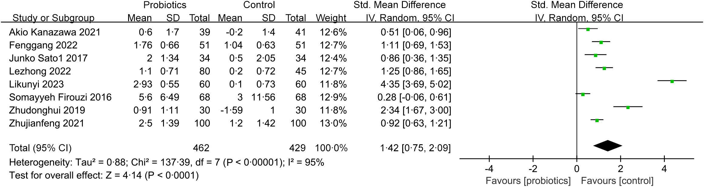
Eight RCTs(Reference Kanazawa, Aida and Yoshida18–Reference Jianfeng and Ying23,Reference Kunyi and Ge25,Reference Donghui26) involving 891 patients evaluated the effect of probiotic intervention on fecal Lactobacillus levels. Pooled data indicated a significant increase in fecal Lactobacillus levels with probiotic supplementation when compared to the control group (SMD (95 % CI): 1.42 (0.75, 2.09), P < 0.0001, I2 = 95 %) (show in Figure 2). Subgroup analyses revealed that probiotics significantly enhanced Lactobacillus levels across all regions (Asia/Europe/America), populations (prediabetes/diabetes), intervention durations (> 3 months/≤ 3 months), and age categories (> 60 years/≤ 60 years) (Table 2). Sensitivity analyses demonstrated that the effect of probiotics on Lactobacillus levels remained consistent and was not influenced by any single study (show in online Supplementary Figure S2a).
Forest plot of Change in Lactobacillus.

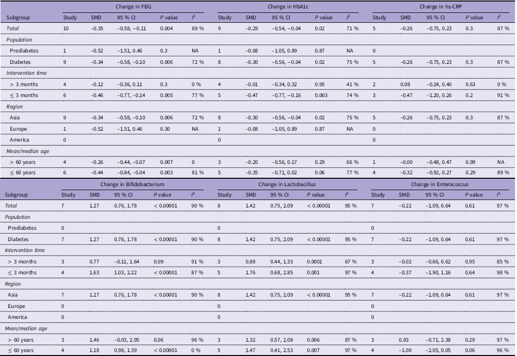
Subgroup analysis of intervention of probiotics in intestinal flora of diabetes mellitus

CI, confidence interval.
Effect on Bifidobacterium
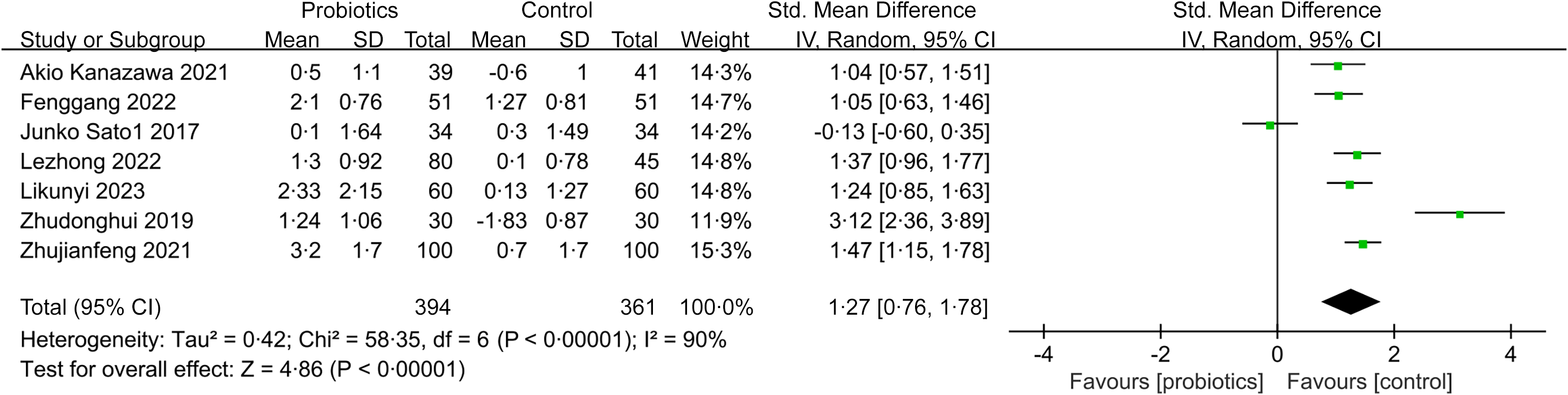
Seven RCTs(Reference Kanazawa, Aida and Yoshida18,Reference Sato, Kanazawa and Azuma20–Reference Jianfeng and Ying23,Reference Kunyi and Ge25,Reference Donghui26) involving 755 participants assessed the influence of probiotic intervention on fecal Bifidobacterium levels. Probiotic supplementation led to a significant increase in fecal Bifidobacterium levels compared to the control group (SMD (95 % CI): 1.27 (0.76, 1.78), P < 0.0001, I2 = 90 %) (show in Figure 3). Subgroup analyses indicated that probiotics did not have a significant effect on Bifidobacterium levels in groups with longer intervention durations (> 3 months) (SMD (95 % CI): 0.77 (–0.11, 1.64), P = 0.09, I2 = 91 %) or in older age groups (> 60 years) (SMD (95 % CI): 1.46 (–0.03, 2.95), P = 0.06, I2 = 96 %) (Table 2). In the younger age category (≤ 60 years), heterogeneity dropped from 90 % to 0 %, suggesting that age may be a primary source of variability. Sensitivity analyses confirmed that the effect of probiotics on Bifidobacterium levels remained stable and was not significantly affected by any single study (show in online Supplementary Figure S3).
Forest plot of Change in Bifidobacterium.

Effect on Enterococcus
Seven RCT(Reference Kanazawa, Aida and Yoshida18,Reference Sato, Kanazawa and Azuma20–Reference Jianfeng and Ying23,Reference Kunyi and Ge25,Reference Donghui26) studies involving 755 patients investigated the effect of probiotic intake on fecal Enterococcus levels. The combined effect of probiotic supplementation showed that compared with the control group, probiotics had no significant effect on the level of fecal Enterococcus (SMD (95 % CI): –0.22 (–1.09, 0.64), P = 0.61, I2 = 97 %) (online Supplementary Figure S4a). Sensitivity analysis showed that the efficacy of probiotic intervention on Enterococcus level was not significantly affected by a study, indicating that this index was relatively stable (online Supplementary Figure S4b). Subgroup analysis showed that the influence of probiotic intervention on Enterococcus level was not affected by population, region, intervention time and age.
Effects on Prevotella, Escherichia coli, enterobacterium and Bacteroides fragilis
In this study, two studies(Reference Kanazawa, Aida and Yoshida18,Reference Sato, Kanazawa and Azuma20) examined the effects of probiotic supplementation on Prevotella, three on Escherichia coli(Reference Stefanaki, Michos and Mastorakos17,Reference Zhonghong, Tingting and Feifei21,Reference Donghui26) and enterobacterium(Reference Gang, Weizhen and Jie22,Reference Jianfeng and Ying23,Reference Kunyi and Ge25) , and four on Bacteroides fragilis(Reference Kanazawa, Aida and Yoshida18,Reference Sato, Kanazawa and Azuma20,Reference Zhonghong, Tingting and Feifei21,Reference Jianfeng and Ying23) , which were included in the meta-analysis. Probiotic supplementation significantly increased Prevotella levels compared to the control group (SMD (95 % CI): 0.39 (0.06, 0.71), P = 0.02, I2 = 0 %), with no heterogeneity (online Supplementary Figure S5). Probiotics might reduce Escherichia coli levels (SMD (95 % CI): –0.65 (–1.18, –0.12), P = 0.02, I2 = 59 %) (online Supplementary Figure S6a). No significant effects were found for enterobacterium (SMD (95 % CI): –1.47 (–2.98, 0.04), P = 0.06, I2 = 98 %) (online Supplementary Figure S7) or Bacteroides fragilis (SMD (95 % CI): 0.15 (–0.86, 1.15), P = 0.78, I2 = 96 %) (online Supplementary Figure S8). While forest plots suggested that probiotics significantly increased Prevotella and reduced Escherichia coli levels, sensitivity analysis revealed that excluding two studies(Reference Stefanaki, Michos and Mastorakos17,Reference Donghui26) significantly impacted the results for Escherichia coli, indicating instability (online Supplementary Figure S6b). No sensitivity analysis was conducted for Prevotella due to insufficient literature. Subgroup analysis was not performed due to insufficient research on these four parameters.
Effects of Probiotics intervention on clinical parameters
Effect on FPG
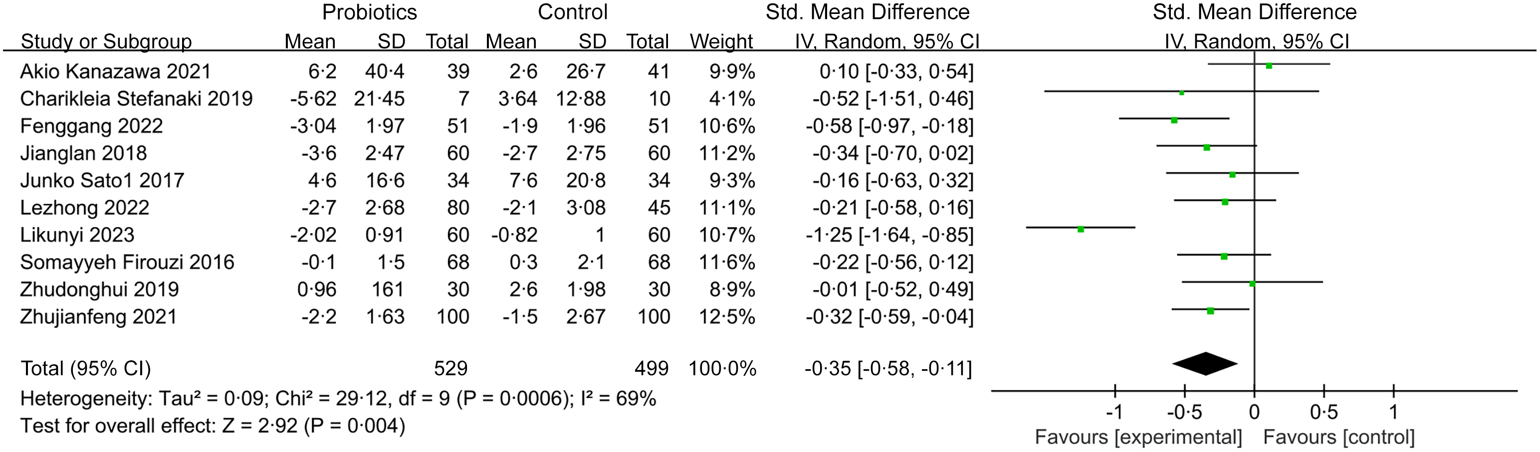
A total of ten studies(Reference Stefanaki, Michos and Mastorakos17–Reference Donghui26) involving 1028 participants investigated the impact of probiotic intervention on fasting plasma glucose (FPG) levels. The results indicated that probiotic supplementation significantly lowered FPG levels when compared to the control group (SMD (95 % CI): –0.35 (–0.58, –0.11), P = 0.004, I2 = 69 %) (show in Figure 4). Subgroup analyses revealed no significant effect of probiotics on FPG levels in long-term interventions (> 3 months) (SMD (95 % CI): –0.12 (–0.36, 0.11), P = 0.3, I2 = 0 %), in studies conducted in Europe (SMD (95 % CI): –0.52 (–1.51, 0.46), P = 0.3, I2 = NA), or among participants with prediabetes (SMD (95 % CI): –0.52 (–1.51, 0.46), P = 0.3, I2 = NA). Notably, heterogeneity decreased to 0 % in subgroup analyses for interventions lasting more than three months and participants over 60 years, indicating that heterogeneity was primarily associated with intervention duration and age (Table 2). Sensitivity analyses demonstrated that no individual study had a substantial impact on the probiotics’ effect on FPG levels, confirming that the observed improvement is both significant and stable (show in online Supplementary Figure S9).
Forest plot of Change in FBG.

Effect on HbA1c
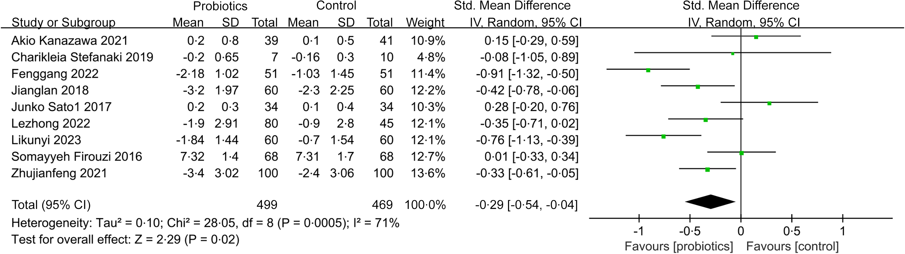
Nine studies(Reference Stefanaki, Michos and Mastorakos17–Reference Kunyi and Ge25) involving 970 participants assessed the impact of probiotic intervention on HbA1c levels. The forest plot indicated that probiotic supplementation led to a reduction in HbA1c levels compared to the control group (SMD (95 % CI): –0.29 (–0.54, –0.04), P = 0.02, I2 = 71 %) (show in Figure 5). Subgroup analyses revealed no significant effects of probiotics on HbA1c levels among participants in Europe (SMD (95 % CI): –0.08 (–1.05, 0.89), P = 0.87, I2 = NA), those with prediabetes (SMD (95 % CI): –0.08 (–1.05, 0.89), P = 0.87, I2 = NA), or in studies with interventions exceeding three months (SMD (95 % CI): –0.01 (–0.34, 0.32), P = 0.95, I2 = 41 %). The heterogeneity for interventions longer than three months decreased from 71 % to 41 %, indicating that duration was a significant contributor (Table 2). Sensitivity analyses showed that removing five studies(Reference Zhonghong, Tingting and Feifei21–Reference Kunyi and Ge25) made the effect on HbA1c levels significant, indicating result instability (show in online Supplementary Figure S10).
Forest plot of Change in HbA1c.

Effects on 1L-6, hsCRP, BMI, 2hPBG, T-CHO, TG and HDL-C
Four trials investigated the impact of probiotic supplementation on levels of IL-6(Reference Kanazawa, Aida and Yoshida18,Reference Sato, Kanazawa and Azuma20,Reference Gang, Weizhen and Jie22,Reference Kunyi and Ge25) , BMI(Reference Stefanaki, Michos and Mastorakos17–Reference Sato, Kanazawa and Azuma20), TG(Reference Kanazawa, Aida and Yoshida18–Reference Sato, Kanazawa and Azuma20,Reference Jianfeng and Ying23) , and HDL-C(Reference Kanazawa, Aida and Yoshida18–Reference Sato, Kanazawa and Azuma20,Reference Jianfeng and Ying23) . Additionally, three studies focused on 2hPBG(Reference Gang, Weizhen and Jie22,Reference Lan, Guo and Chenlin24,Reference Kunyi and Ge25) and T-CHO(Reference Kanazawa, Aida and Yoshida18–Reference Sato, Kanazawa and Azuma20), while five trials examined hsCRP levels(Reference Kanazawa, Aida and Yoshida18–Reference Sato, Kanazawa and Azuma20,Reference Gang, Weizhen and Jie22,Reference Kunyi and Ge25) . The forest plot indicated a significant reduction in IL-6 levels with probiotics compared to controls (SMD (95 % CI): –0.31 (–0.57, –0.06), P = 0.02, I2 = 34 %), showing low heterogeneity (show in online Supplementary Figure S11a). However, no significant effects were observed for hsCRP (SMD (95 % CI): –0.26 (–0.75, 0.23), P = 0.30, I2 = 87 %) (show in online Supplementary Figure S12a), BMI (SMD (95 % CI): –0.45 (–1.19, 0.29), P = 0.24, I2 = 88 %) (show in online Supplementary Figure S13), 2hPBG (SMD (95 % CI): –0.05 (–1.34, 1.24), P = 0.94, I2 = 97 %) (show in online Supplementary Figure S14), T-CHO (SMD (95 % CI): –0.17 (–0.40, 0.07), P = 0.16, I2 = 0 %) (show in online Supplementary Figure S15), TG (SMD (95 % CI): –0.12 (–0.42, 0.17), P = 0.42, I2 = 61 %) (show in online Supplementary Figure S16), or HDL-C (SMD (95 % CI): –0.13 (–0.31, 0.05), P = 0.15, I2 = 0 %) (show in online Supplementary Figure S17). Sensitivity analysis revealed that omitting three studies(Reference Stefanaki, Michos and Mastorakos17,Reference Gang, Weizhen and Jie22,Reference Kunyi and Ge25) significantly impacted the effect of probiotics on IL-6, indicating result instability (show in online Supplementary Figure S11b). No individual study had a substantial impact on the probiotics’ effect on hsCRP levels showed in Sensitivity analyses,confirming that the result is stable (show in online Supplementary Figure S12b).
Subgroup analyses based on region (Asia/Europe/America), duration of intervention (> 3 months/≤ 3 months), and age (> 60/≤ 60 years) found no significant effect of probiotics on hsCRP levels. Notably, heterogeneity for hsCRP in interventions exceeding three months dropped to 0 %, suggesting a potential link to duration (Table 2). Due to a lack of sufficient studies, subgroup analysis for other indicators was not conducted.
Publication bias analysis
The evaluation of publication bias was conducted using a funnel plot alongside Egger’s test in studies examining the effects of probiotics on gut microbiota and clinical parameters related to diabetes. The funnel plot indicated a presence of publication bias for Lactobacillus (see online Supplementary Figure S2b), while Egger’s test yielded a P-value of 0.047, suggesting mild bias. For other indicators, no evidence of publication bias was found: Bifidobacterium (P = 0.668), Enterococcus (P = 0.476), Escherichia coli (P = 0.445), Enterobacterium (P = 0.290), Bacteroides fragilis (P = 0.386), FPG (P = 0.927), HbA1c (P = 0.633), IL-6 (P = 0.301), hsCRP (P = 0.974), BMI (P = 0.458), 2hPBG (P = 0.240), T-CHO (P = 0.486), TG (P = 0.332), and HDL-C (P = 0.836).
Discussion
The relationship between changes in gut microbiota and the pathogenesis of type 2 diabetes mellitus (T2DM) has garnered considerable attention in recent research. Dysbiosis of gut microbiota is strongly associated with the pathophysiology of diabetes(Reference Kena and Hanmei27–Reference Cani and Delzenne30). A review by Gurung M et al. (Reference Gurung, Li and You7) analyzed 42 studies and revealed that approximately half of T2DM patients exhibited a reduction in at least one of the following species: Bacteroides, Bifidobacterium, Roseburia, Faecalibacterium, and Akkermansia muciniphila. The gut microbiota plays a critical role in regulating inflammation, intestinal permeability, and glucose metabolism, with Bifidobacterium, Bacteroides, and Actinobacteria being particularly relevant to T2DM. Furthermore, studies using animal models support the association between gut microbiota and T2DM(Reference Kieler, Osto and Hugentobler31). Mocanu V et al. (Reference Mocanu, Zhang and Deehan32) found that modulating gut microbiota can enhance insulin sensitivity in individuals with metabolic dysfunction.
Probiotics, when administered in sufficient quantities, are beneficial microorganisms that promote host health. Their use to modulate gut microbiota and influence glucose metabolism has been extensively applied in managing and preventing type 2 diabetes mellitus (T2DM) and its associated complications. Probiotics generally help manage diabetes by altering the composition of gut microbiota and their metabolic byproducts(Reference Gurung, Li and You7,Reference Cani33) . Kanazawa A(Reference Kanazawa, Aida and Yoshida18) reported that administering a synthetic probiotic blend, consisting of Lacticaseibacillus paracasei strain Shirota, Bifidobacterium breve strain Yakult, and galactooligosaccharides, over a period of 24 weeks led to significant increases in Bifidobacterium, Prevotella, total lactic acid bacteria, Lactobacillus, and fecal organic acid concentrations in obese T2DM patients, while Akkermansia levels decreased. These changes in the gut environment contribute to improvements in glucose metabolism in individuals with obese T2DM. A study by Hsieh MC et al. (Reference Hsieh, Tsai and Jheng34) demonstrated that supplementation with Lactobacillus reuteri strains ADR-1 and ADR-3 led to significant reductions in HbA1c levels and at least an eight-fold increase in fecal Lactobacillus reuteri. The findings also showed a positive correlation between increased Lactobacillus reuteri and the abundance of Bifidobacterium and Lactobacillus. Zhang C et al. (Reference Zhang, Jiang and Wang11) conducted a meta-analysis, concluding that probiotic supplementation did not significantly lower HbA1c levels in individuals with type 2 diabetes (T2DM). However, they observed significant reductions in fasting plasma glucose (FPG) and fasting insulin levels. The improvements in glycemic control were more substantial with multi-strain and higher-dose probiotic formulations compared to single-strain or lower-dose probiotics. Similarly, Naseri K et al. (Reference Naseri, Saadati and Ashtary-Larky9) performed a meta-analysis of 46 randomized controlled trials involving 3067 participants, with 1644 receiving the intervention and 1423 serving as controls. Their findings indicated that both probiotics and synbiotics were linked to better metabolic outcomes in patients with prediabetes and T2DM, notably reducing FPG, HbA1c, and fasting insulin levels. Furthermore, a meta-analysis by Rittiphairoj et al. (Reference Rittiphairoj, Pongpirul and Janchot35), which included 28 studies, demonstrated that probiotics may lower blood glucose levels in T2DM patients, although they do not appear to influence HbA1c levels. The reasons behind these inconsistencies remain uncertain, but they may be influenced by factors such as patient age, ethnicity, dietary patterns, and the specific conditions under which probiotics are administered. Earlier meta-analyses mostly concentrated on how probiotic supplementation affected metabolic markers associated with diabetes and certain clinical parameters, such as inflammatory mediators and intestinal permeability, with less focus on the direct influence of probiotics on gut microbiota composition. To better understand how probiotics regulate the gut microbiota, we examined data from 12 randomized controlled trials, encompassing 1113 participants (586 receiving probiotics and 527 in the control group), focusing on probiotic-induced changes in gut microbiota.
Subgroup analysis showed that short-term interventions (≤ 3 months) had a more substantial impact on improving fasting plasma glucose (FPG), HbA1c, and Bifidobacterium levels. Furthermore, younger individuals (≤ 60 years) exhibited a more notable increase in Bifidobacterium levels following probiotic treatment. This finding echoes the molecular mechanism by which bifidobacteria inhibit DPP-4 activity reported in previous studies, which can prolong the half-life of GLP-1, thereby partially explaining the better blood glucose control in young patients. A significant reduction in FPG was also seen with short-term interventions (≤ 3 months), whereas no significant difference was noted in the long-term intervention group (> 3 months), suggesting that extended treatment might not yield better results. This raises the possibility that negative effects of probiotics may be involved, a point that requires further validation through large-scale, multicenter clinical trials. In regional and population subgroups, no significant reduction in FPG levels was observed following probiotic use in Europe or among prediabetic individuals. However, as this finding is based on only one study, its reliability is questionable, and additional clinical research across different regions and prediabetic populations is necessary for confirmation. Examining methodological variability across included studies, this study found heterogeneity primarily due to probiotic strain diversity (seven different Lactobacillus species), dose variations, dosage form variations (powder/capsule/drink), and a wide range of intervention durations (8 d to 24 weeks). Despite the use of a random-effects model and subgroup-stratified analyses, heterogeneity remained high, suggesting the need for standardized strain selection and dosing regimens in future studies.
For HbA1c, probiotic intervention resulted in significant improvement within the subgroups of Asia, diabetes, and short-term interventions of ≤ 3 months (Table 2). However, no significant change was observed in the subgroups of Europe, prediabetes, and long-term interventions lasting > 3 months. These results help clarify discrepancies in previous research, suggesting that the efficacy of probiotic treatment may depend on factors such as geographic region, specific populations, and intervention duration. Therefore, identifying the most suitable population and conditions for probiotic therapy is essential for its effective clinical application and achieving precise treatment outcomes. Nonetheless, the limited number of studies focusing on European and prediabetic populations is inadequate to establish firm conclusions, and In the future, multicenter RCTs targeting European populations (with particular focus on patients with a Mediterranean diet background) and the expansion of dedicated studies on prediabetic populations are needed to make up for the representativeness deficiencies of current subgroup analyses. The heterogeneity between studies was substantial (I2 = 71 %). Subgroup analysis indicated that in the long-term intervention group (> 3 months), I2 decreased to 41 %, suggesting that heterogeneity is mainly attributed to the intervention duration. However, sensitivity analyses revealed that HbA1c results are unstable, suggesting that this indicator is susceptible to differences in testing methods and variability in follow-up time. In future clinical applications, more stable indicators such as FPG should be prioritized. Future studies also need to standardize HbA1c testing standards and establish core follow-up timelines. A meta-analysis by Wang X et al. (Reference Xiaofang, Xin and Huiqin29) found that probiotics had a beneficial impact on blood glucose regulation in patients with T2DM, particularly by improving HbA1c within 6–8 weeks and regulating insulin and HOMA-IR between 6–8 weeks and 12–24 weeks, which aligns with our results.
Research on the molecular mechanisms behind probiotic interventions in T2DM remains somewhat limited. Mechanistically, probiotics help mitigate T2DM-related pathologies by repairing the intestinal barrier, reducing inflammation, alleviating oxidative stress, restoring energy metabolism, and producing beneficial metabolites such as short-chain fatty acids (SCFAs) and bile acids (BAs)(Reference Zhai, Wu and Lam36). Bajinka O et al. (Reference Bajinka, Sylvain Dovi and Simbilyabo37) reviewed experimental studies from 2016 to 2022 and concluded that probiotics may regulate gut microbiota, enhance SCFA and glucagon-like peptide production, inhibit α-glucosidase, raise SIRT1 levels, decrease fetuin-A, and modulate inflammatory cytokines. Lactobacillus and Bifidobacterium, among other probiotics, have been thoroughly investigated in diabetic animal models for their influence on glucose intolerance and insulin resistance (IR). Supplementing with Lactobacillus species helps reshape the gut microbiota, correcting dysbiosis and promoting a balanced microbial environment with a greater abundance of beneficial bacteria(Reference Rastogi and Singh38). For example, Lactobacillus plantarum Ln4 enhances oral glucose tolerance tests (OGTT), insulin tolerance tests (ITT), and the homeostasis model for assessing insulin resistance (HOMA-IR) in mice fed a high-fat diet, helping to reduce both weight gain and insulin resistance(Reference Lee, Jung and Lee39).Yan F(Reference Yan, Li and Shi40) explored the anti-diabetic properties of Lactobacillus acidophilus in mice with type 2 diabetes mellitus (T2DM) and its effects on the gut microbiota. The findings revealed that six weeks of oral administration of Lactobacillus acidophilus markedly enhanced epithelial barrier integrity, lowered inflammatory markers such as IL-8, TNF-α, and IL-1β in liver and colon tissues, and influenced the expression of genes involved in glucose and lipid metabolism. More specifically, it reduced glycogen synthase kinase 3β (GSK-3β), fatty acid synthase (FAS), and sterol regulatory element-binding transcription factor 1c (SREBP-1c), while increasing the activity of protein kinase B (Akt). Further studies indicated that L. acidophilus KLDS1.0901 can remodel the gut microbiota, increasing the prevalence of short-chain fatty acid (SCFA)-producing bacteria (Blautia, Roseburia, and Anaerotruncus) and SCFA concentrations, while simultaneously reducing the abundance of Gram-negative bacteria (Desulfovibrio, Alistipes, and Bacteroides). Selenium-enriched Bifidobacterium longum has been demonstrated to delay the onset of STZ-induced diabetes and alleviate renal damage caused by hyperglycemia(Reference Lin, Ren and Zhang41). Longgum DD98 and selenium-fortified B. longum DD98 were found to lower fasting blood glucose (FBG) and HbA1c levels in high-fat diet (HFD) and STZ-induced diabetic mice, while also improving glucose tolerance(Reference Zhao, Zhu and Gao42). Moreover, inactivated Clostridium butyricum BR-108 has been reported to reduce blood glucose levels in the Tsumura Suzuki obese diabetic (TSOD) mouse model. B. animalis 01 enhances OGTT and HOMA-IR indices in HFD and STZ-induced diabetic rats and suppresses pro-inflammatory factors(Reference Ben Othman and Sakamoto43).
This investigation represents the first systematic review and meta-analysis assessing the actual impacts of probiotic interventions on gut microbiome composition in diabetes. It also seeks to identify the most suitable target populations and conditions for probiotic therapy in this context. By conducting a comprehensive analysis of sources of heterogeneity, this study offers evidence-based support for subsequent clinical trials and aims to enhance precision treatment via probiotic interventions. Nevertheless, several limitations exist within this study. Our analysis revealed notable statistical heterogeneity, likely resulting from differences in methodologies (varied study designs), treatment protocols (types, doses, viability, administration routes of probiotics, and duration of interventions), as well as the types of interventions (probiotics and/or synbiotics). While most studies exhibited a lack of transparency regarding blinding and allocation methods, as shown in the quality assessment charts, this did not considerably influence our findings. Furthermore, the limited number of studies focusing on prediabetic individuals and European populations, along with several small-sample, single-center studies, may introduce publication bias and regional selection bias. These factors could impact the reliability of the results from the current meta-analysis. Therefore, future investigations should aim to include well-designed, multicenter, large-sample randomized controlled trials to furnish more robust evidence. In addition, future research should first develop high-potential strains (such as B. adolescentis with DPP-4 inhibitory activity), secondly optimize strain-prebiotic synergistic formulas, and ultimately establish personalized intervention strategies based on intestinal flora typing.
Conclusion
In conclusion, our investigation indicated that probiotic interventions lead to significant improvement in fecal Lactobacillus and Bifidobacterium levels, as well as fasting plasma glucose (FPG) in individuals with diabetes. Subgroup analyses highlighted that probiotics significantly improved gut microbiota and FPG in Asian patients with diabetes aged ≤ 60 years. However, intervention for more than three months did not significantly improve FPG/HbA1c, and Bifidobacterium did not significantly increase in those aged > 60 years. A short-term intervention strategy of ≤ 3 months is recommended for young and middle-aged Asian patients. However, long-term efficacy and strain-specific effects require further verification. Due to the small sample size, the predominance of Asian populations, and the variability among studies, further large-scale, prospective research is necessary to validate our results.
Supplementary material
For supplementary material/s referred to in this article, please visit https://doi.org/10.1017/S0007114525105709
Acknowledgements
Not applicable. National Natural Science Foundation of China (Grant No. U22A20376). National Natural Science Foundation of China (82204977). All authors contributed to the study conception and design. L. H. and Y. Z.: Conceptualization, Methodology, Software, Writing- Original draft, Data curation, Visualization were performed; W. Y., M. J., K. Z., S. C. and H. H.: Investigation, Writing - Original Draft, Writing - Reviewing and Editing were performed; S. X., Z. Y. and C. L.: Conceptualization, Supervision, Project administration were performed. All authors read and approved the final manuscript. The authors declare that there are no conflicts of interest. The data used to support the findings of this study are included within the article. Ethics approval and consent to participate: Not applicable. Consent for publication: Not applicable.